


BIOBRIEF
Ridge Augmentation and Delayed Implant Placement on an Upper Lateral Incisor
THE SITUATION
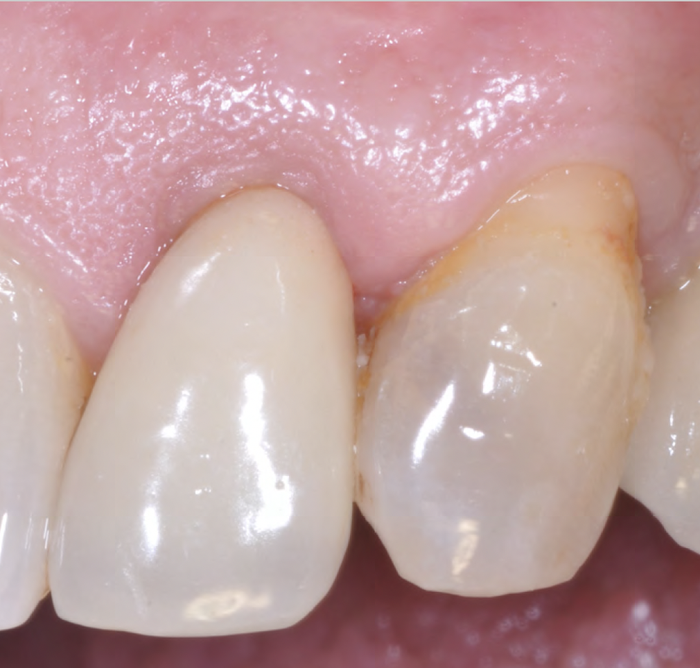
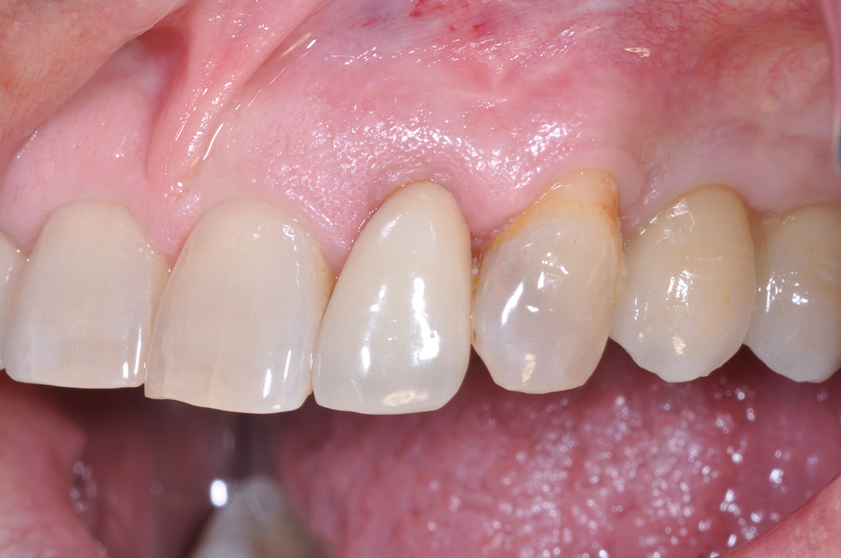
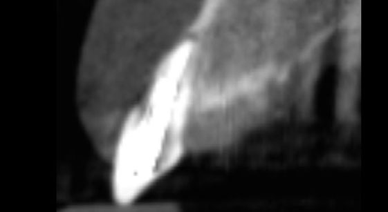
An adult female patient presented with an endodontic/prosthetic failure on the maxillary left lateral incisor. The patient‘s request was to have a definitive implant-supported single crown. The clinical situation revealed recession of the free gingival margin, while the CBCT evaluation showed the missing buccal bone plate, which contra-indicated an immediate implant placement. The treatment plan included a staged approach with a ridge augmentation procedure at the time of tooth extraction, in order to recreate the buccal bone plate and reduce the gingival recession. By moving the free gingival margin, keratinized tissue was gained through an open-healing approach.
THE RISK PROFILE
| Low Risk | Medium Risk | High Risk | |
|---|---|---|---|
| Patient’s health | Intact immune system Non-smoker | Light smoker | Impaired immune system |
| Patient’s esthetic requirements | Low | Medium | High |
| Height of smile line | Low | Medium | High |
| Gingival biotype | Thick – “low scalloped” | Medium – “medium scalloped” | Thin – “high scalloped” |
| Shape of dental crowns | Rectangular | Triangular | |
| Infection at implant sight | None | Chronic | Acute |
| Bone height at adjacent tooth site | ≤ 5 mm from contact point | 5.5 – 6.5 mm from contact point | ≥ 7 mm from contact point |
| Restorative status of adjacent tooth | Intact | Restored | |
| Width of tooth gap | 1 tooth (≥ 7 mm) | 1 tooth (≤ 7 mm) | 2 teeth or more |
| Soft-tissue anatomy | Intact | Compromised | |
| Bone anatomy of the alveolar ridge | No defect | Horizontal defect | Vertical defect |
THE APPROACH
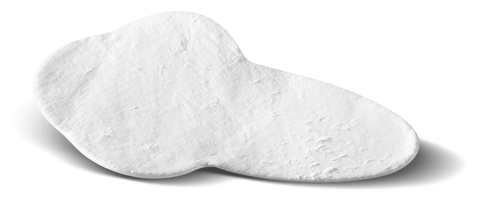
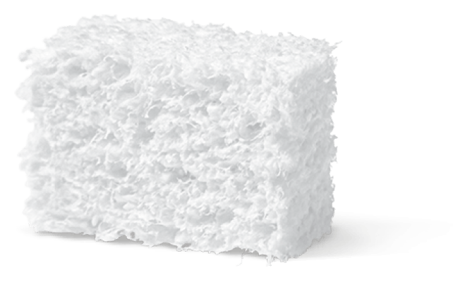
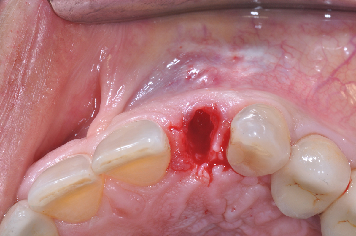
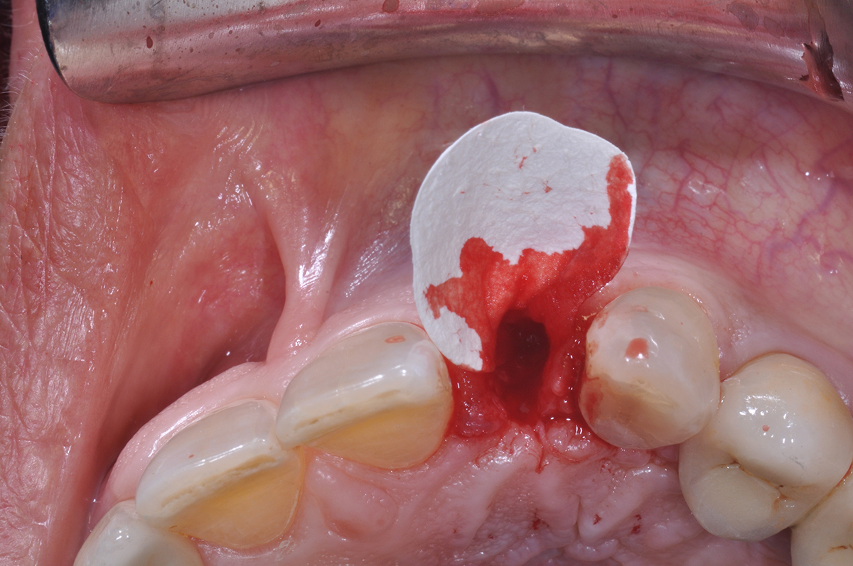
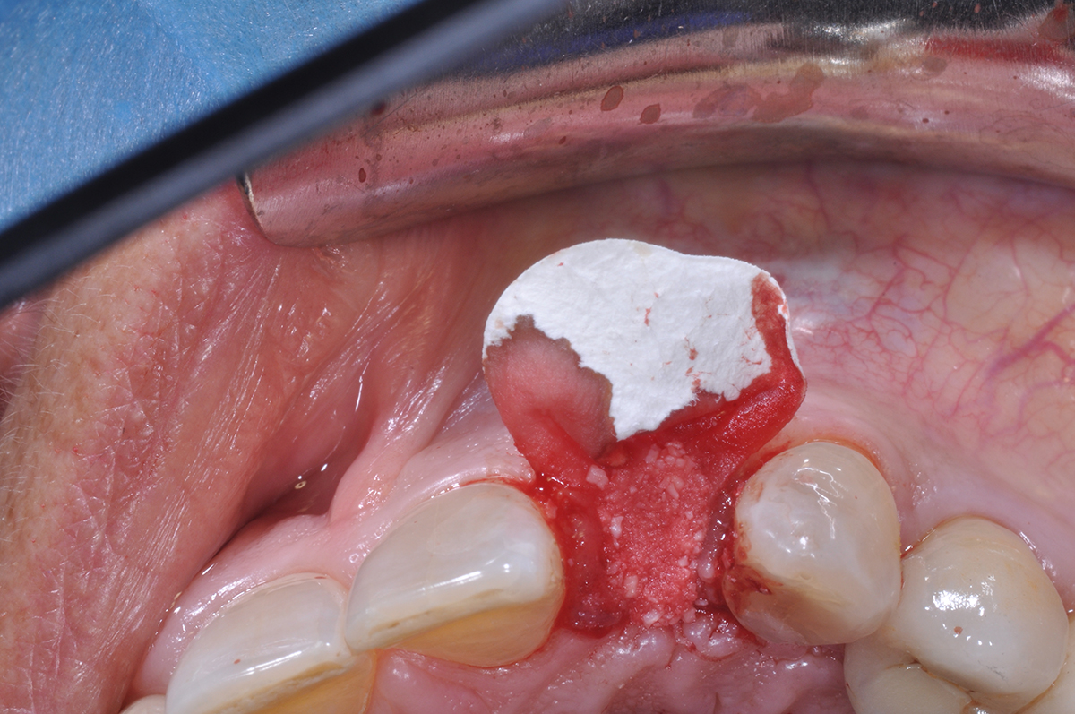
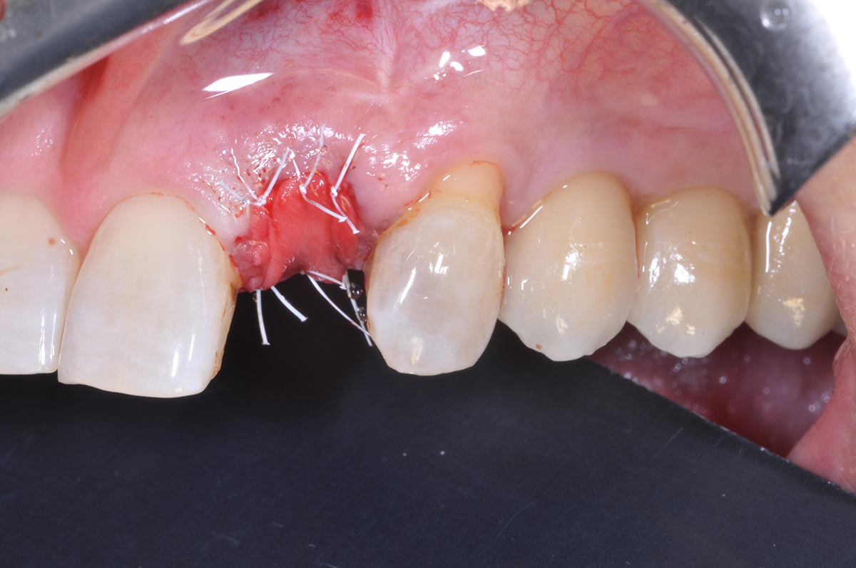
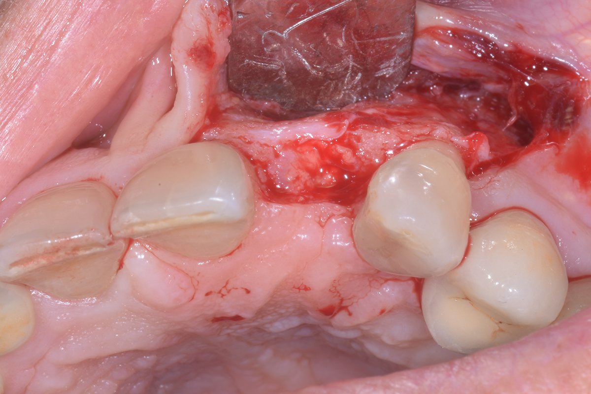
The treatment goals were to improve the soft-tissue levels and regenerate the buccal bone plate. After performing a flapless extraction procedure, a specifically designed resorbable bilayer collagen membrane, Geistlich Bio-Gide® Shape, was inserted into the socket with the long wing in contact with the buccal surface and the smooth, compact upper layer facing outward. The alveolus was then grafted with Geistlich Bio-Oss Collagen®. The three smaller wings of the membrane were folded on top of the graft material and sutured to the surrounding soft-tissue, allowing for open-healing.
“The patient had a failing crown with compromised soft tissue and requested a single crown rehabilitation with improved esthetics.”
THE OUTCOME
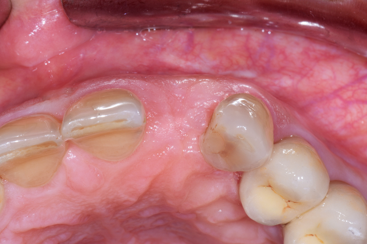
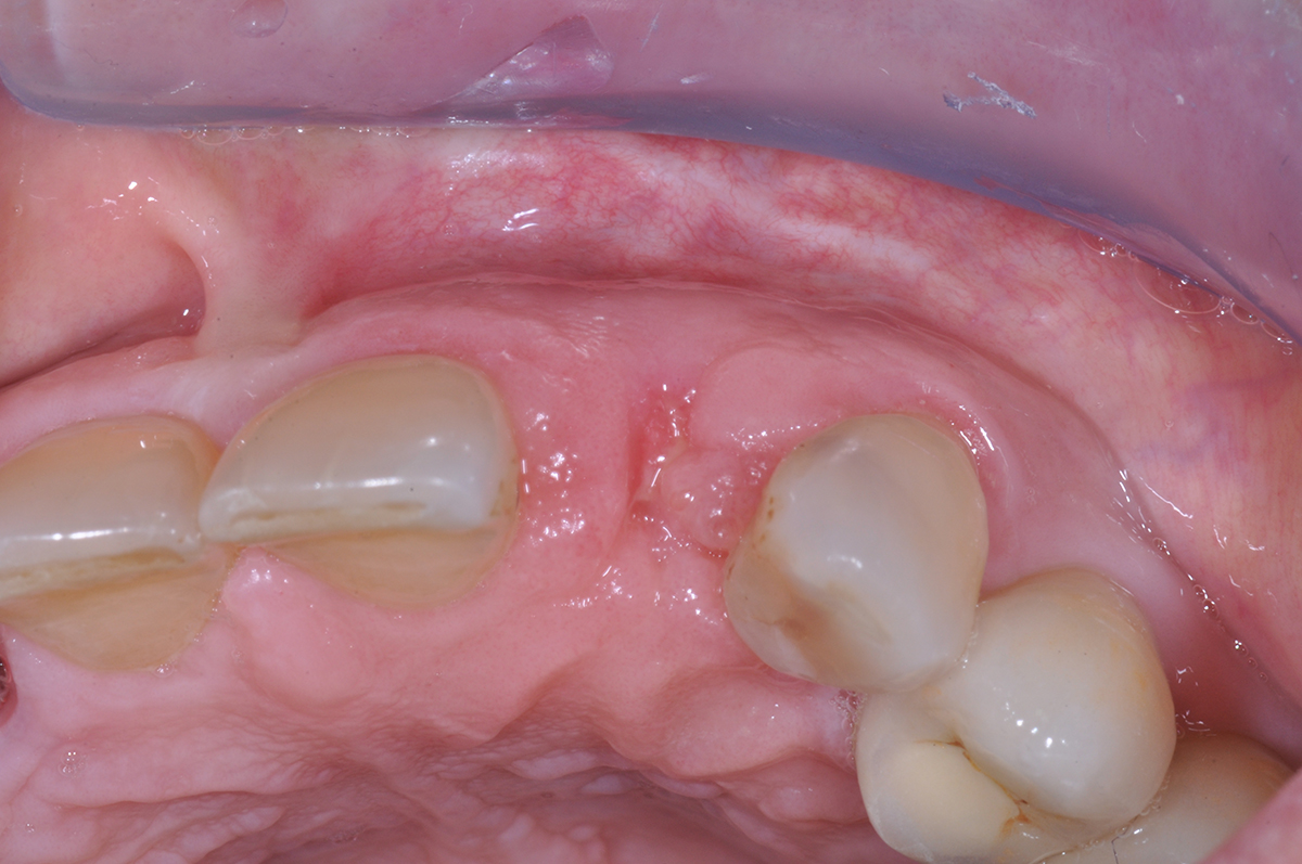
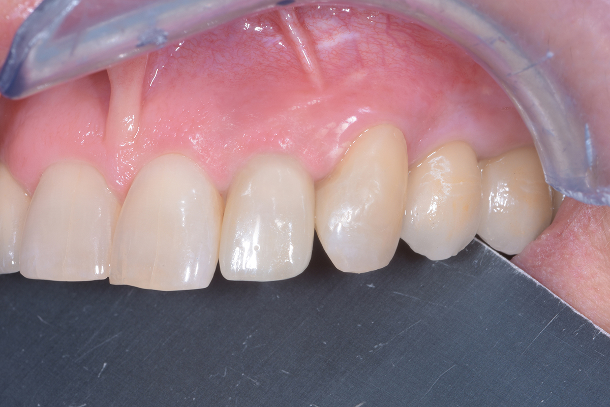
This case demonstrates how it is possible to improve the clinical and esthetic situation that was presented at baseline. Despite missing the buccal bone plate and the recession of the free gingival margin, the ridge augmentation procedure performed with the combination of Geistlich Bio-Gide® Shape and Geistlich Bio-Oss Collagen® was able to create a positive volume of the ridge, allowing for a prosthetically guided implant placement.
Dr. Daniele Cardaropoli
Periodontist – PRoED, Institute for Professional Education in Dentistry, Torino
Doctor of Dentistry and Certificate in Periodontology from the University of Torino, Italy.
Active member of the Italian Society of Periodontology, European Federation of Periodontology, Italian Academy of osseointegration and Academy of osseointegration. International member of the American Academy of Periodontology. Scientific Director of Institute for Professional Education in Dentistry (PRoED), Torino. Member of the Editorial Board of The International Journal of Periodontics and Restorative Dentistry. Private practice in Torino, Italy.