Buy products 5, get 1 free. Use code b5g1. Check it out!
Sale-a-Bration Promotion: Geistlich Biomaterials – Buy 4: Get 1, Buy 20: Get 6, Buy 50: Get 20, Buy 100: Get 45 | Use code Q4FLASH26 Check it out!
Clinical Resources
Use the search and filter form below to find informative clinical content by format, therapeutic areas, or product.

FEATURED RESOURCE
BIOBRIEF
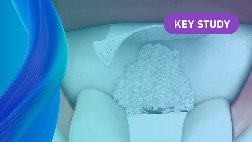
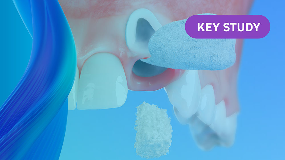
Alveolar Ridge Preservation with vallos® Mineralized Cortico-Cancellous Allograft
Hanae Saito, DDS, MS, CCRC
, Andrew Tong, DDS
On-Demand Webinars
Learn on your own Time and Earn CE
Geistlich is committed to educating our clinician partners in regenerative dentistry through in-person and virtual sessions. We look forward to growing with you on this journey.
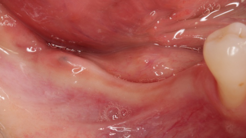
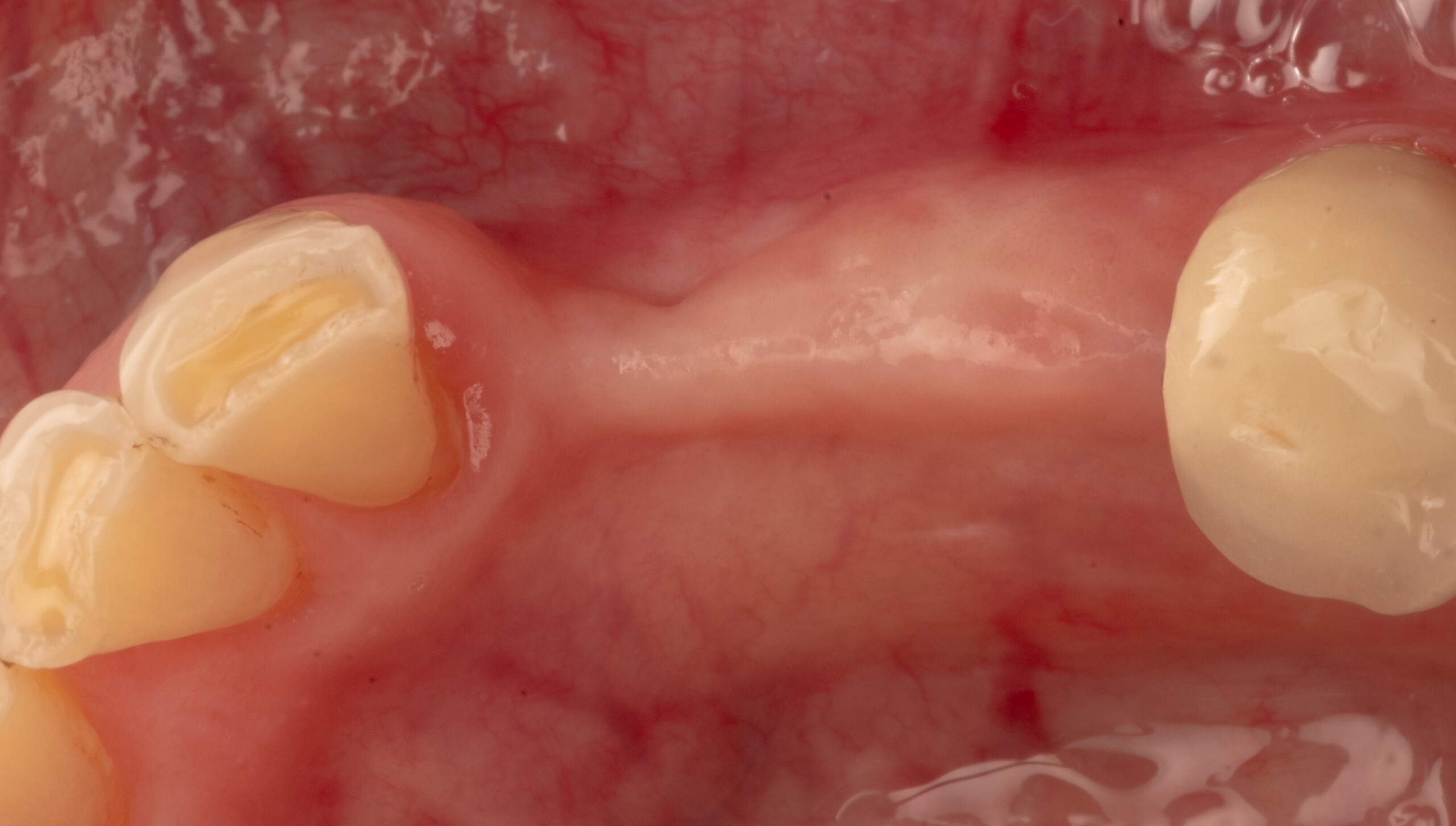
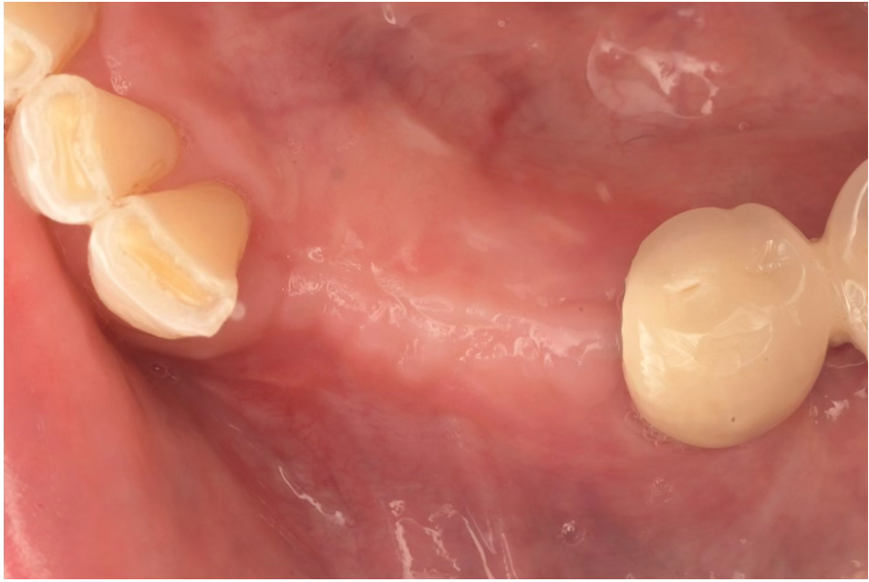
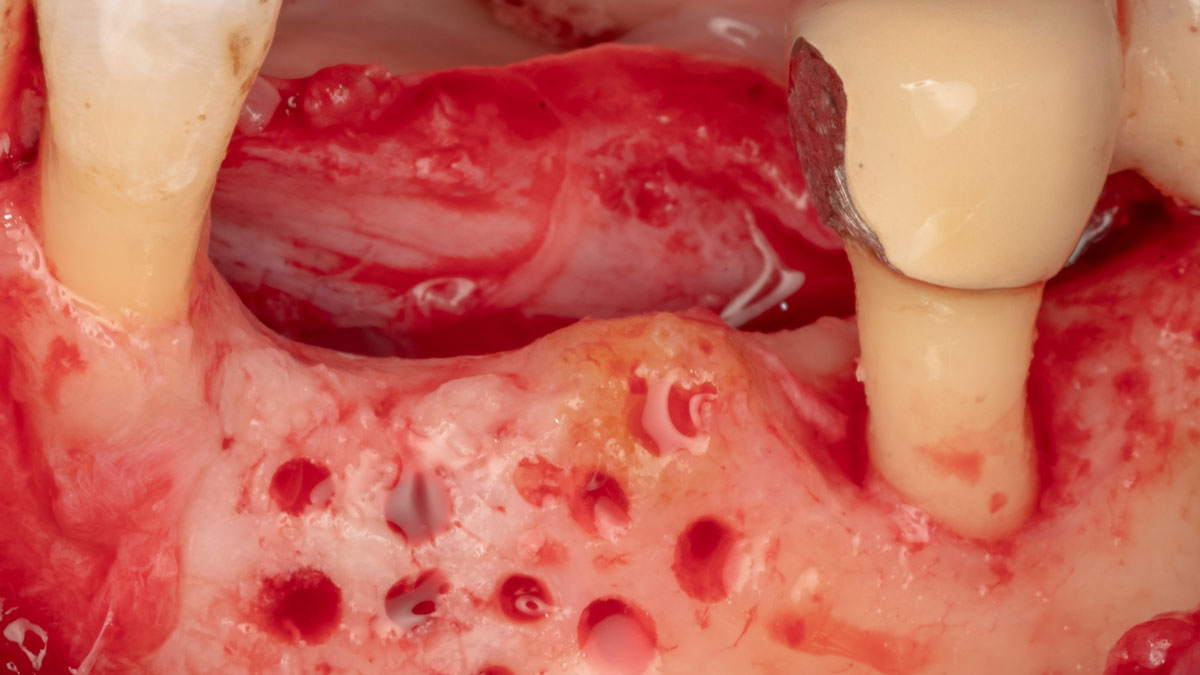
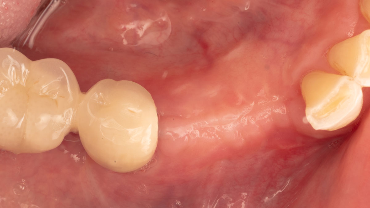
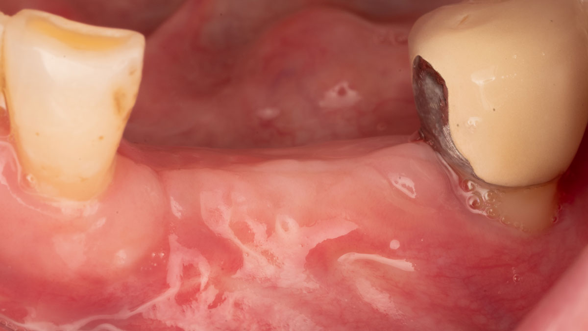
Initial clinical presentation showing the reduced ridge width.
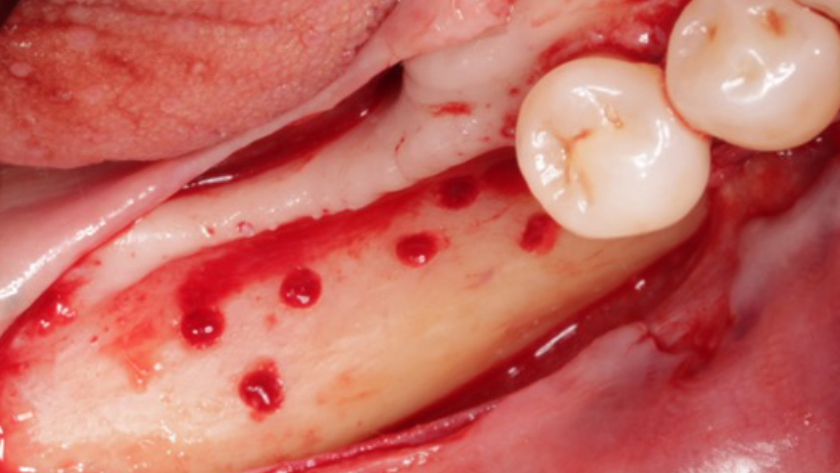
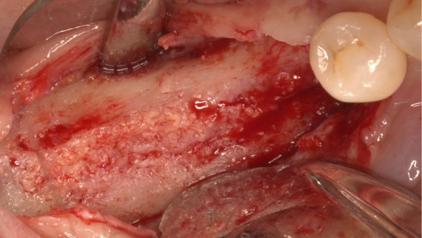
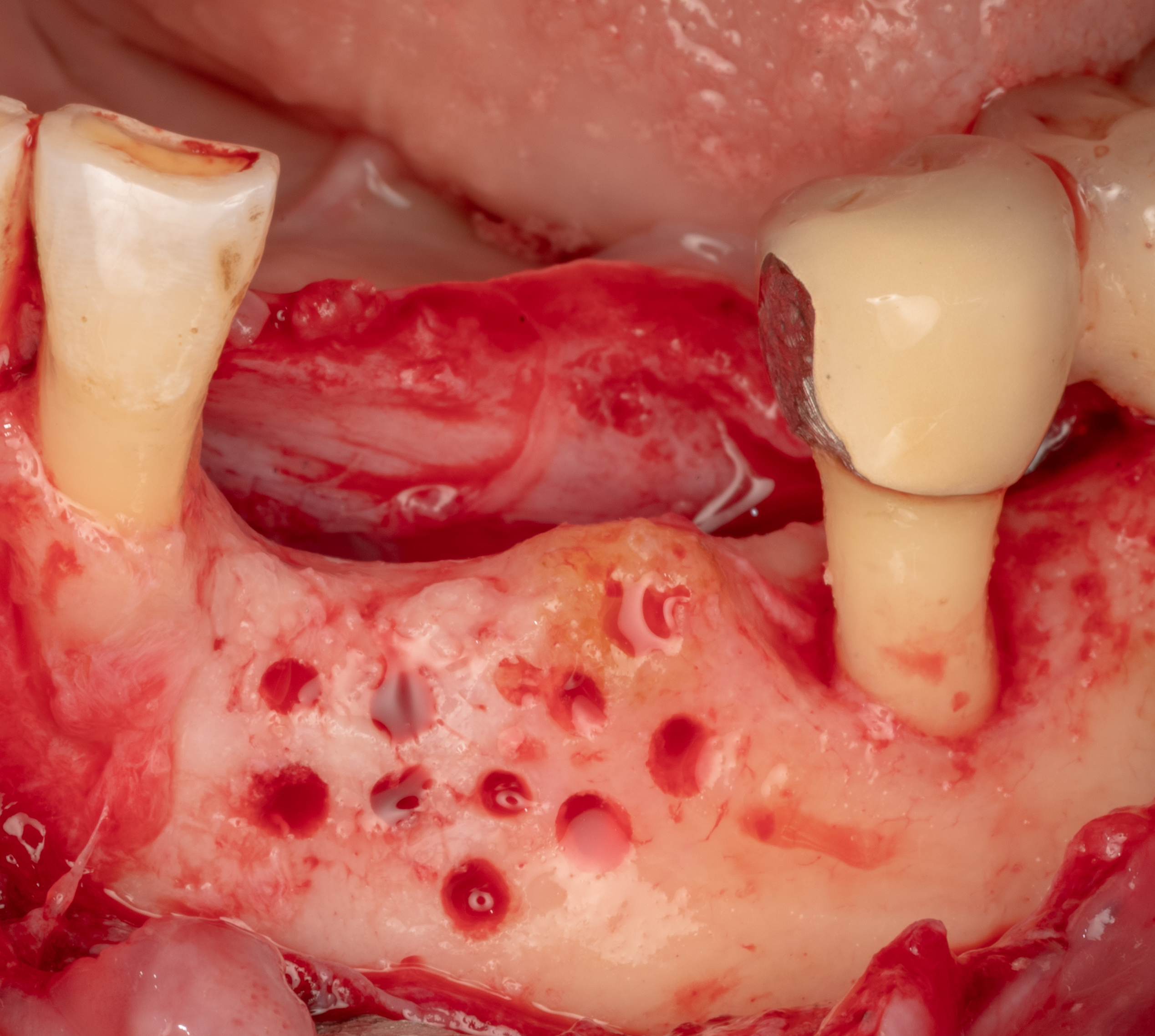
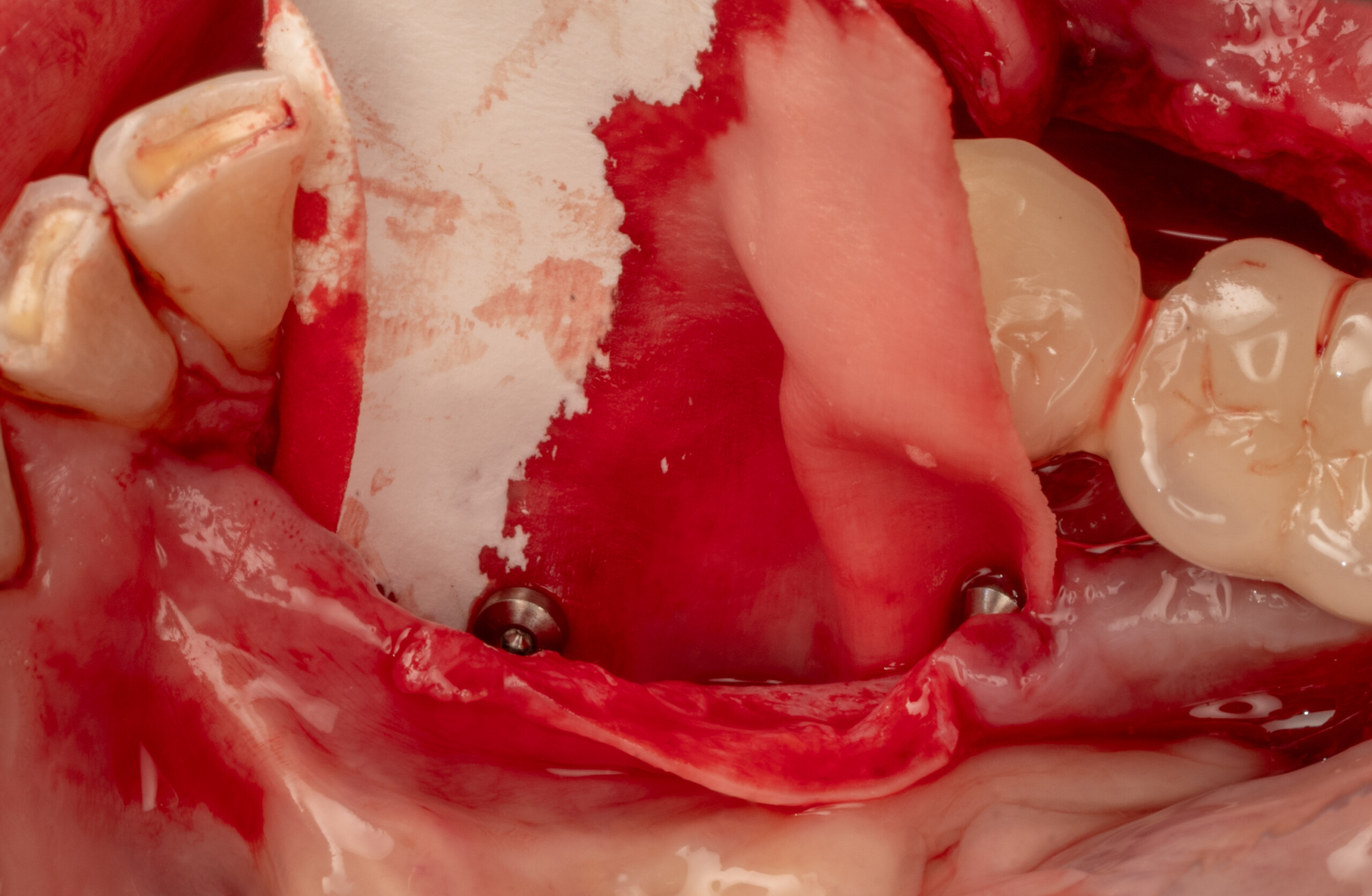
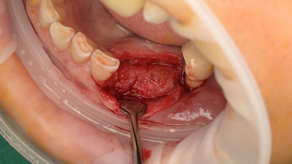
Decortication was performed and the flap was mobilized.
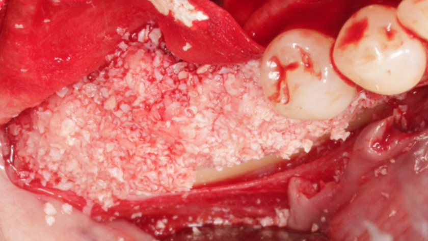
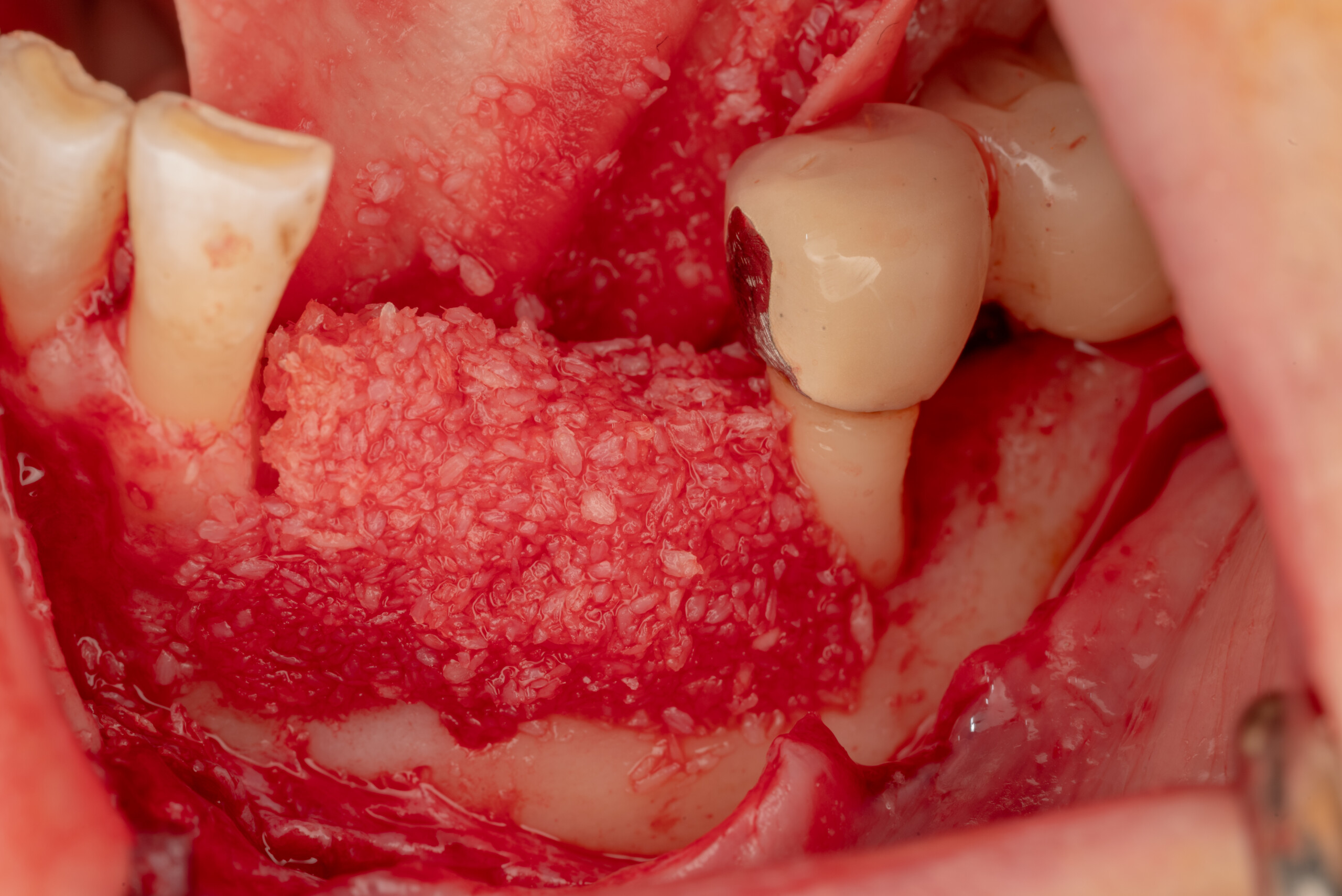
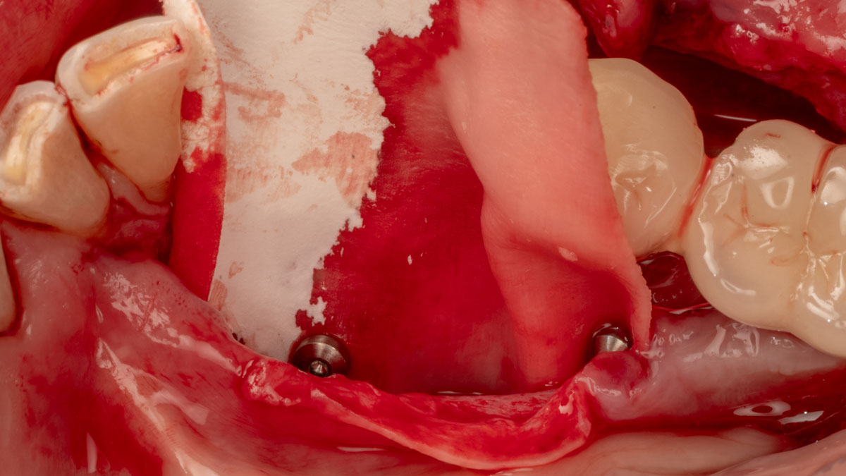
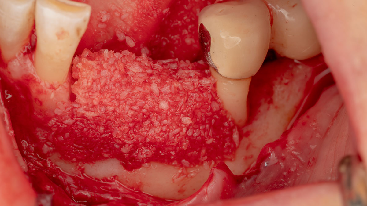
Geistlich Bio‑Gide® Forte was fixed on the lingual aspect, and the defect was filled with Geistlich Bio‑Oss® and vallos® mineralized cortical‑cancellous granules.
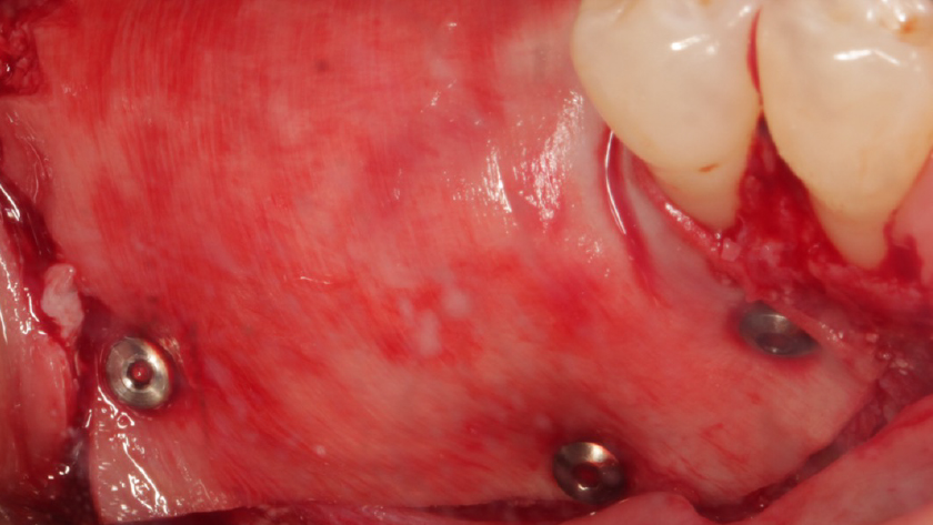
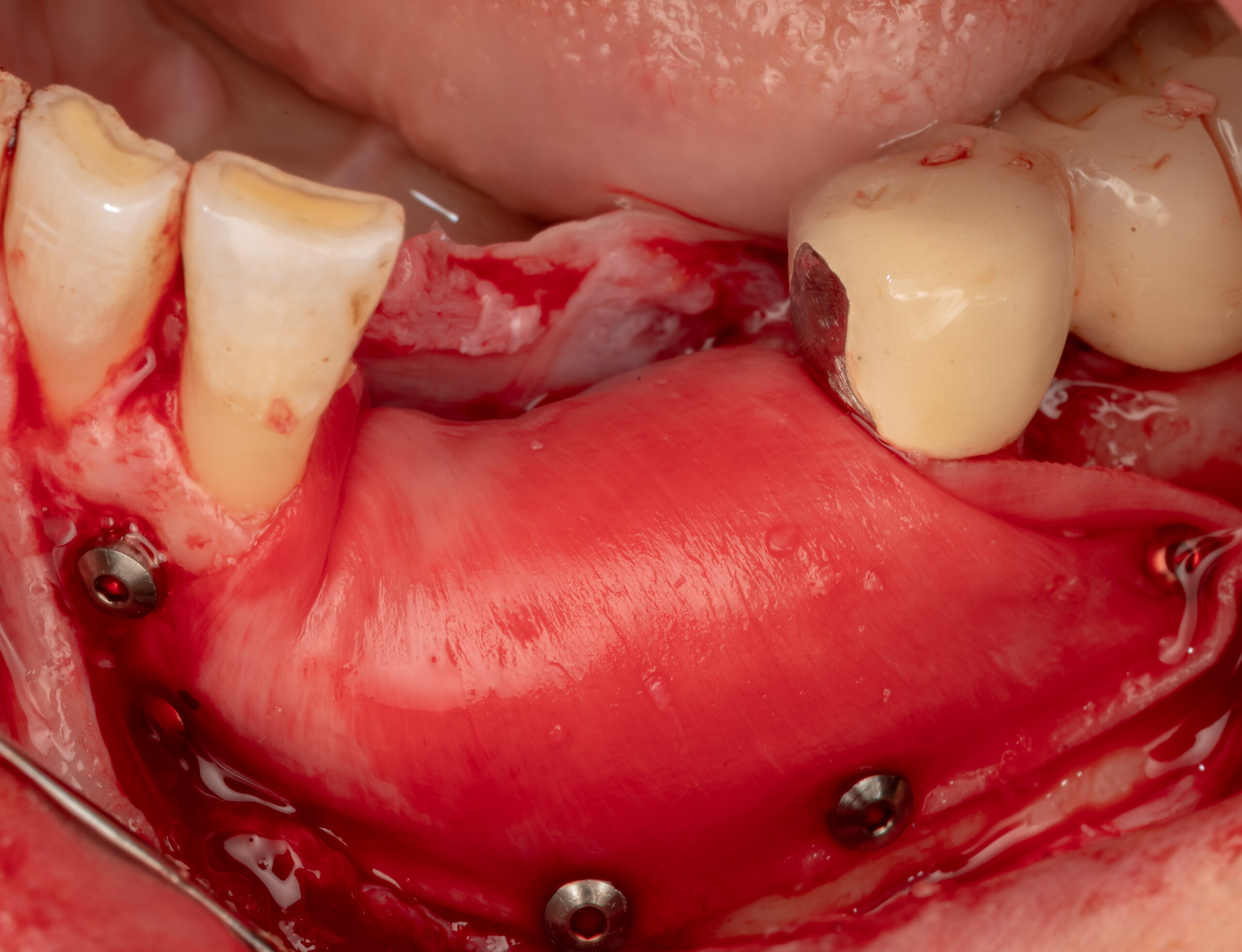
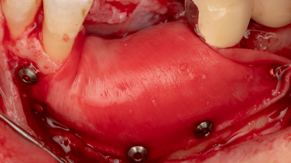
The densely packed Geistlich Bio‑Gide® Forte membrane was secured with pins on the buccal aspect.
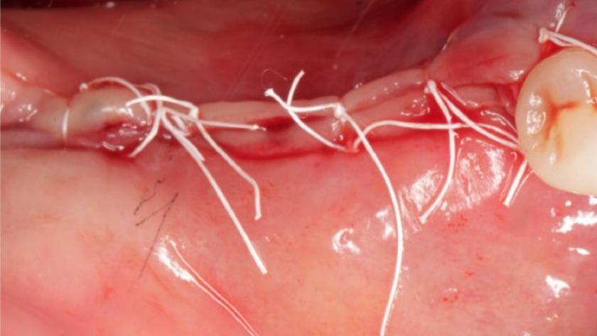
Primary wound closure was achieved with single interrupted sutures.
Six‑month follow‑up showing successful bone regeneration.
Andrea Ravidà, DDS, MS, PhD (Periodontist)
Jasper Wu, DDS, MS (Periodontist)
2-Stage Implant Placement in the Posterior Mandible using the Sausage Technique
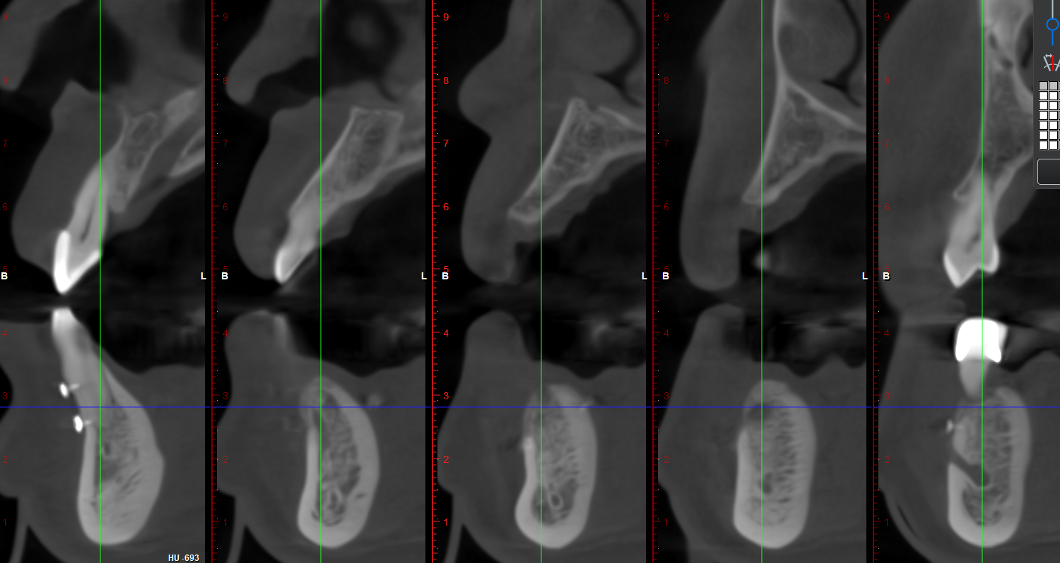
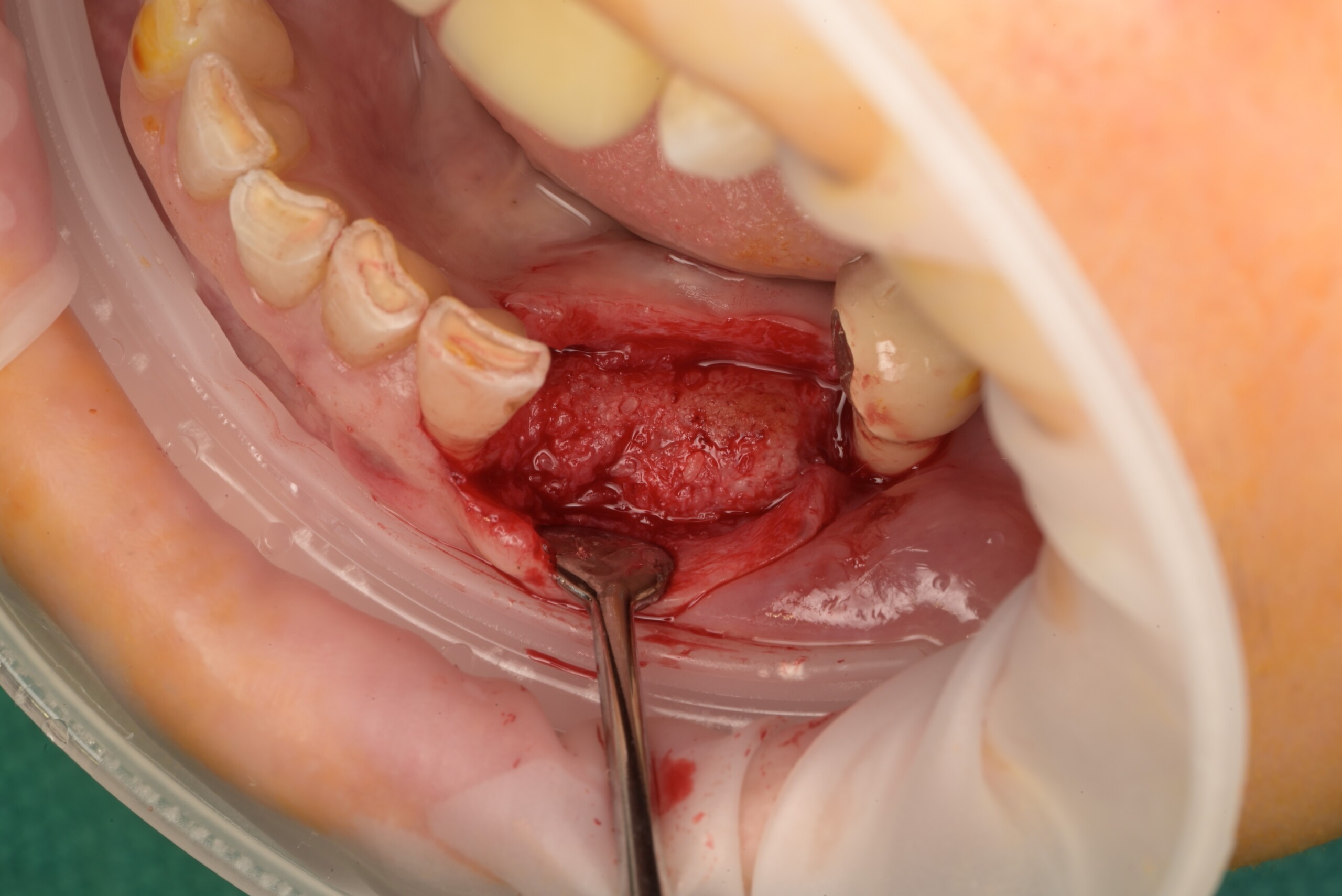
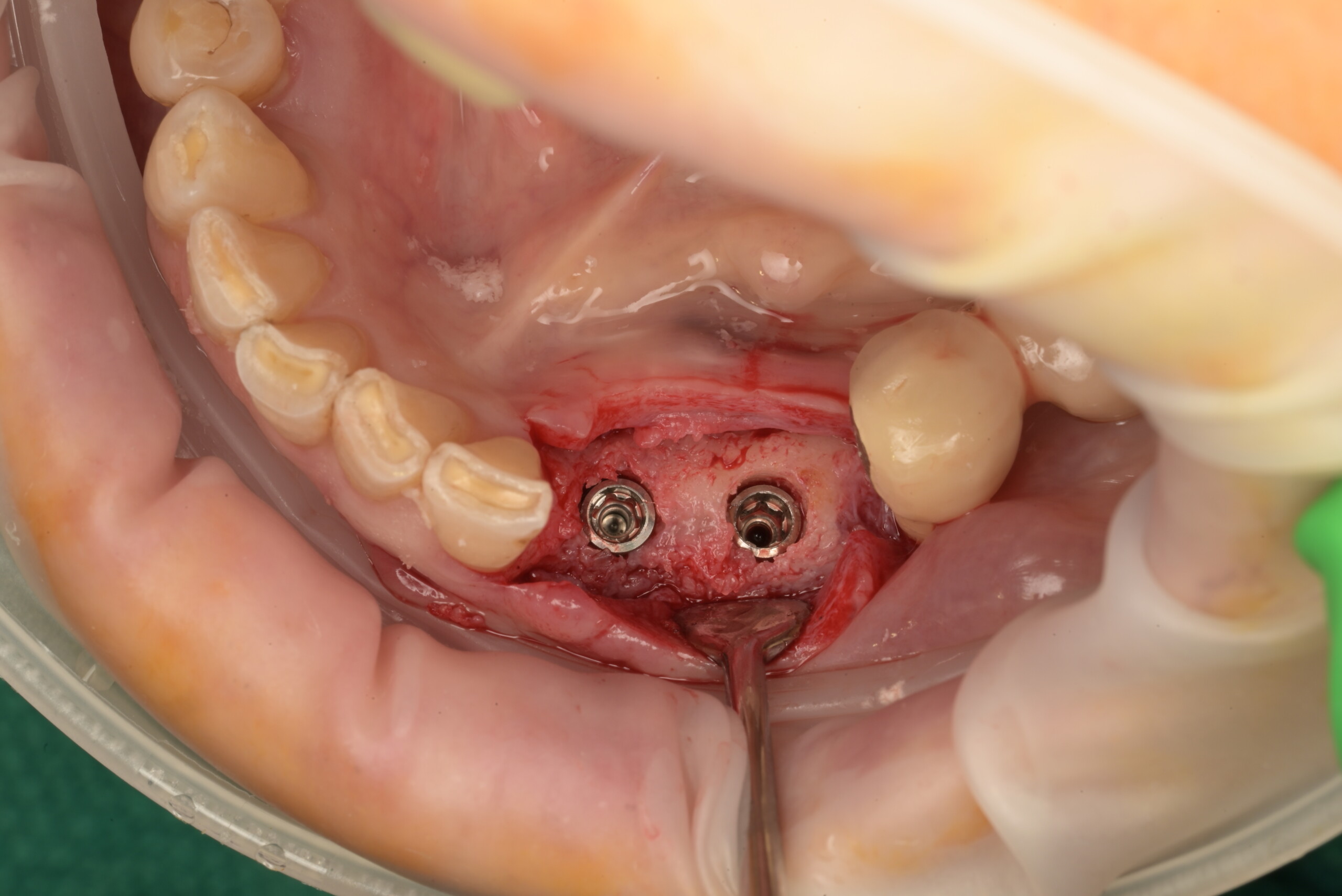
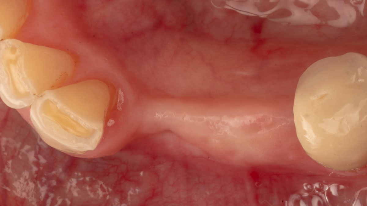
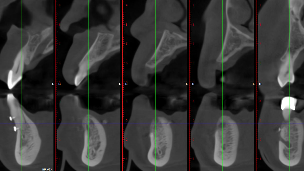
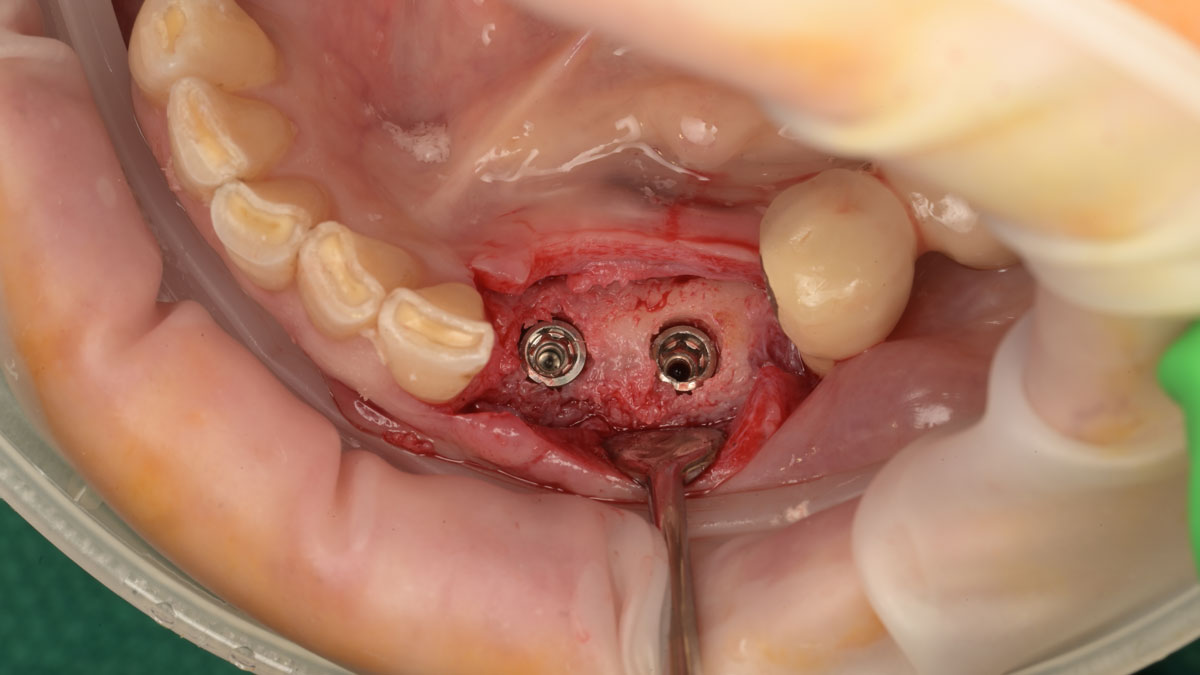
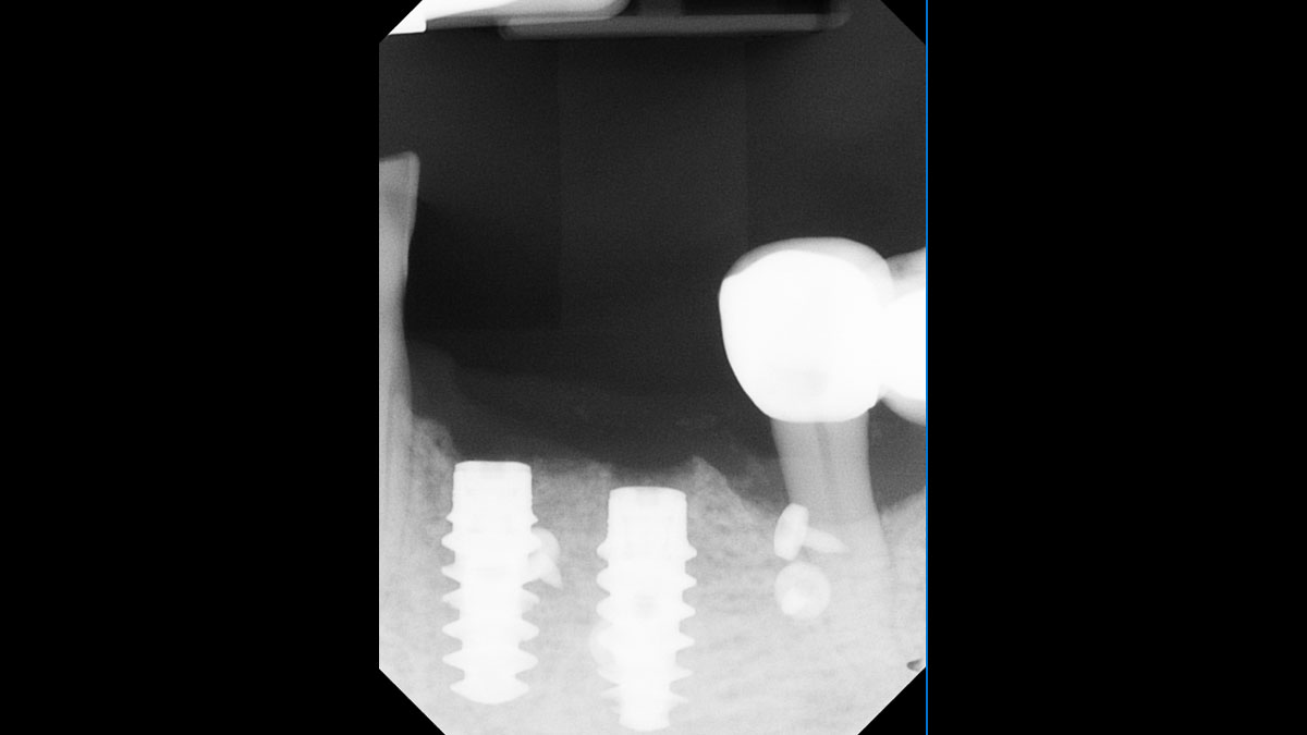
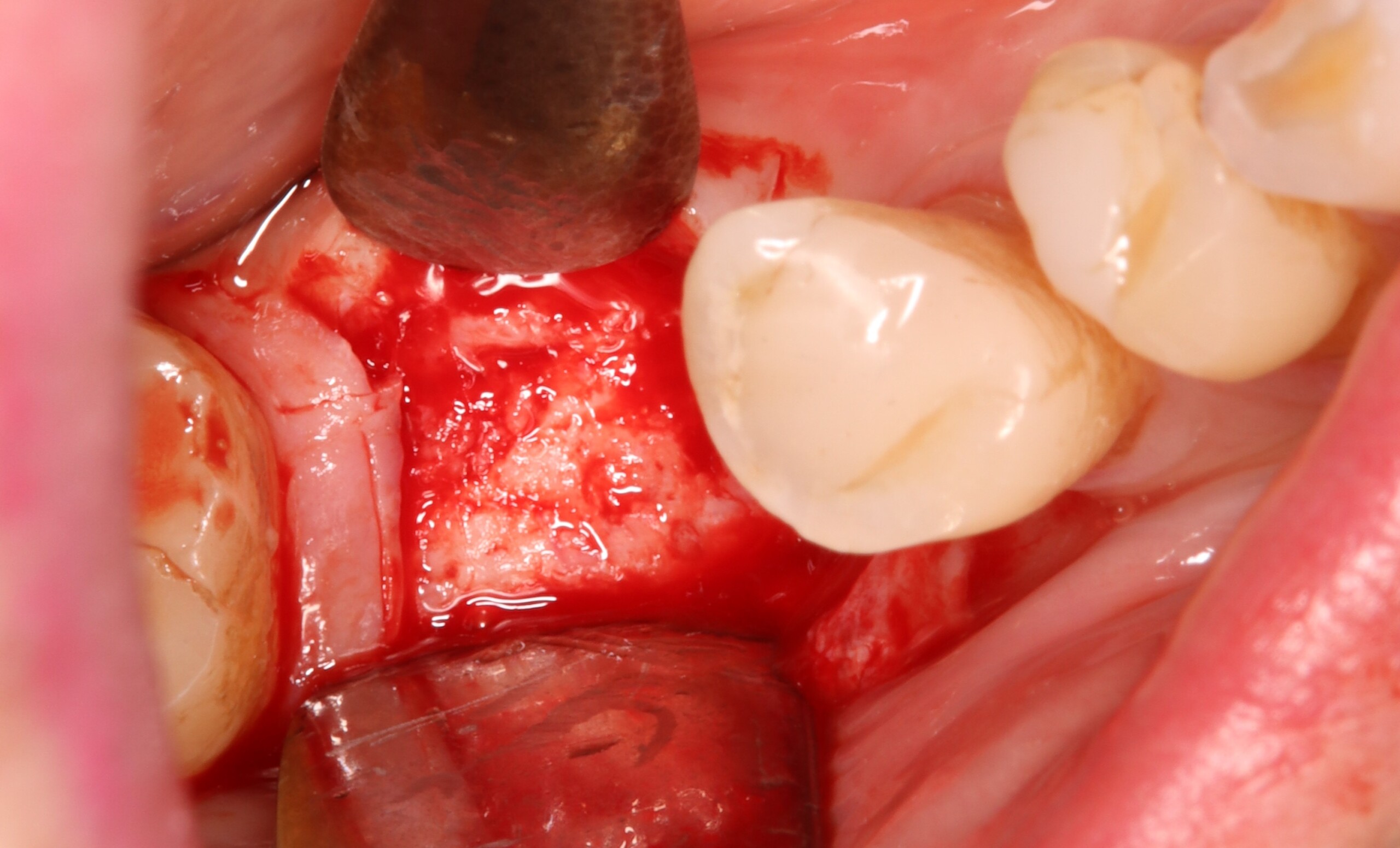
Initial clinical presentation showing the reduced ridge width.Decortication is performed and the flap is mobilized.Fixation of Geistlich Bio‑Gide® Forte using the Sausage Technique™ was initiated on the lingual aspect.The space beneath the membrane is filled with a 50:50 mixture of Geistlich Bio-Oss® and autologous bone.The densely packed membrane is secured with pins on the buccal aspect (Sausage Technique™).3-month follow-up shows visible horizontal bone gain and favorable soft-tissue conditions.Five month CBCT cross sections showing consistent horizontal ridge augmentation and implant ready bone.Six‑month re‑entry showing a stable, well‑integrated graft ready for implant placement, with a Geistlich Panorama retractor used to enhance visibility and access.Six-month occlusal view showing implant placement following successful graft maturation.
Andrea Ravidà, DDS, MS, PhD
Jasper Wu, DDS, MDS
2-Stage Implant Placement in the Posterior Mandible Using the Sausage Technique™
Initial clinical presentation showing the reduced ridge width.Initial clinical presentation showing the reduced ridge width: Post extraction of #22 with bone loss to apex.Initial clinical presentation showing the reduced ridge width: #22 calculus involving entire length of root.Initial clinical presentation showing the reduced ridge width: #22 with 100% bone loss with no buccal or lingual plate.Pre-operative intraoral view of the edentulous ridge demonstrating horizontal deficiency prior to flap elevation.Decortication is performed and the flap is mobilized.Fixation of Geistlich Bio Gide® Forte using the Sausage Technique™ was initiated on the lingual aspect.The space beneath the membrane is filled with a 50:50 mixture of Geistlich Bio-Oss® and autologous bone.The densely packed membrane is secured with pins on the buccal aspect (Sausage TechniqueTM).Three-month follow-up shows visible horizontal bone gain and favorable soft-tissue conditions.Three-month occlusal view demonstrating increased ridge width and favorable soft-tissue maturation following horizontal augmentation.Five month CBCT cross sections showing consistent horizontal ridge augmentation and implant ready bone.Six-month re-entry showing a stable, well-integrated graft ready for implant placement, with a Geistlich Panorama retractor used to enhance visibility and access.Six-month occlusal view showing implant placement following successful graft maturationProsthetically driven implant placement awaiting uncovery and crown fabrication.
3-Year Results Show that Yxoss CBR® is a Predictable Technique for Bone Gain in Advanced Defects
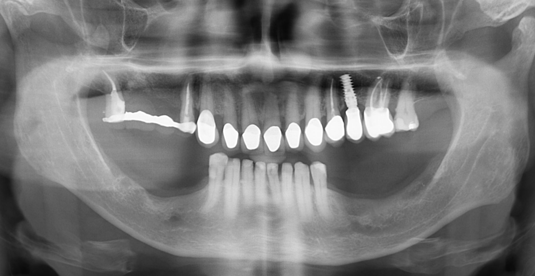
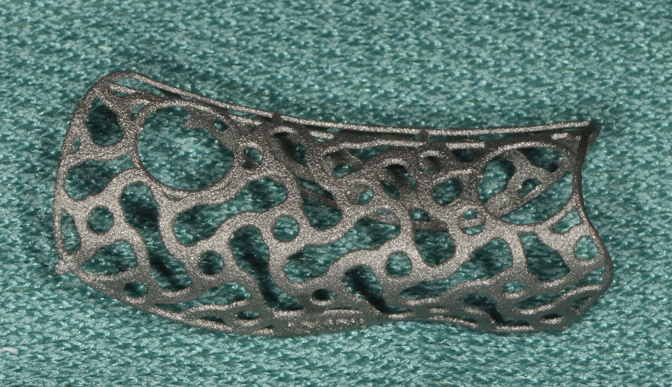
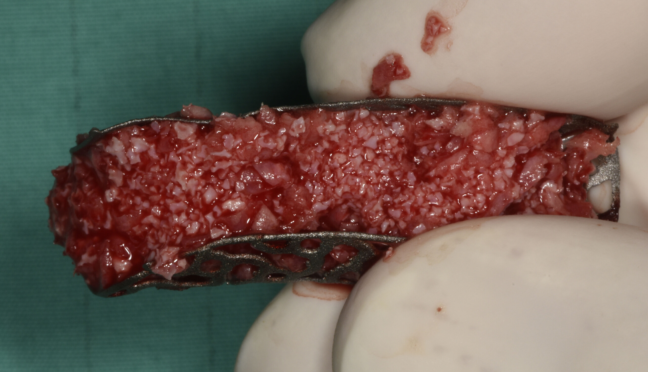
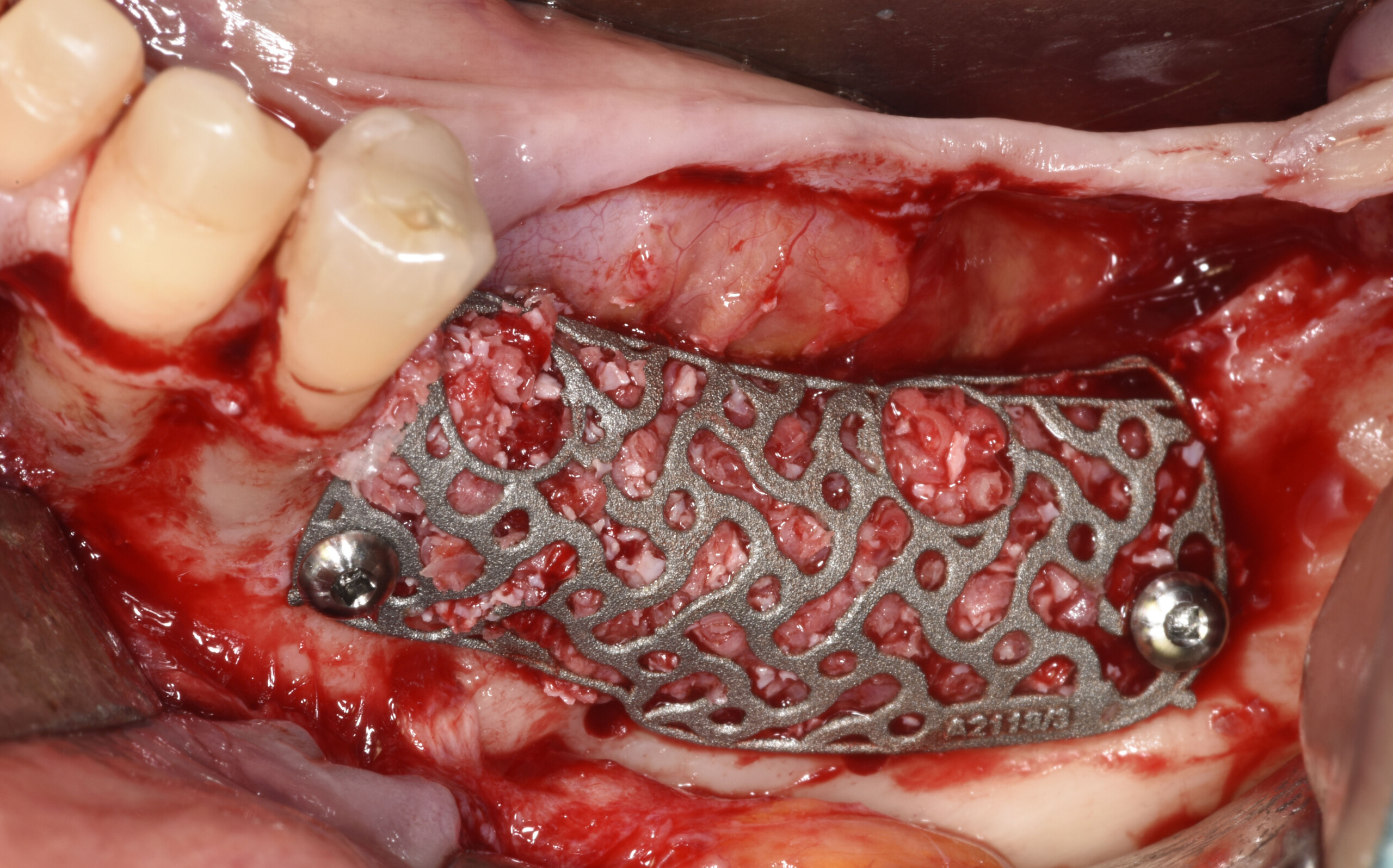
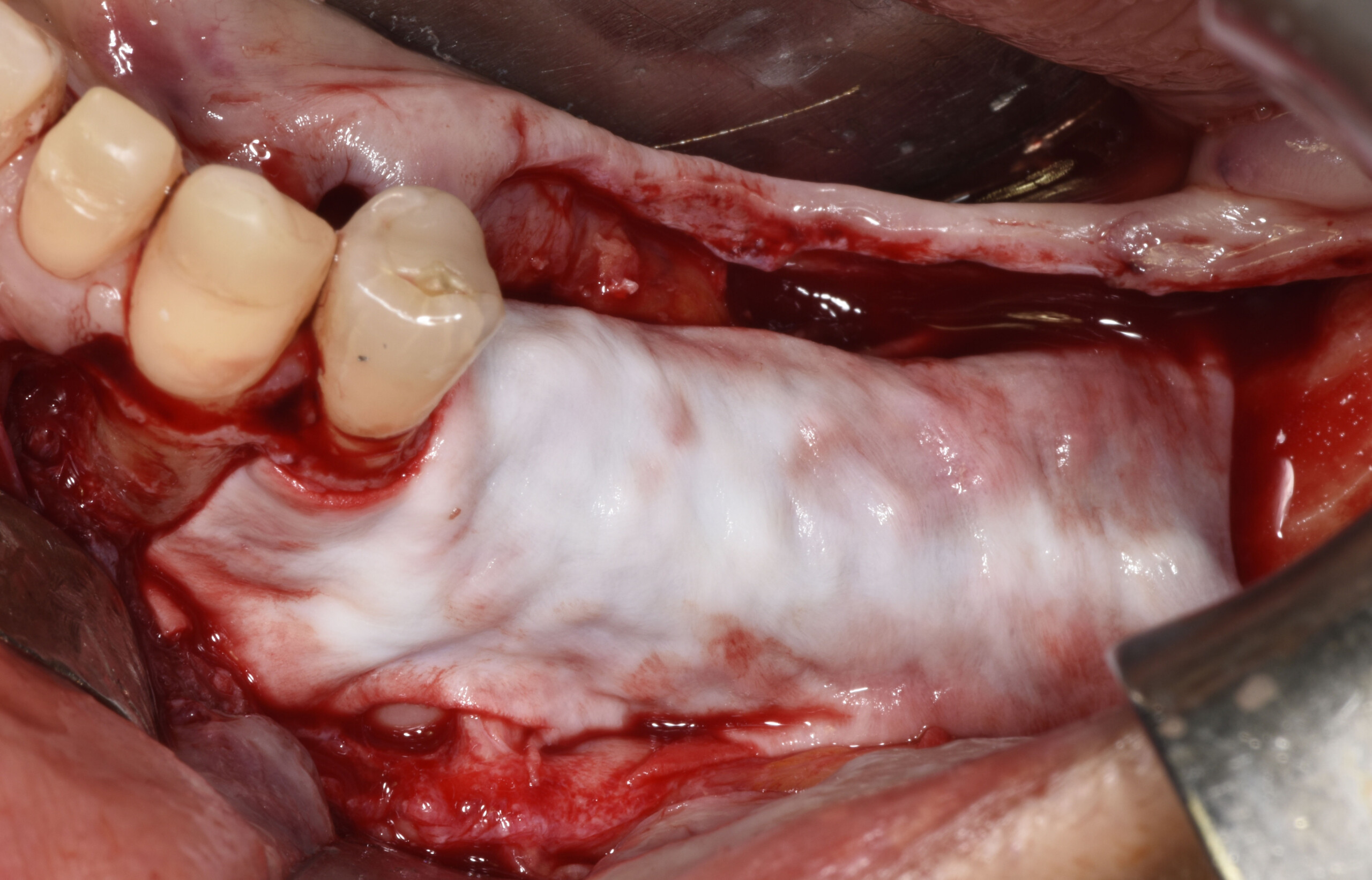
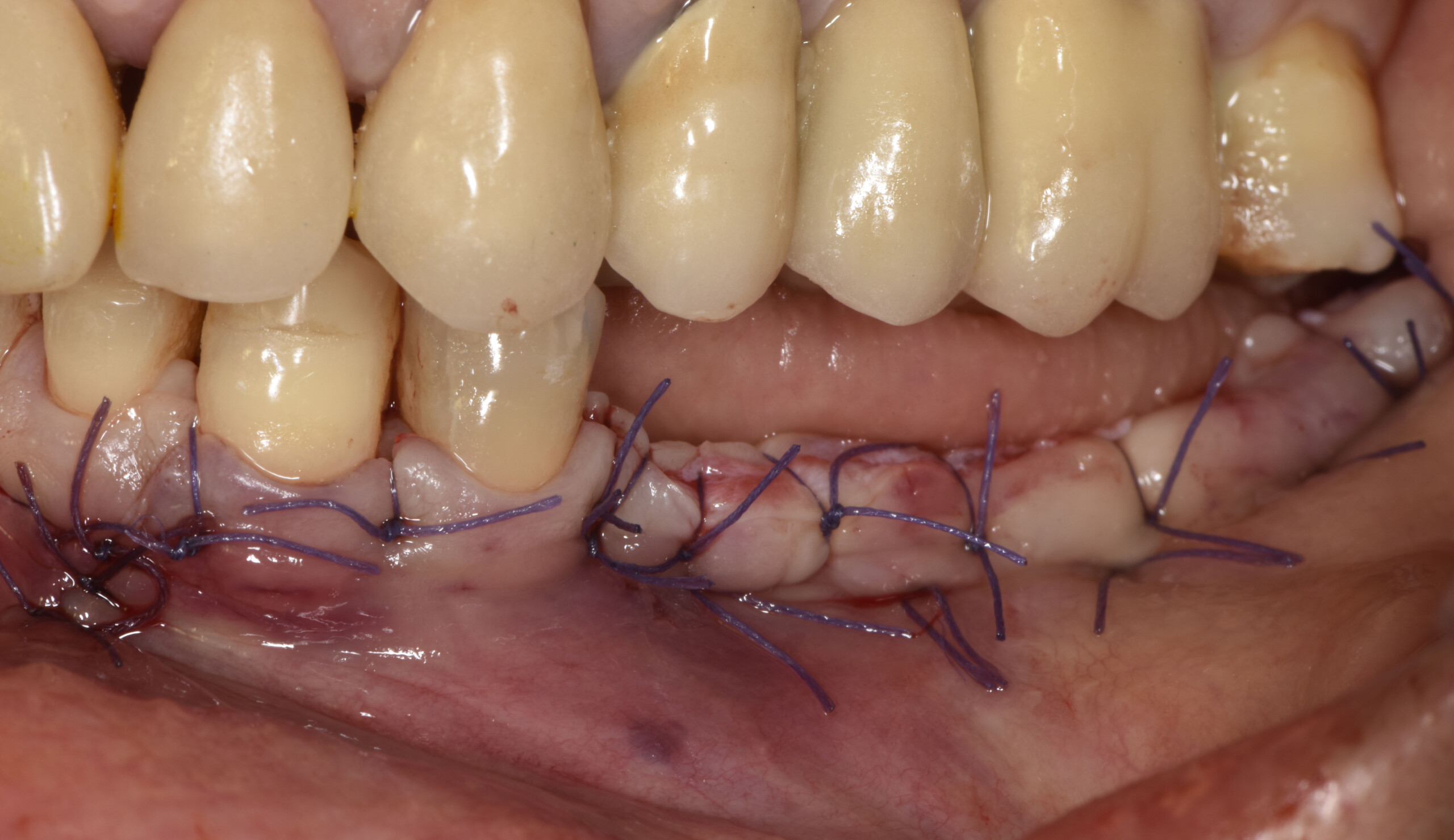
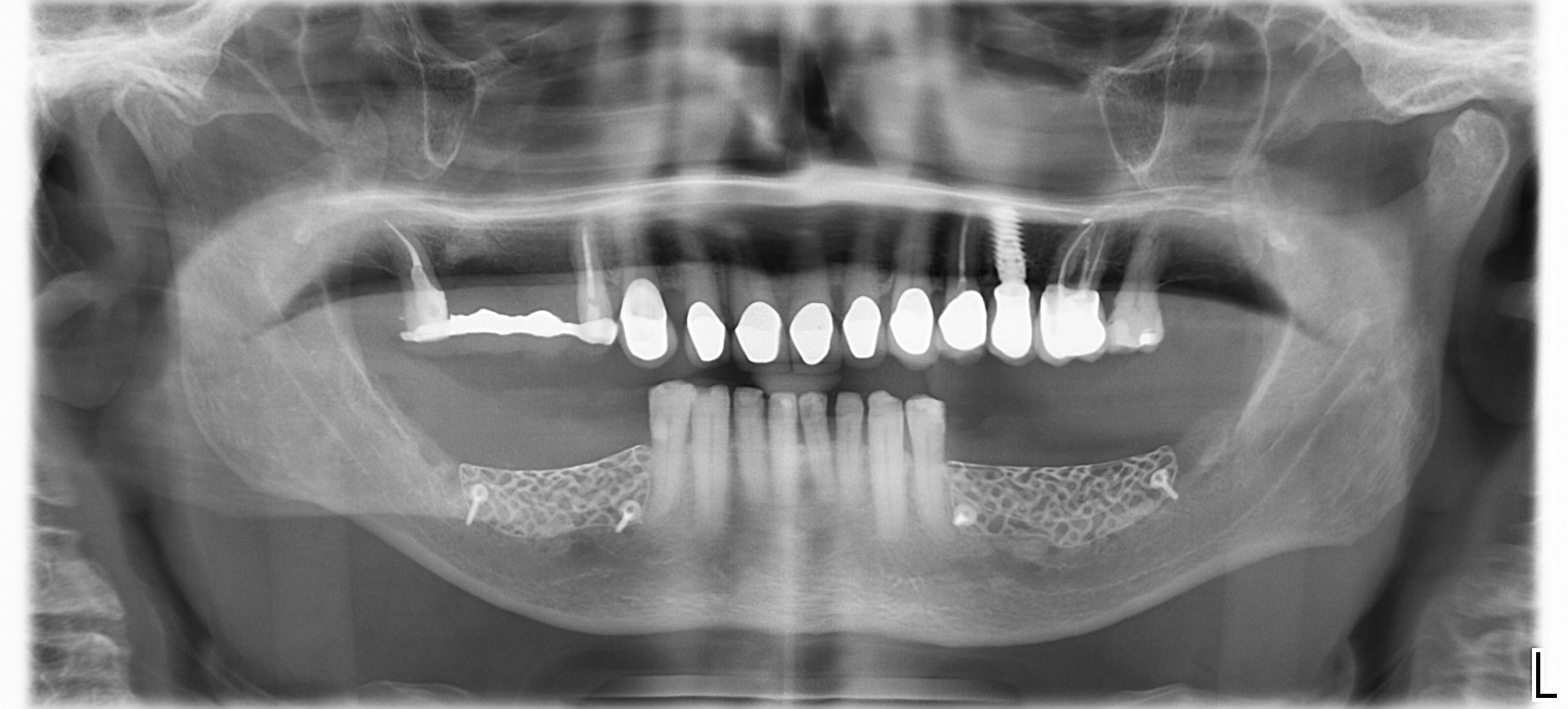
Panoramic radiograph of initial situation showing the atrophic mandibular areas.The final Yxoss CBR® ready for use.The customized Ti-mesh is filled with the autologous bone chips mixed with Geistlich Bio-Oss® granules in a 50:50 ratio.Intra-operative view at the end of the reconstruction showing the bone augmentation: the customized mesh was stabilized with 2 fixation screws.A Geistlich Bio-Gide® membrane is used to cover the customized mesh in order to increase the barrier effect.Intra-operative view after primary closure of the surgical wound.Panoramic radiograph after surgery.Clinical control 3 months later showing favorable healing of the soft tissue and correction of the defect.
A 4- to 5-year retrospective clinical and radiographic study of Neoss implants placed with or without GBR procedures
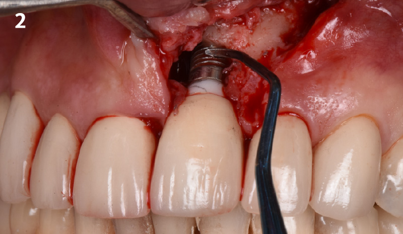
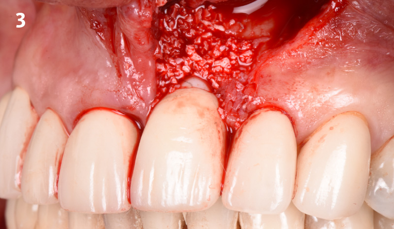
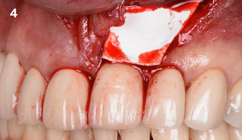
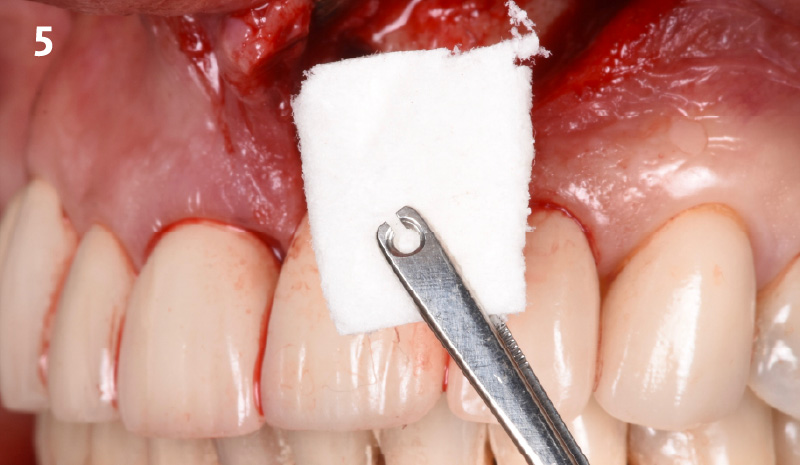
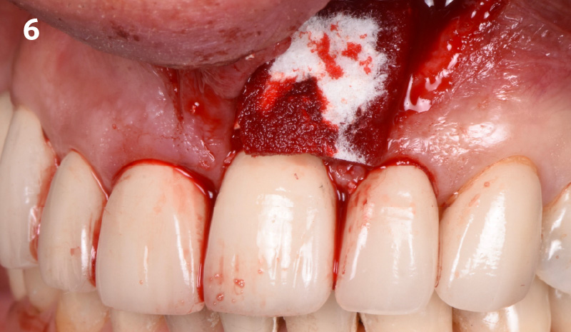
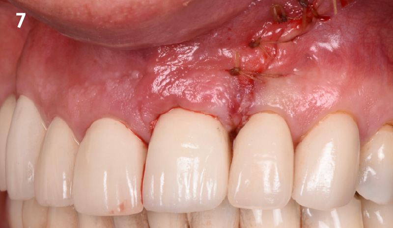
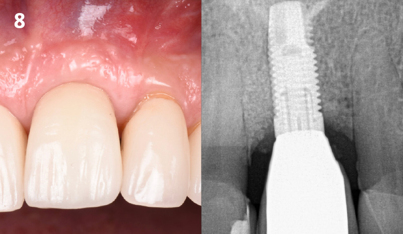
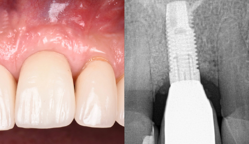
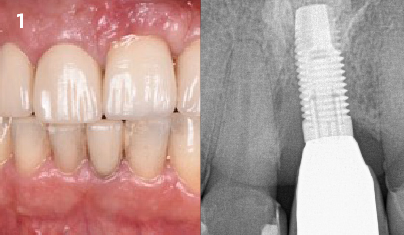
Initial situation, patient presented with radiographic and clinically traditional signs of peri-implantitis, including bleeding on probing, suppuration, radiographic progressive bone loss and clinical pathologic probing depths. Mechanical debridement was achieved using titanium scalers, an ultrasonic device with an implant protective cap and titanium brushes to remove all of the visible contaminants of the implant surface. Citric acid was then placed on shreds of a non-woven gauze and applied to the surface for approx. 1min. Copious irrigation was done using saline solution and the surface was ablated using the Er:YAG laser at 20pps/50mj. After the surface was prepped and no signs of residual granulation tissue was noted, the defect was grafted with Geistlich Bio-Oss®. Attention was given towards not augmenting beyond the bony envelope. A protective Geistlich Bio-Gide® membrane was placed over Geistlich Bio-Oss®.Geistlich Fibro-Gide® was placed over Geistlich Bio-Gide® to enhance soft-tissue volume and quality. Geistlich Fibro-Gide® was trimmed and adapted to the defect site ensuring a tension free closure. Geistlich Fibro-Gide® was place on the top of the bone graft to enhance soft-tissue thickness. Geistlich Fibro-Gide® is porous. We can observe the rapid penetration of blood through the matrix. Closure with a tension-free flap was achieved by releasing incisions and secured using 4-0 chromic gut sutures.1.5 year post-operative photo and radiograph show the healing of the soft-tissues with no signs of peri-implantitis and adequate tissue thickening. Radiographic bone levels have maintained stable over the course of the year.
Hector L. Sarmiento, D.M.D., MSc.
Manhattan, New York, USA
Initial situation, patient presented with radiographic and clinically traditional signs of peri-implantitis, including bleeding on probing, suppuration, radiographic progressive bone loss and clinical pathologic probing depths. Mechanical debridement was achieved using titanium scalers, an ultrasonic device with an implant protective cap and titanium brushes to remove all of the visible contaminants of the implant surface. Citric acid was then placed on shreds of a non-woven gauze and applied to the surface for approx. 1min. Copious irrigation was done using saline solution and the surface was ablated using the Er:YAG laser at 20pps/50mj. After the surface was prepped and no signs of residual granulation tissue was noted, the defect was grafted with Geistlich Bio-Oss®. Attention was given towards not augmenting beyond the bony envelope. A protective Geistlich Bio-Gide® membrane was placed over Geistlich Bio-Oss®.Geistlich Fibro-Gide® was placed over Geistlich Bio-Gide® to enhance soft-tissue volume and quality. Geistlich Fibro-Gide® was trimmed and adapted to the defect site ensuring a tension free closure. Geistlich Fibro-Gide® was place on the top of the bone graft to enhance soft-tissue thickness. Geistlich Fibro-Gide® is porous. We can observe the rapid penetration of blood through the matrix. Closure with a tension-free flap was achieved by releasing incisions and secured using 4-0 chromic gut sutures.1.5 year post-operative photo and radiograph show the healing of the soft-tissues with no signs of peri-implantitis and adequate tissue thickening. Radiographic bone levels have maintained stable over the course of the year.
Alecsandru Ionescu, DDS, PhD
Alveolar Ridge Augmentation Using the Open Healing Concept