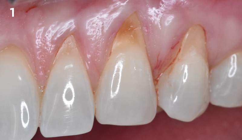
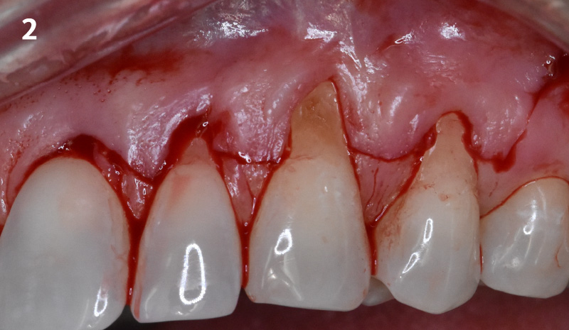
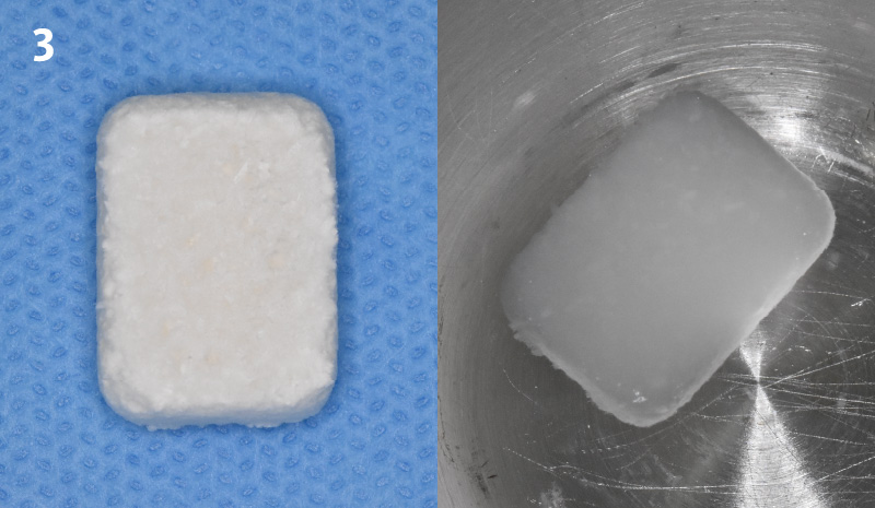
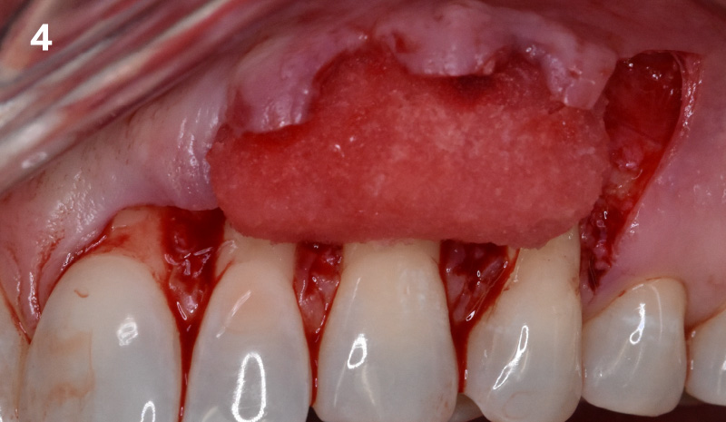
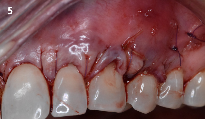
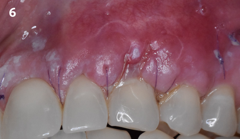
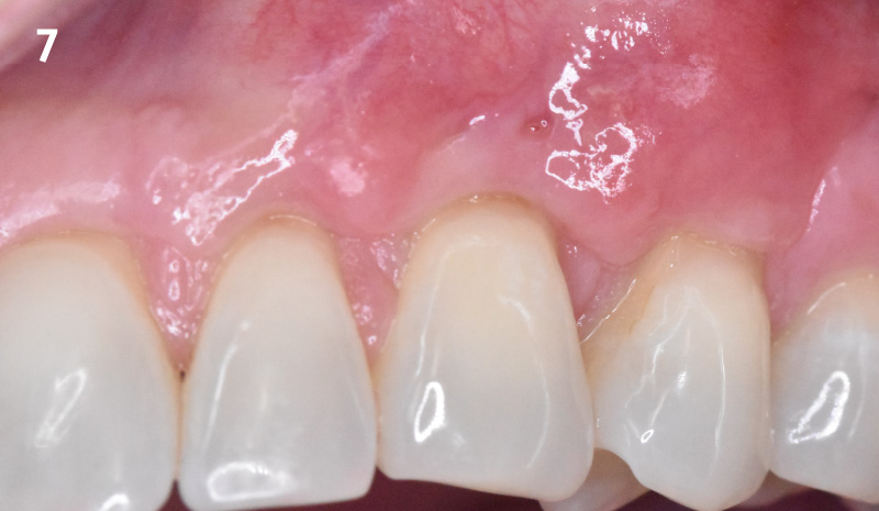
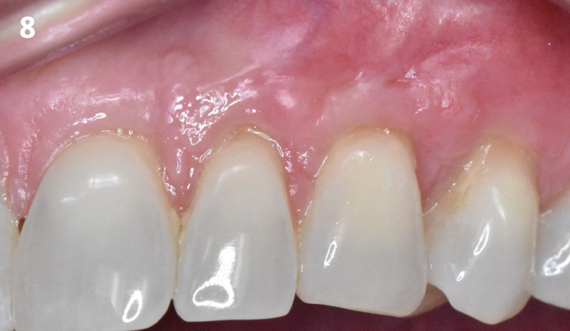
CLINICAL CASE Pre-operative condition: Note that the gingival recession of 3-4 mm is evident as is the cervical root abrasions. The interdental papillae completely fills the embrasure space. Incision design showing the sulcular incisions with horizontal incisions across the interdental regions ending with a remote oblique vertical releasing incision distal to the first bicuspid tooth. Geistlich Fibro-Gide® is trimmed in a dry state to 10 x 15 x 6mm. The corners were trimmed for better adaptation and the matrix was then hydrated in sterile saline solution. The interdental papillae were de-epithelialized and Geistlich Fibro-Gide®was placed over the exposed roots extending onto the bone. Geistlich Fibro-Gide® was not secured with sutures. Internal periosteal releasing incision was made to allow tension-free coronal advancement of the buccal flap to completely cover Geistlich Fibro-Gide®.The flap was secured with 5-0 Monocryl® sutures. 1-week post-operative visit: the healing looks good and sutures are intact. There was a small soft-tissue dehiscence at the buccal margin of the canine tooth. Healing progressed well at 2 months post-operatively and the dehiscence defect seen at 1 week appears to be healing. Soft-tissue thickness is also evident at this stage. At 6 months, 100% root coverage has been achieved. Note the increase in keratinized gingiva at the canine tooth where there was previously delayed healing. The patient is happy with the esthetic and functional outcome.