Mix & Match! Buy 5 Products, Get 1 Free! Use code B5G1. Get Started!

Product: Yxoss CBR®

BIOBRIEF
Lateral and Vertical Bone Regeneration with Simultaneous Soft Tissue Augmentation
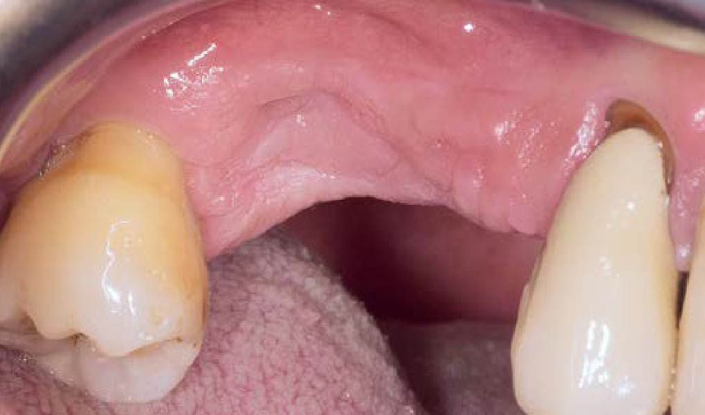
THE SITUATION
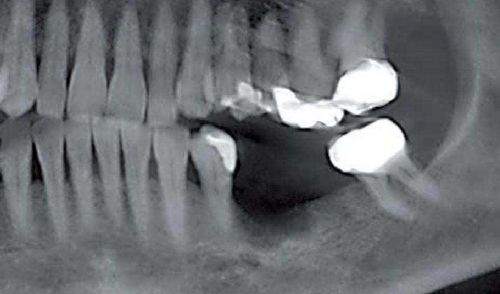
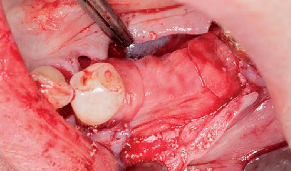
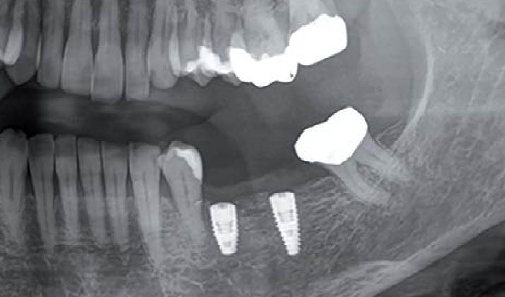
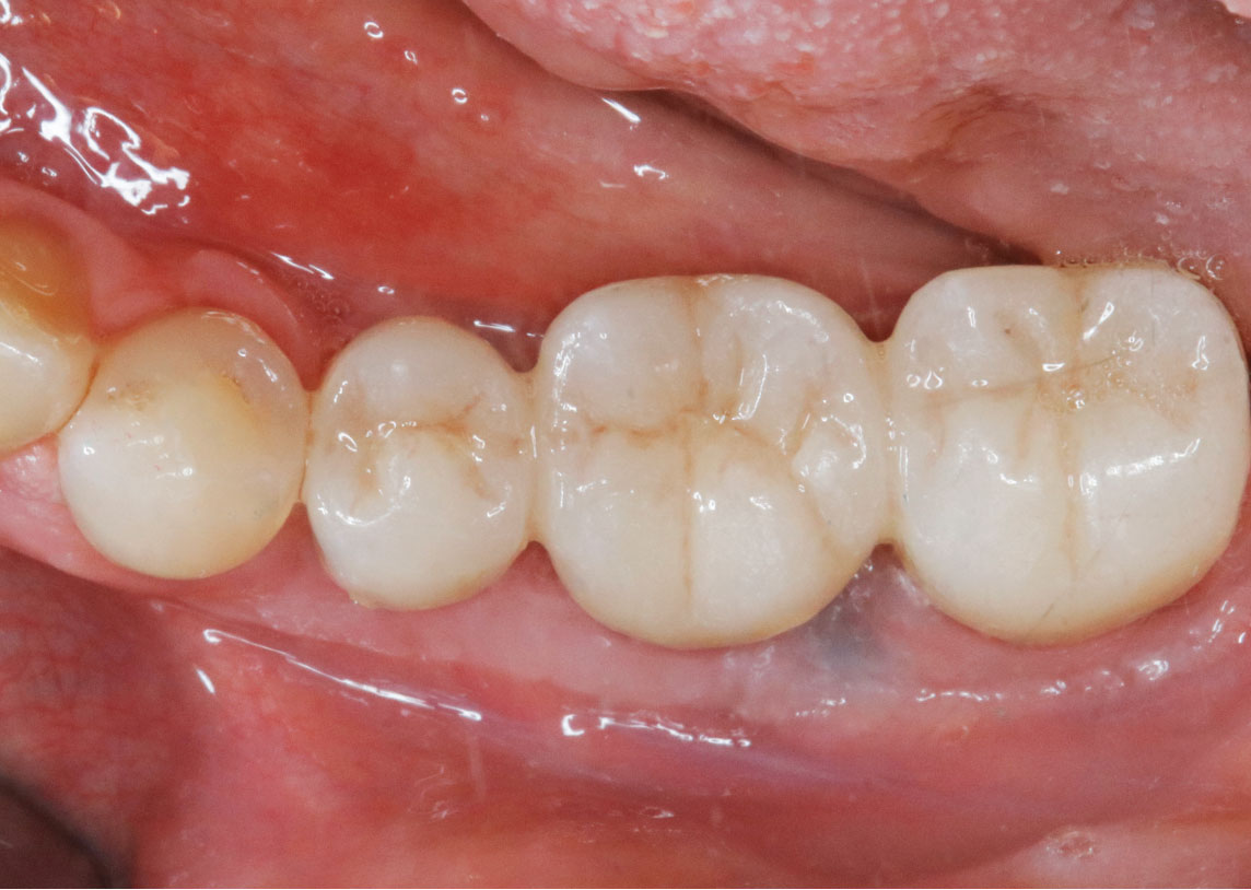
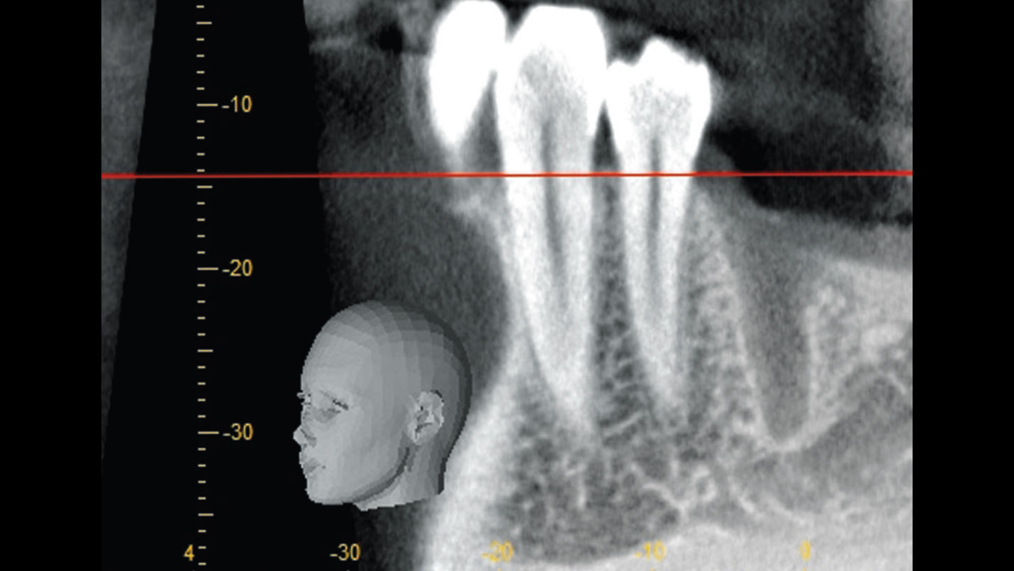
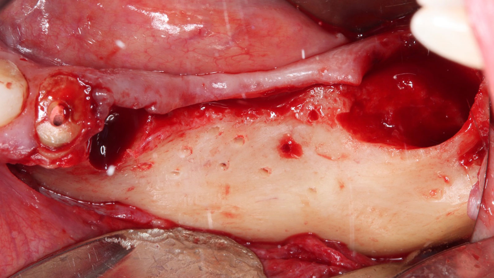
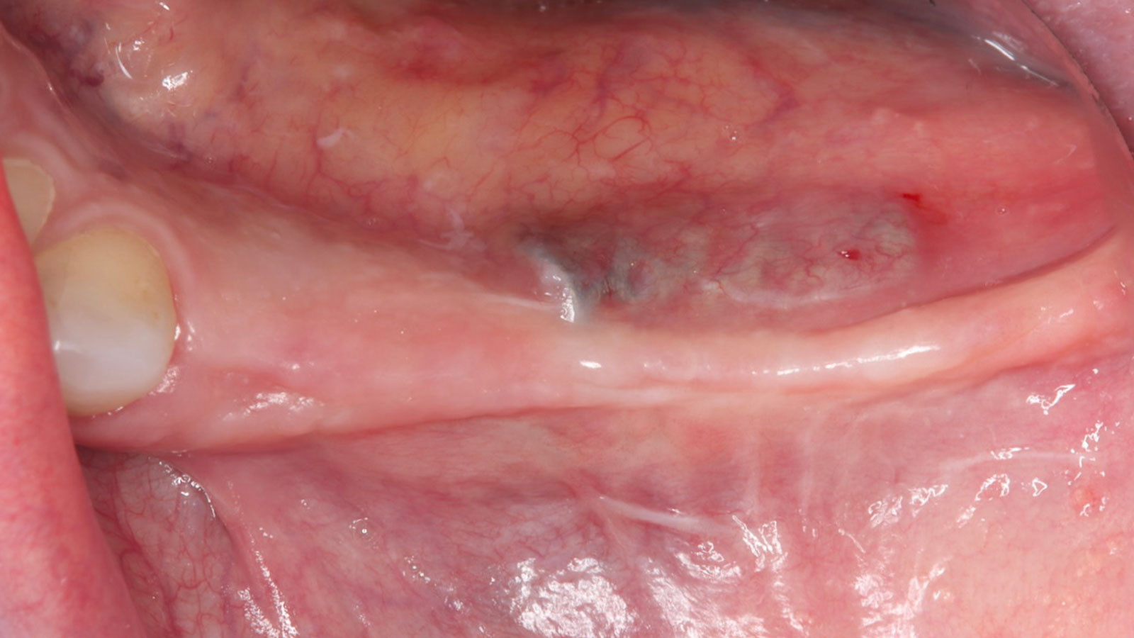
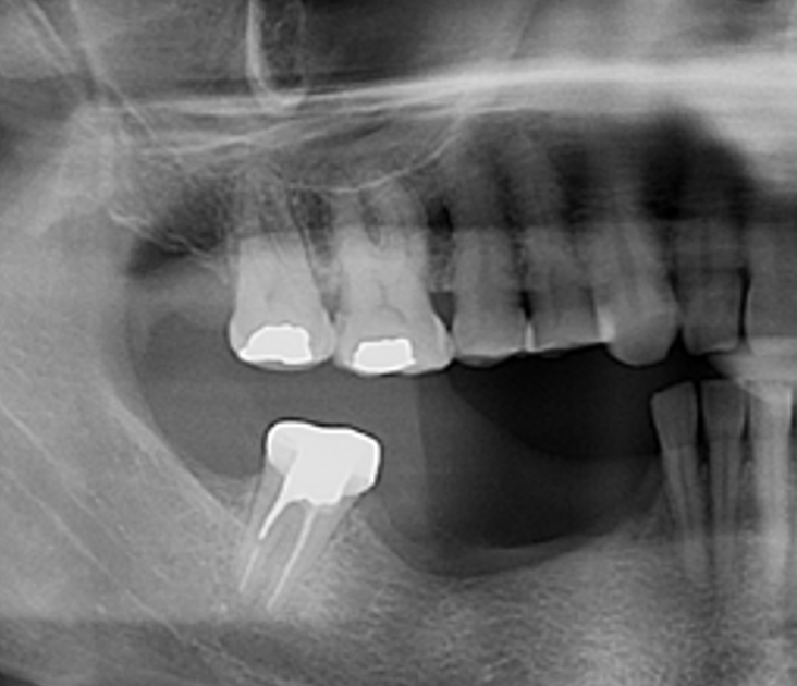
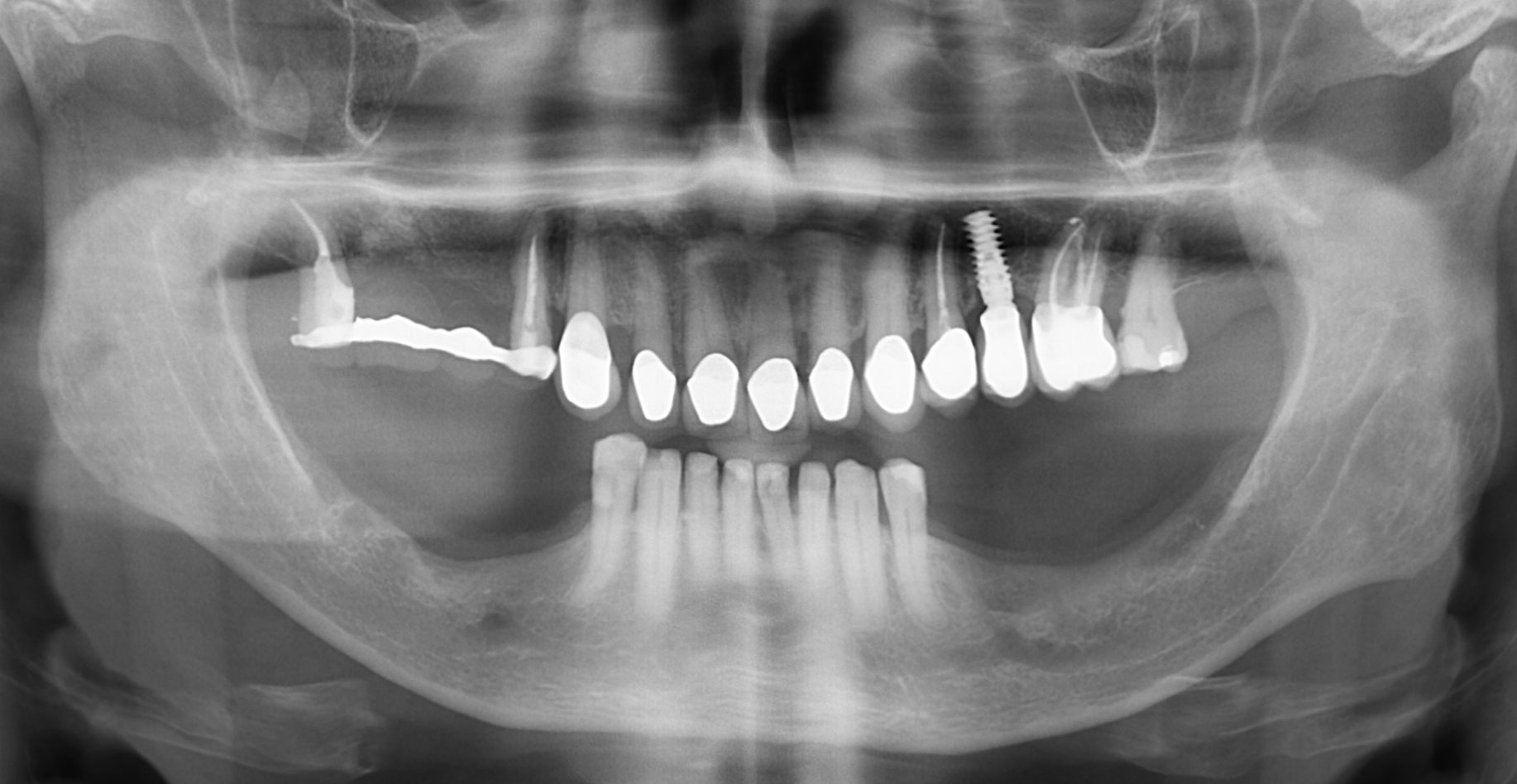
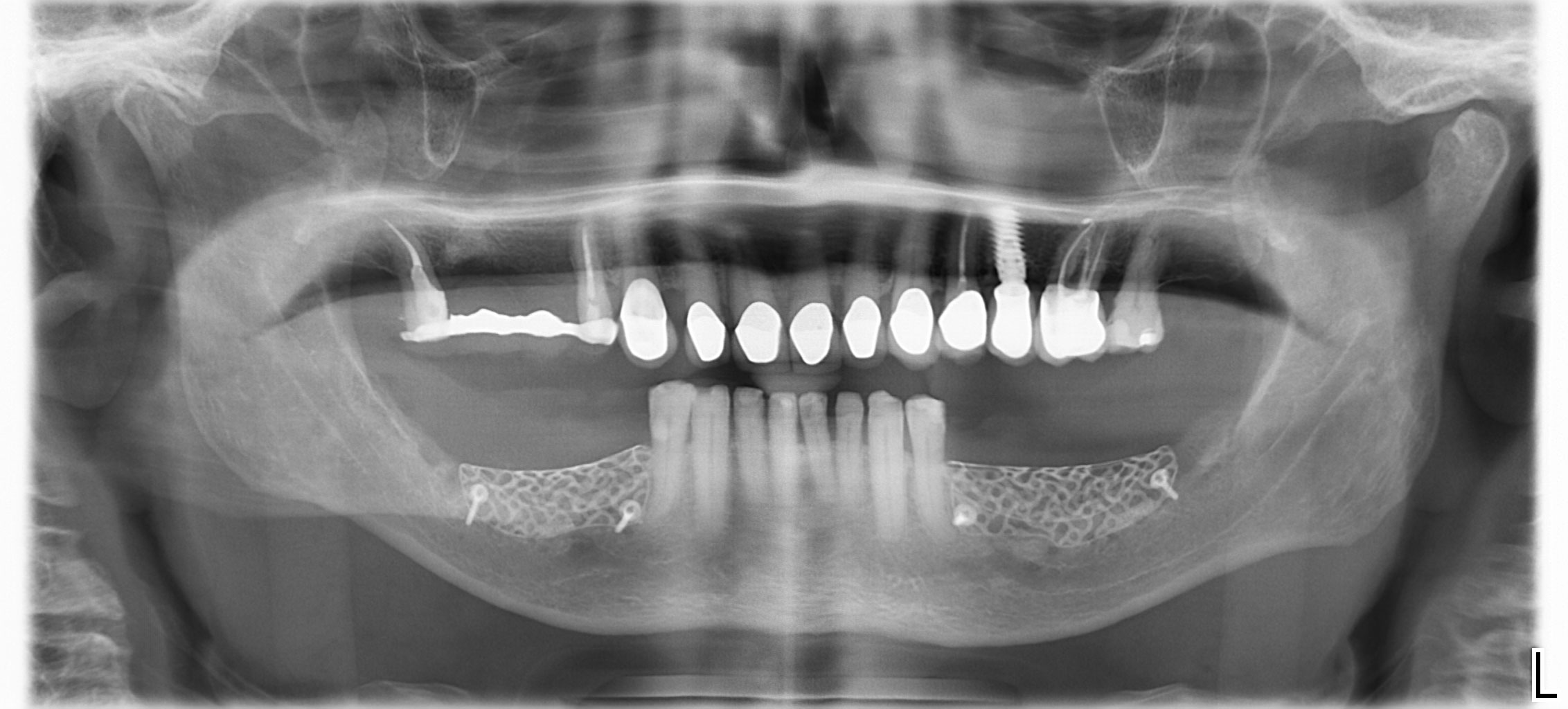
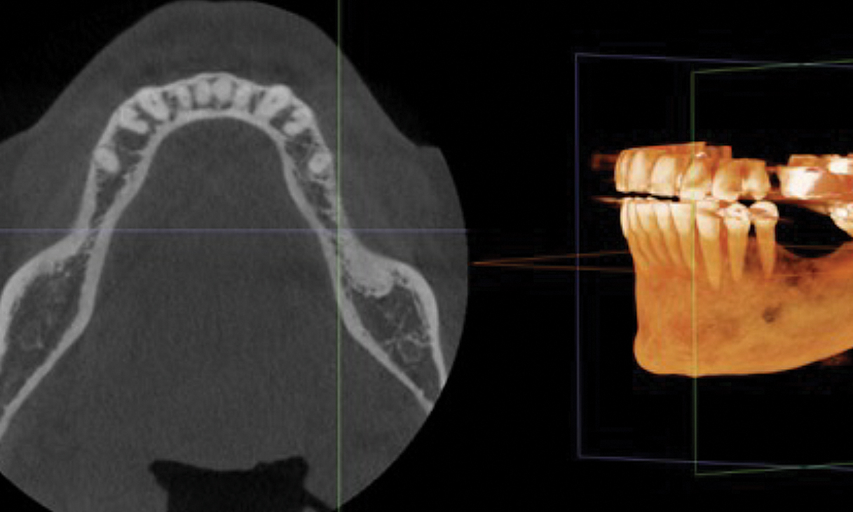
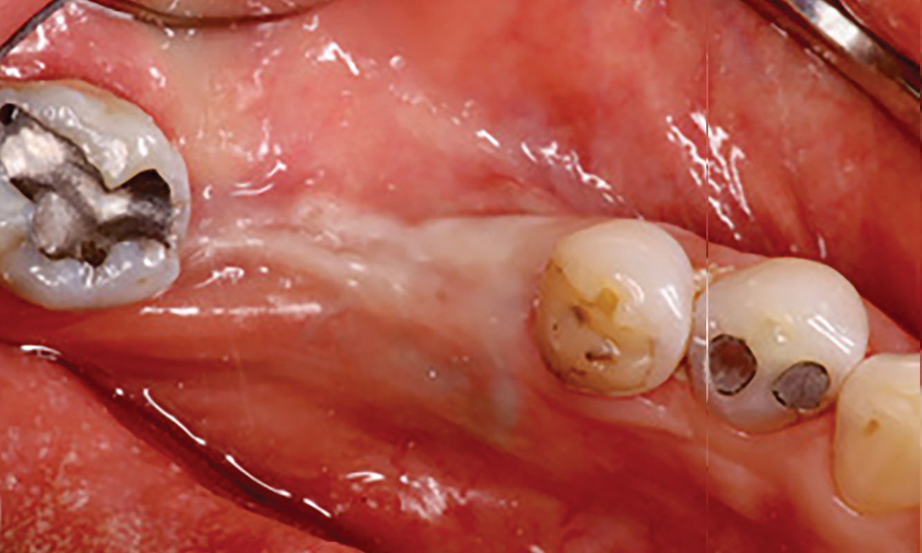
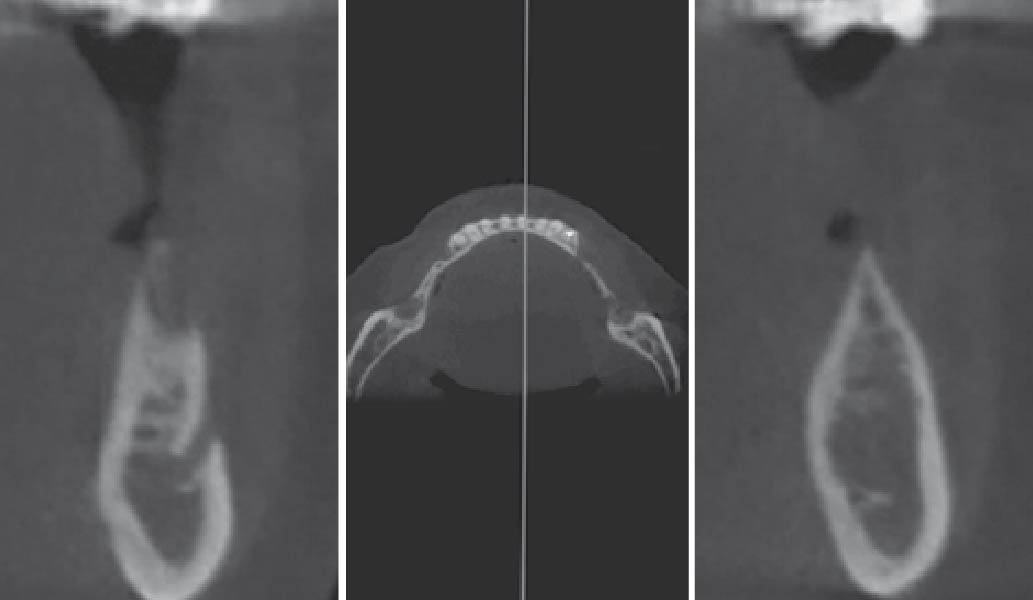
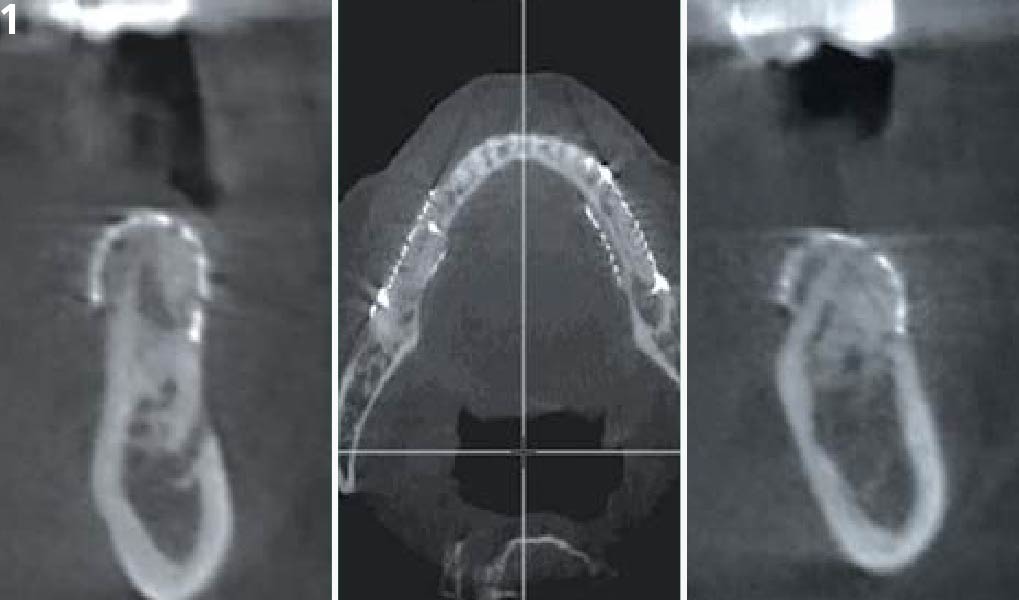
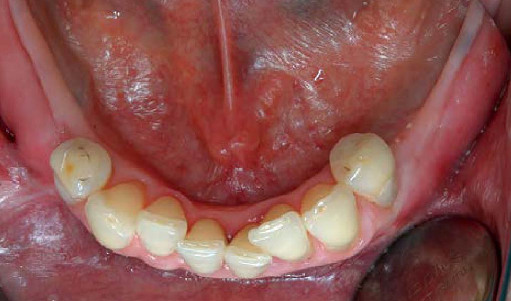
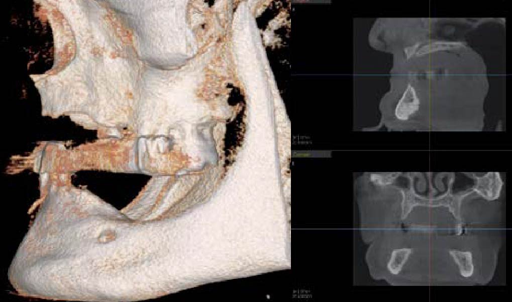
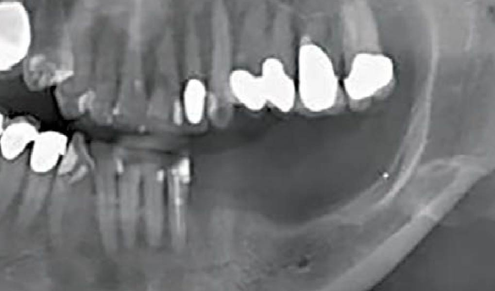
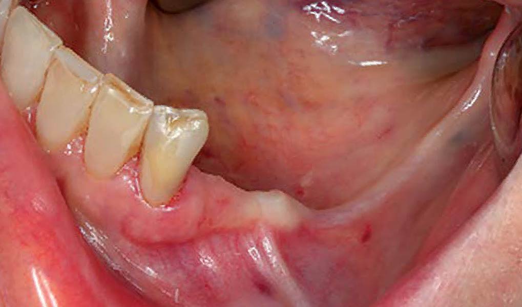
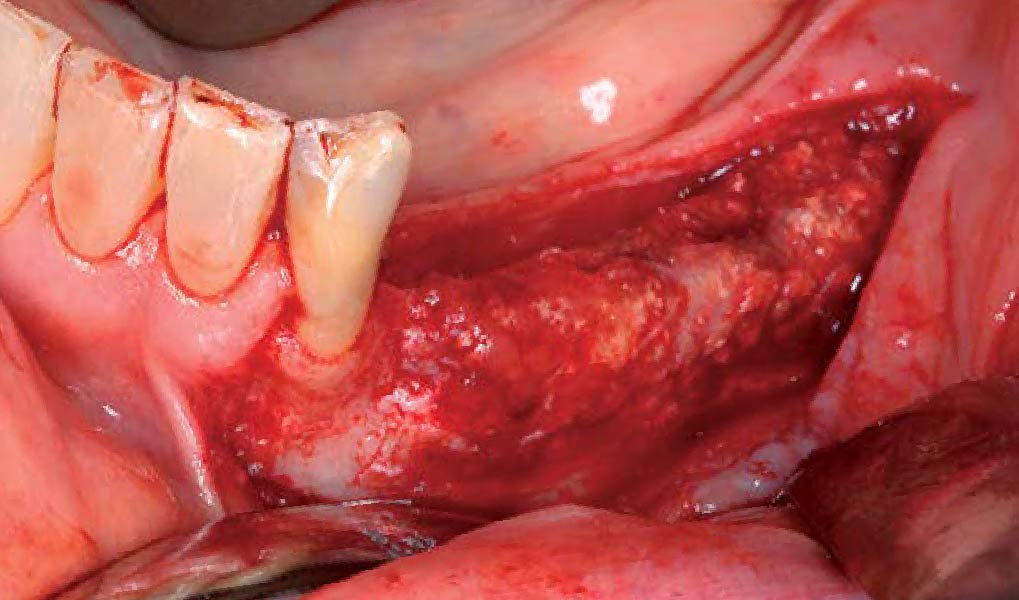
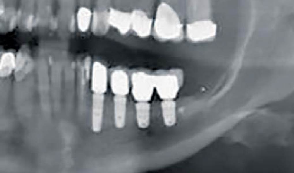
After extraction of the periodontally damaged tooth #20 the preoperative Cone-Beam Computed Tomography (CBCT) imaging shows reduced vertical bone volume in the area of tooth #s 18 – 20. A lateral and vertical bone regeneration was necessary.
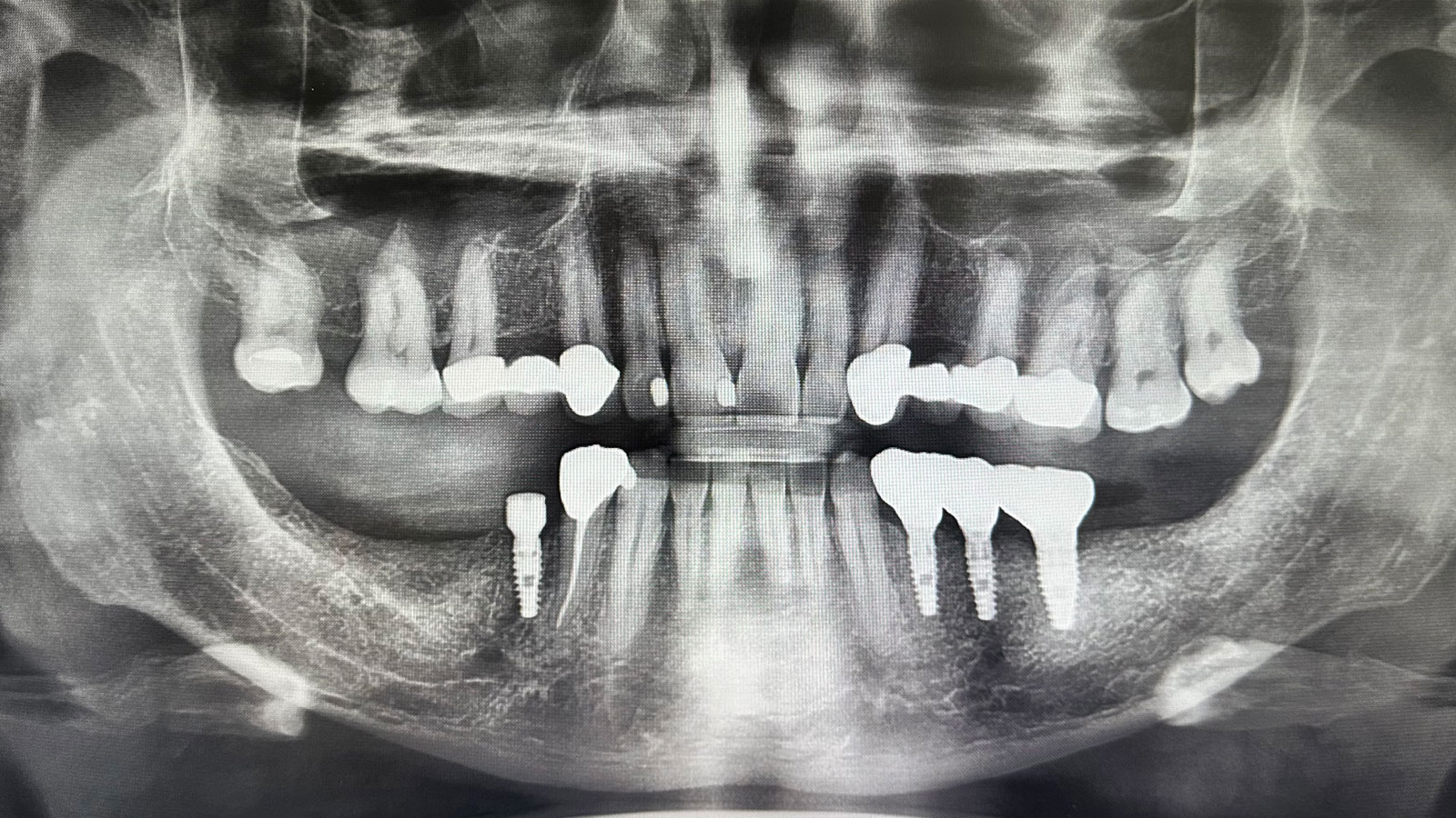
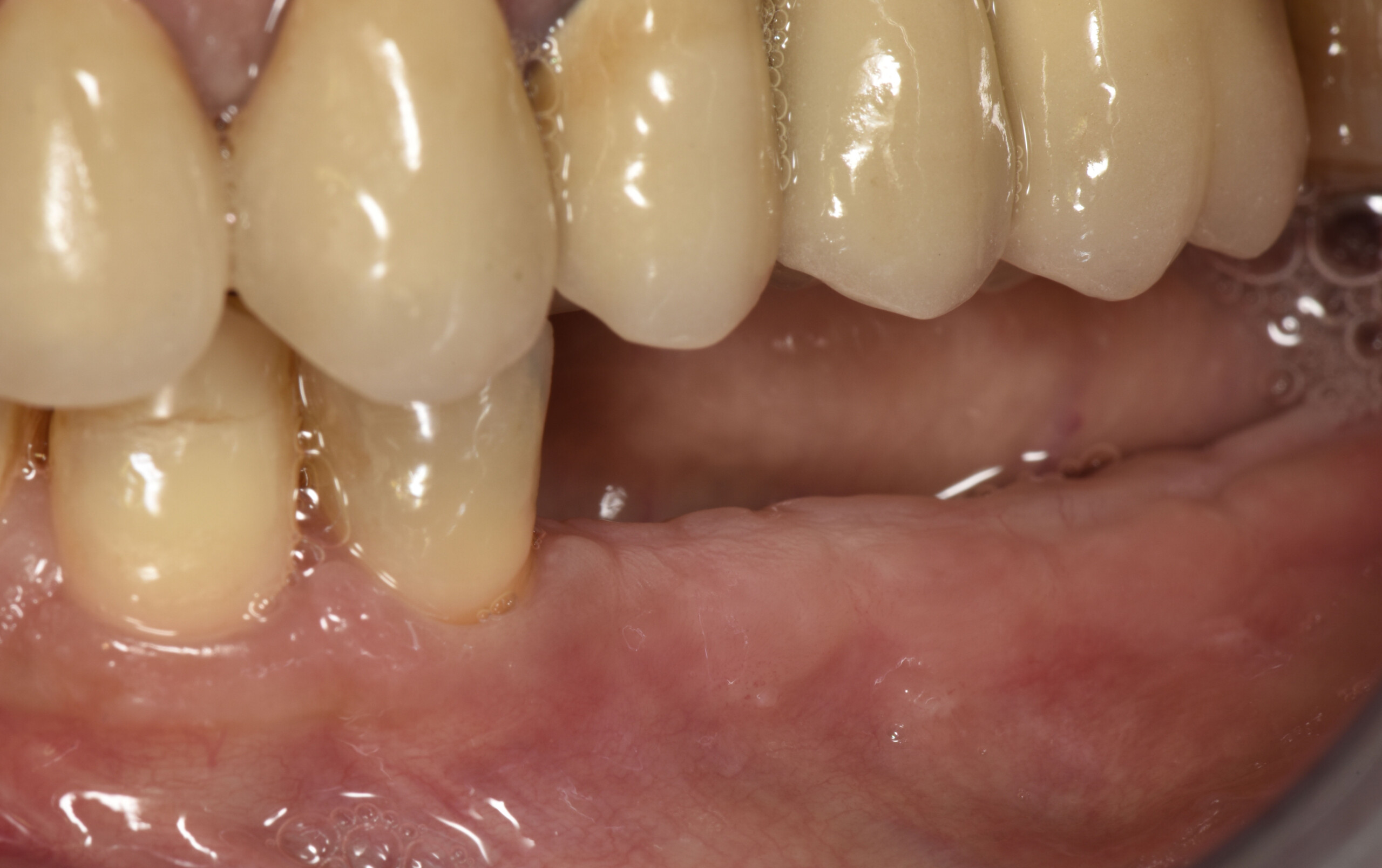
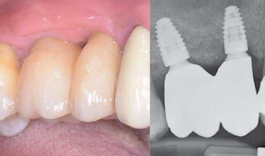
The goal of treatment was a late implant placement after bone regeneration and creation of stable periimplant soft tissue for long-term implant preservation.
THE RISK PROFILE
| Low Risk | Medium Risk | High Risk | |
|---|---|---|---|
| Patient’s health | Intact immune system | Light smoker | Impaired immune system |
| Patient’s esthetic requirements | Low | Medium | High |
| Gingival biotype | Thick – “low scalloped” | Medium – “medium scalloped” | Thin – “high scalloped” |
| Infection at implant sight | None | Chronic | Acute |
| Bone height at adjacent tooth | ≤ 5 mm from contact point | 5.5 – 6.5 mm from contact point | ≥ 7 mm from contact point |
| Width of tooth gap | 1 tooth (≥ 7 mm) | 1 tooth (≤ 7 mm) | 2 teeth or more |
| Soft-tissue anatomy | Intact | Compromised | |
| Bone anatomy of the alveolar ridge | No defect | Horizontal defect | Vertical defect |
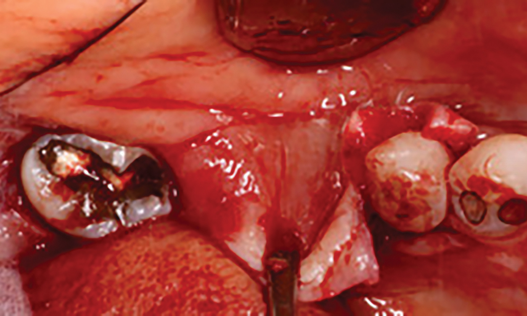
Additional Risk Factors: Roots were divergent, and intra-radicular bone (septal bone) was excellent, with more than 5 mm of remaining apical bone to achieve optimal primary stability.
THE APPROACH
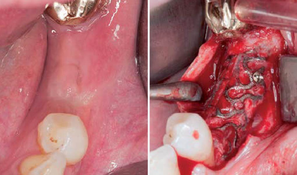
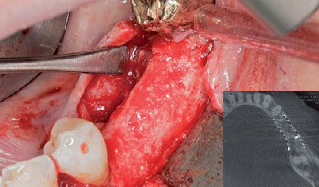
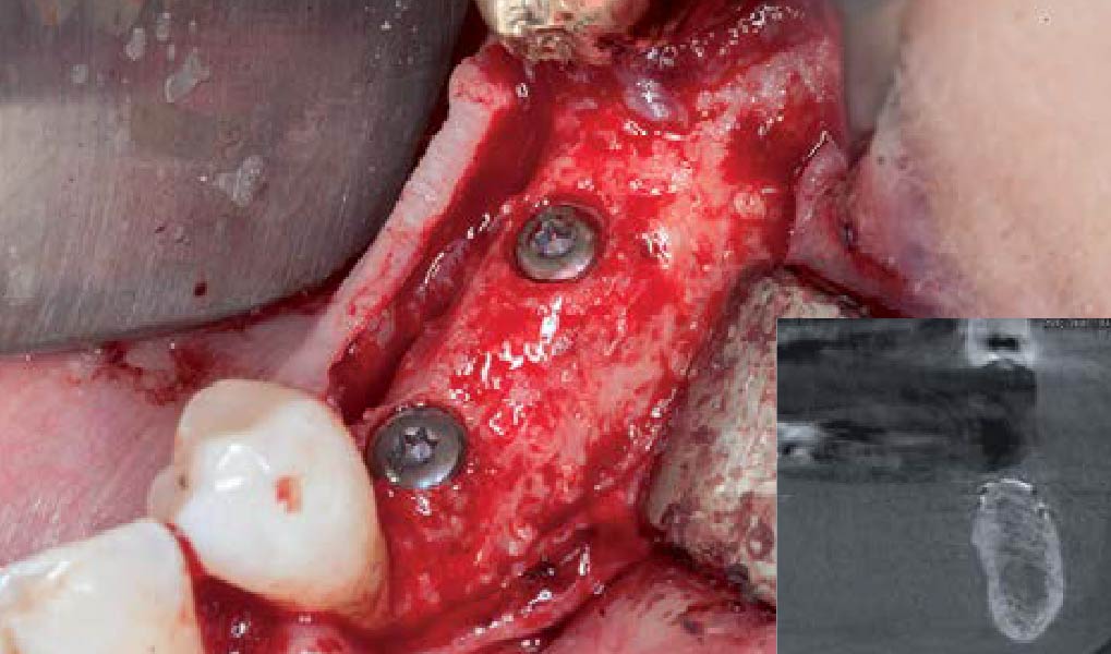
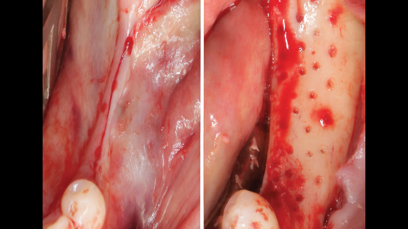
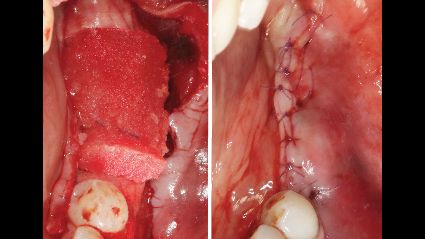
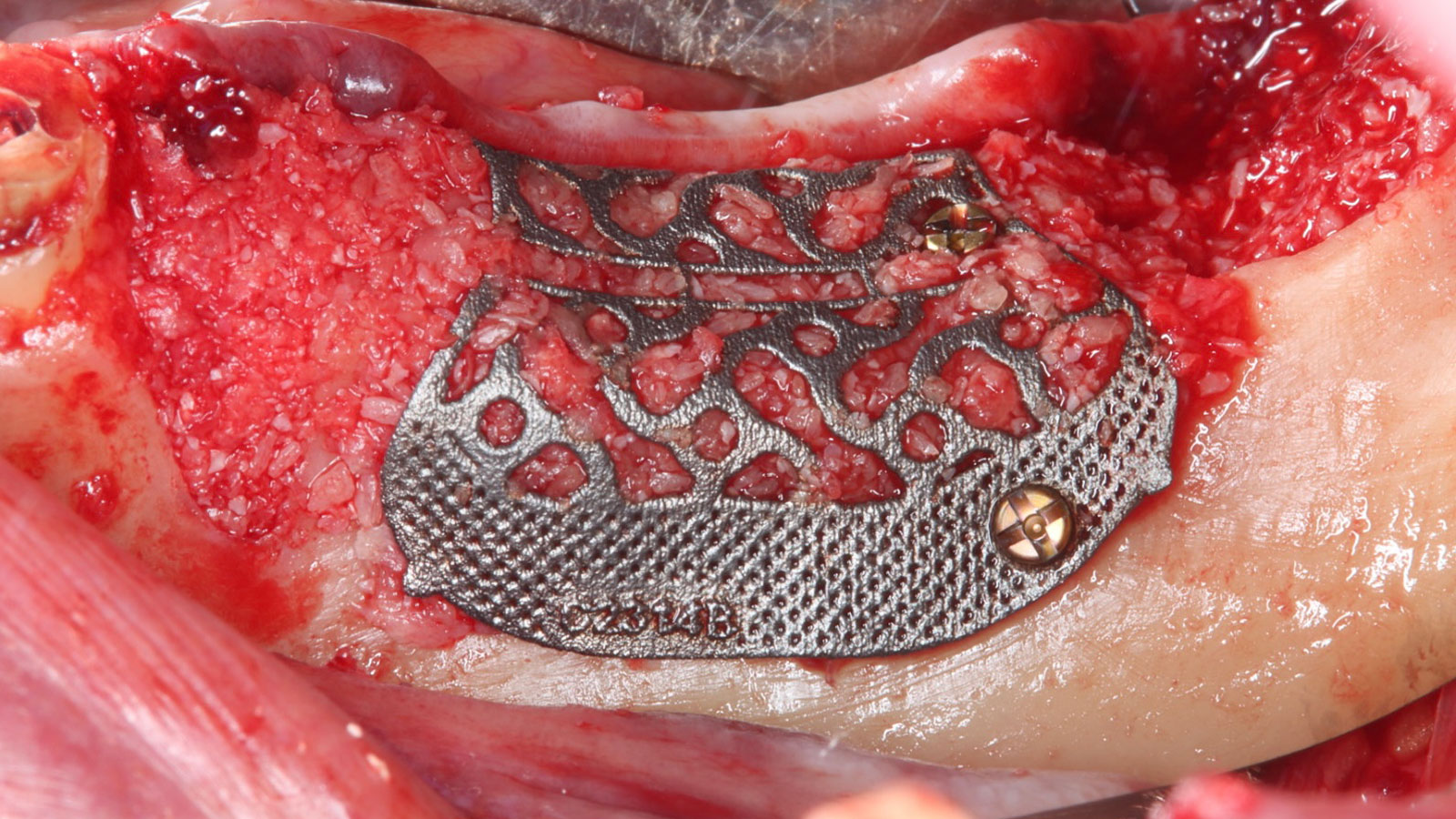
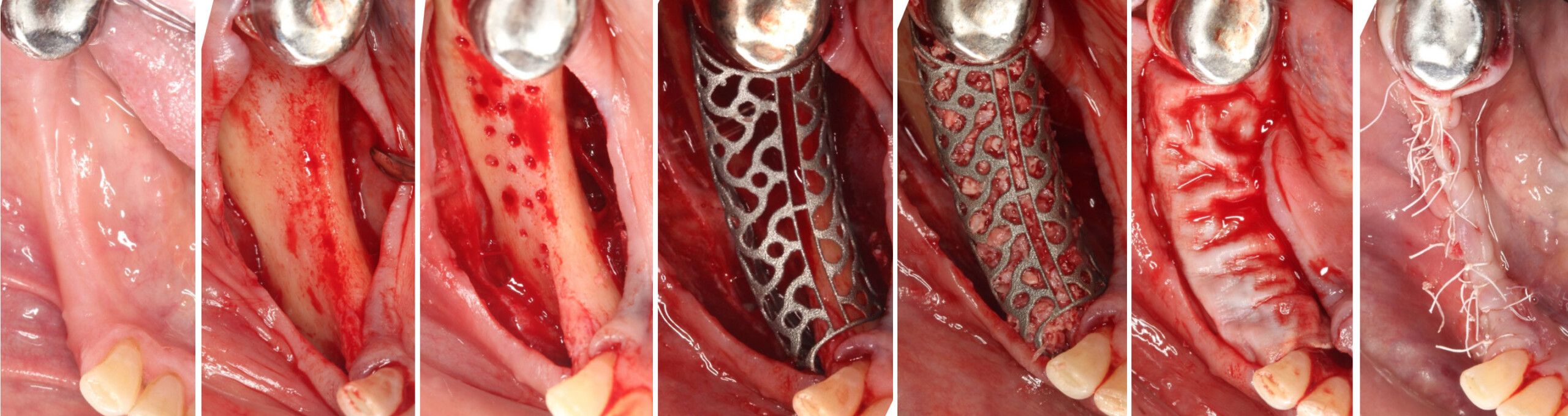
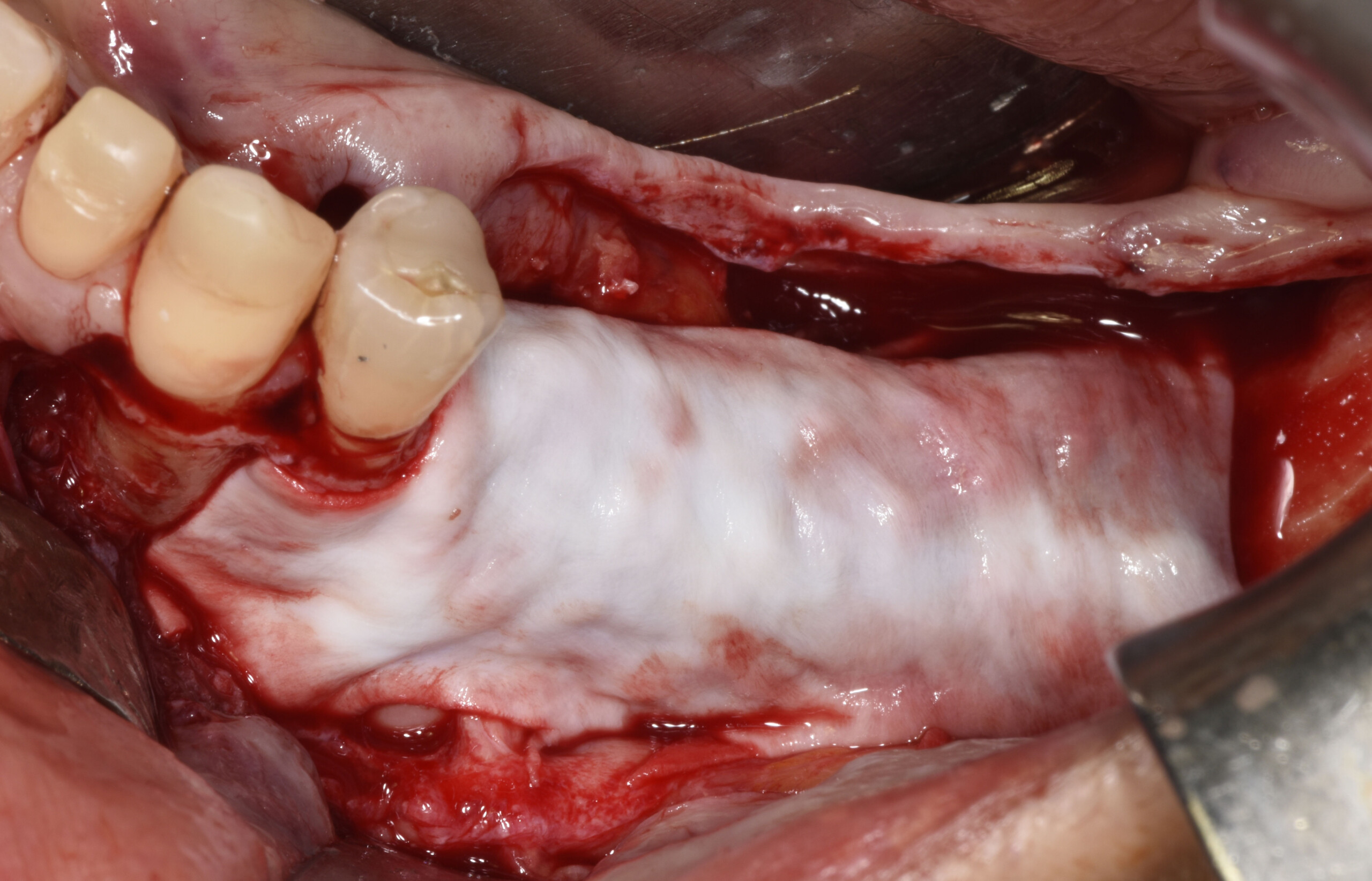
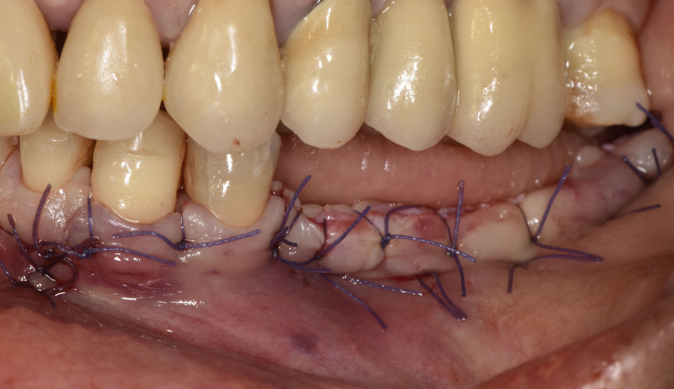
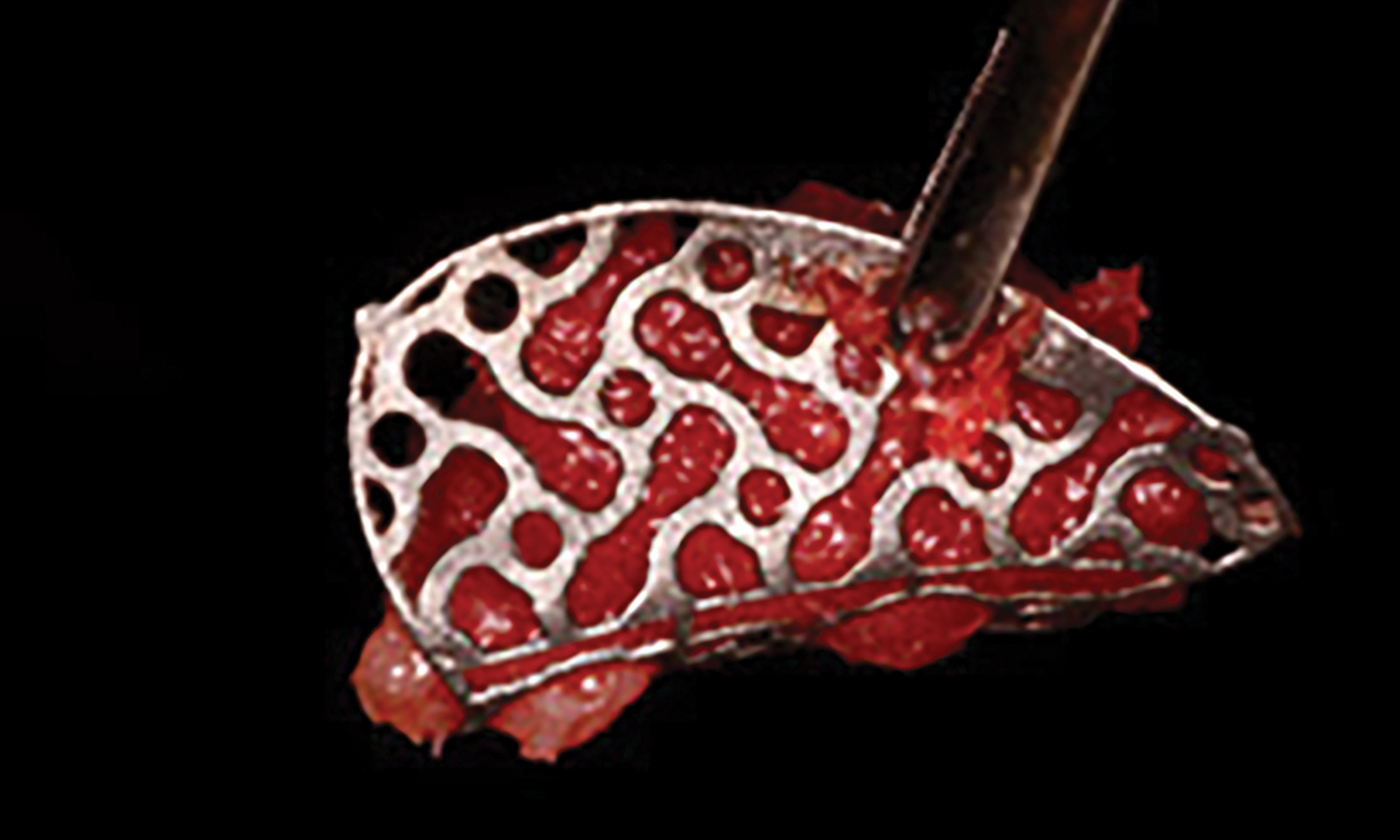
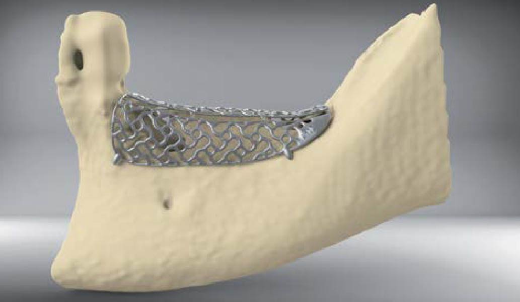
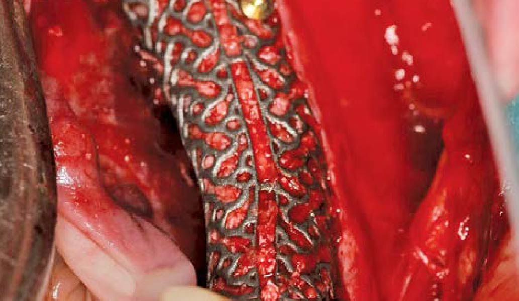
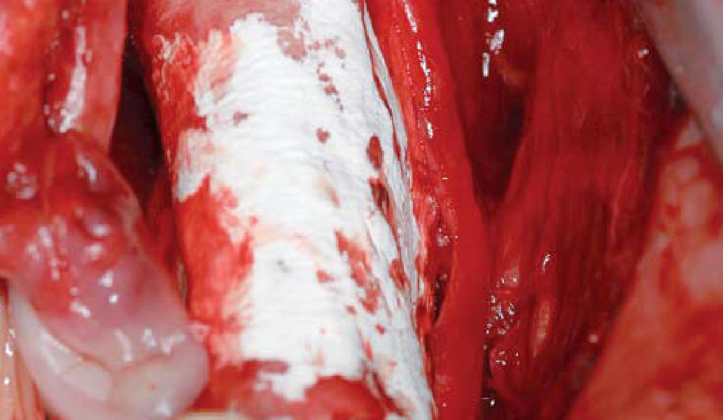
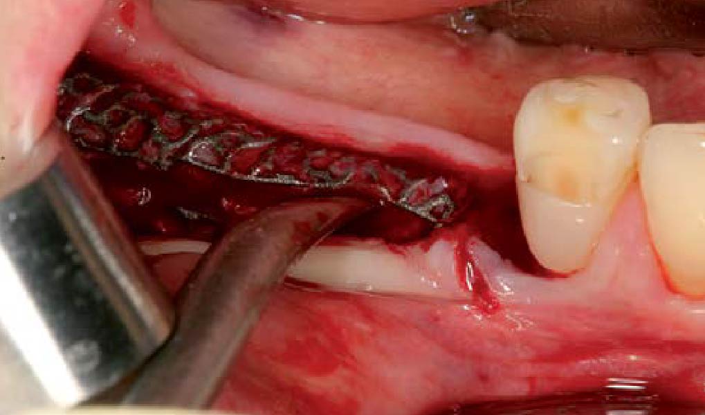
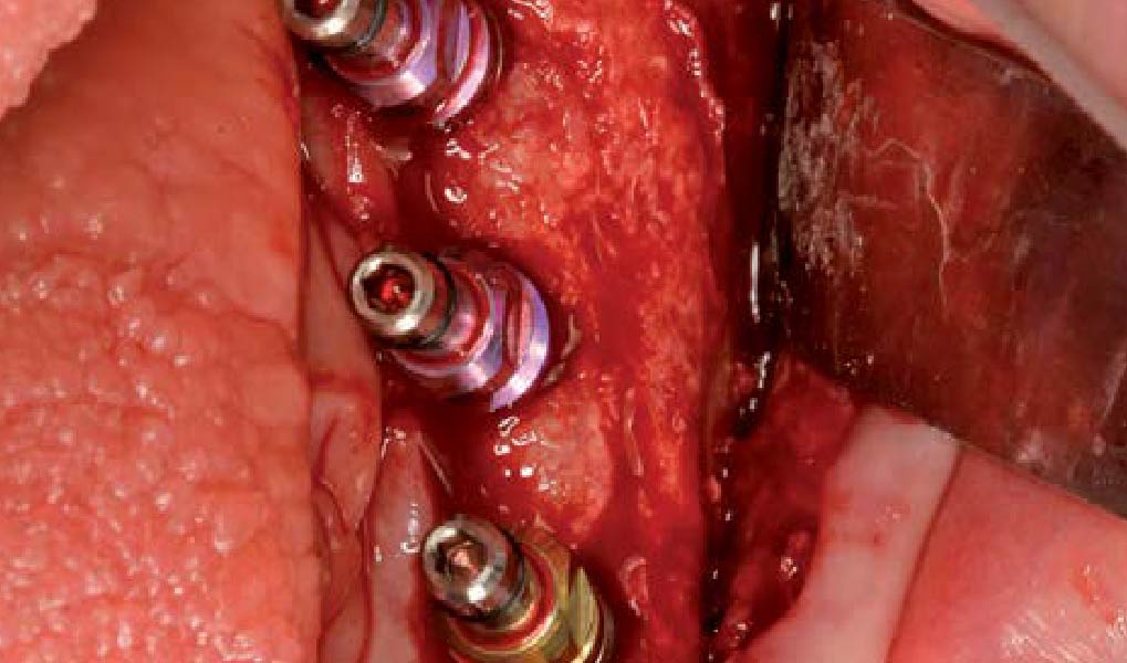
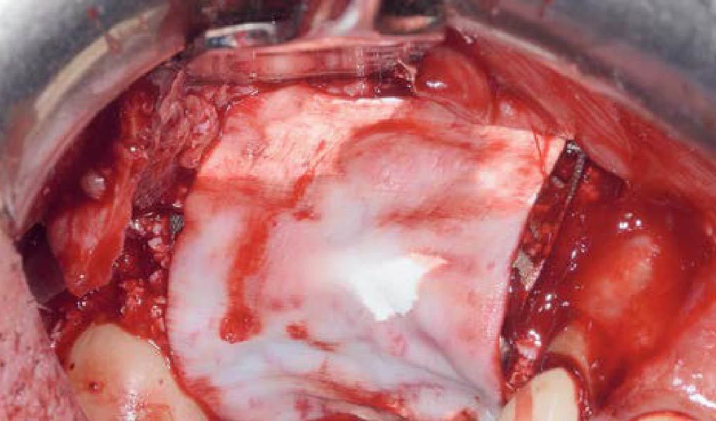
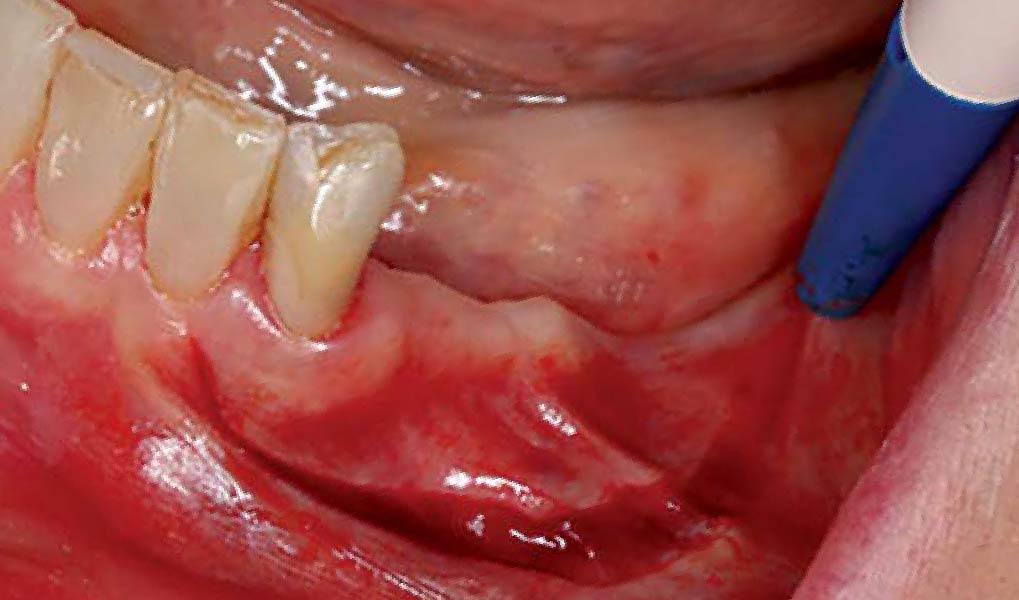
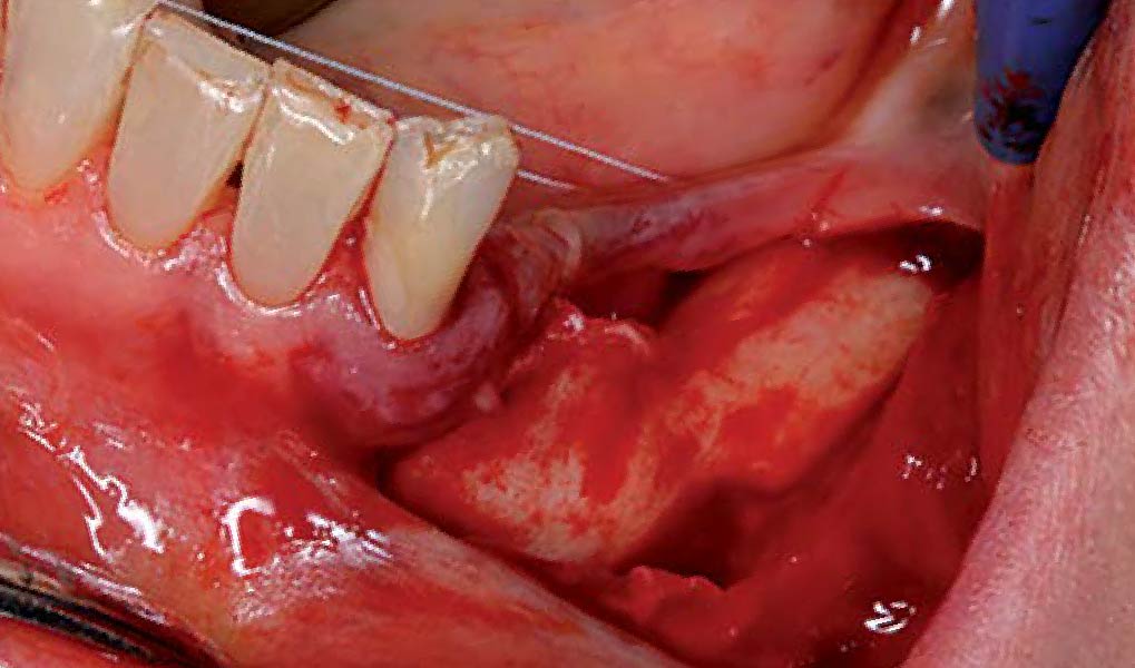
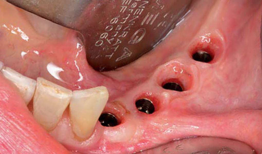
A customized bone regeneration procedure utilizing Yxoss CBR®. Followed by coverage of the graft with Geistlich Bio-Gide® for the purpose of Guided Bone Regeneration (GBR). Soft tissue thickening using Geistlich Fibro-Gide®. Delayed implantation into the augmented tissue. A vestibuloplasty with Geistlich Mucograft® for the regeneration of keratinized mucosa.
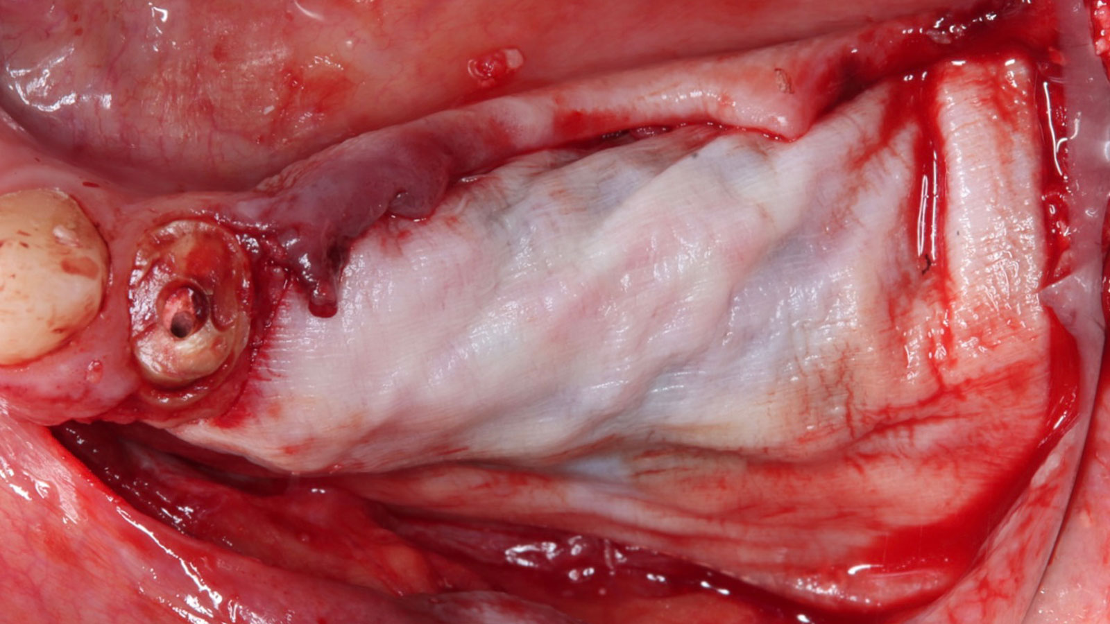
“Using the Geistlich Fibro-Gide® matrix enabled concurrent augmentation of hard
— Arnd Lohmann, MSc
and soft tissues without any postoperative complications. At the same time, the soft
tissue thickening facilitated floor of the mouth surgery and vestibuloplasty.”
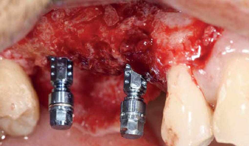
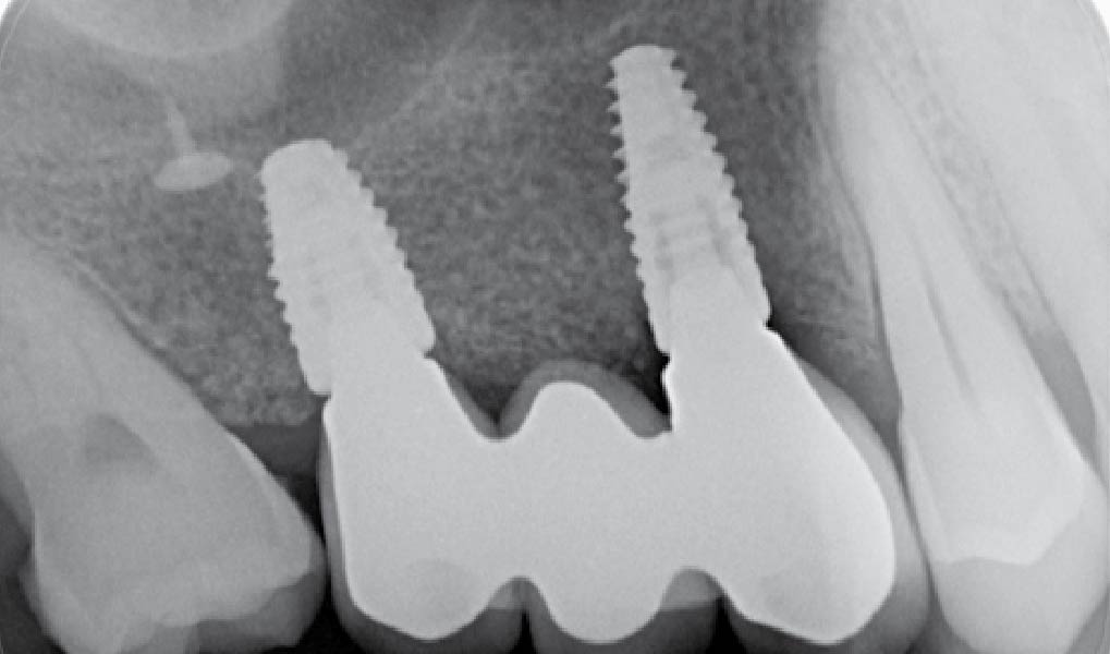
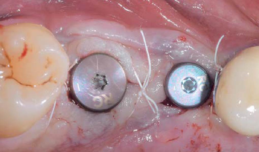
THE OUTCOME
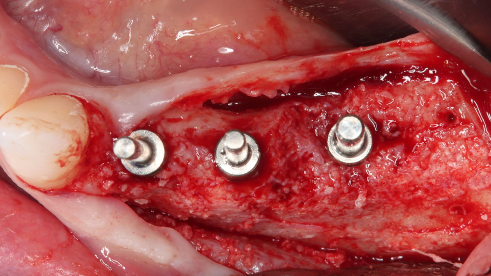
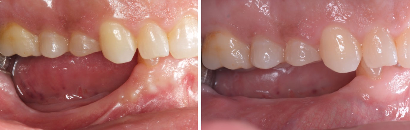
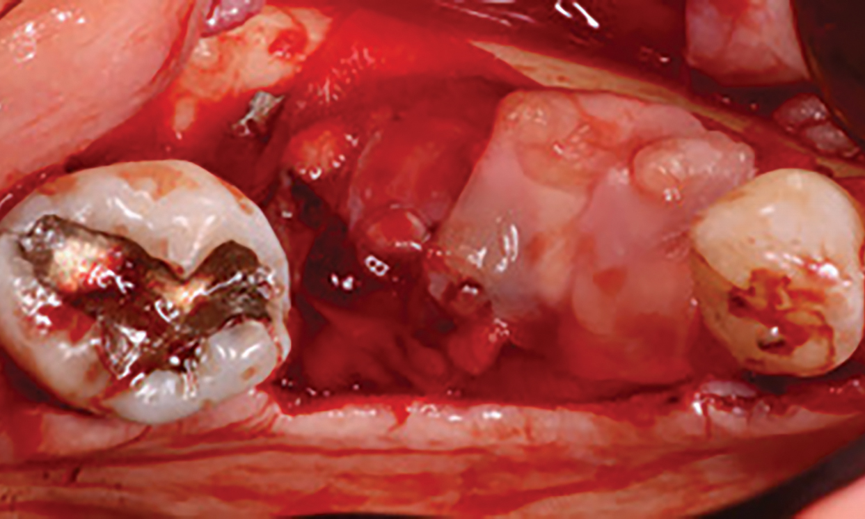
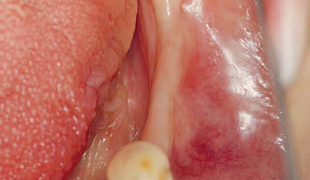
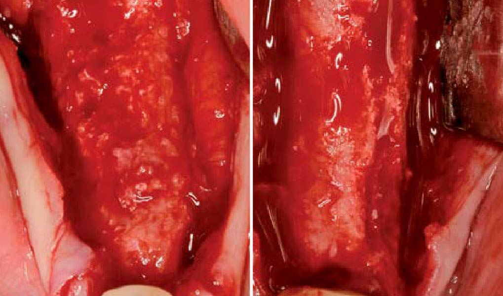
Treatment resulted in approximately 5 mm of vertical bone regeneration. The potential occurrence of a dehiscence associated with a wound opening and exposure of Yxoss CBR® was able to be prevented with Geistlich Fibro-Gide®.
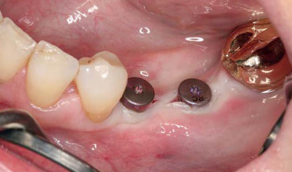
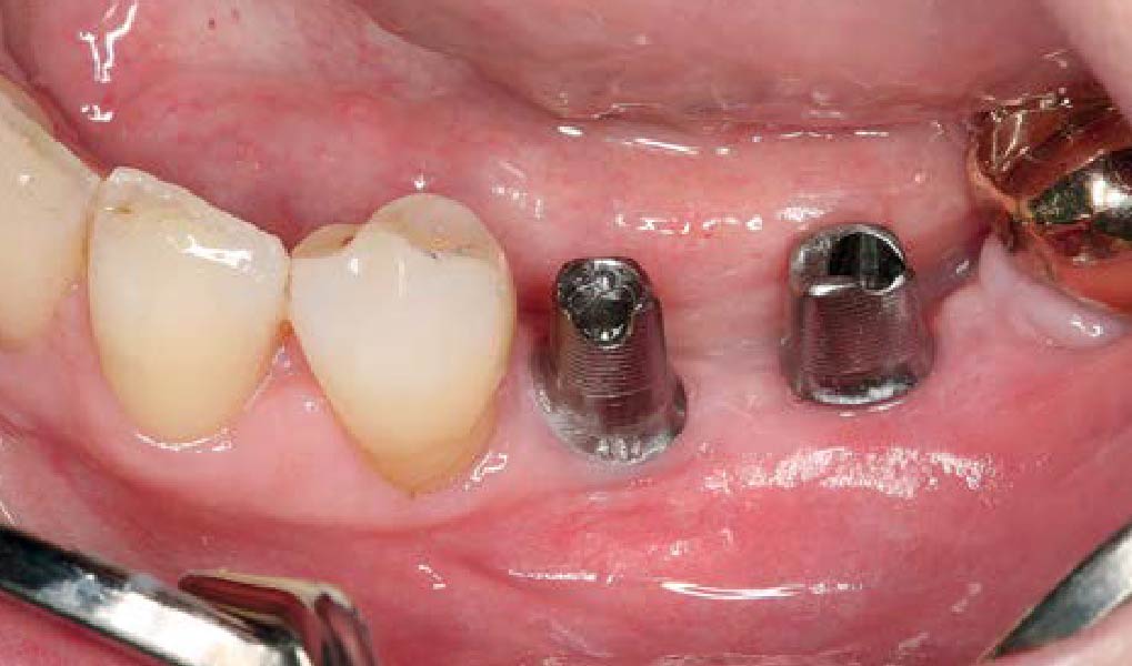
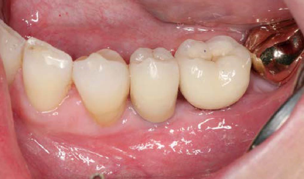
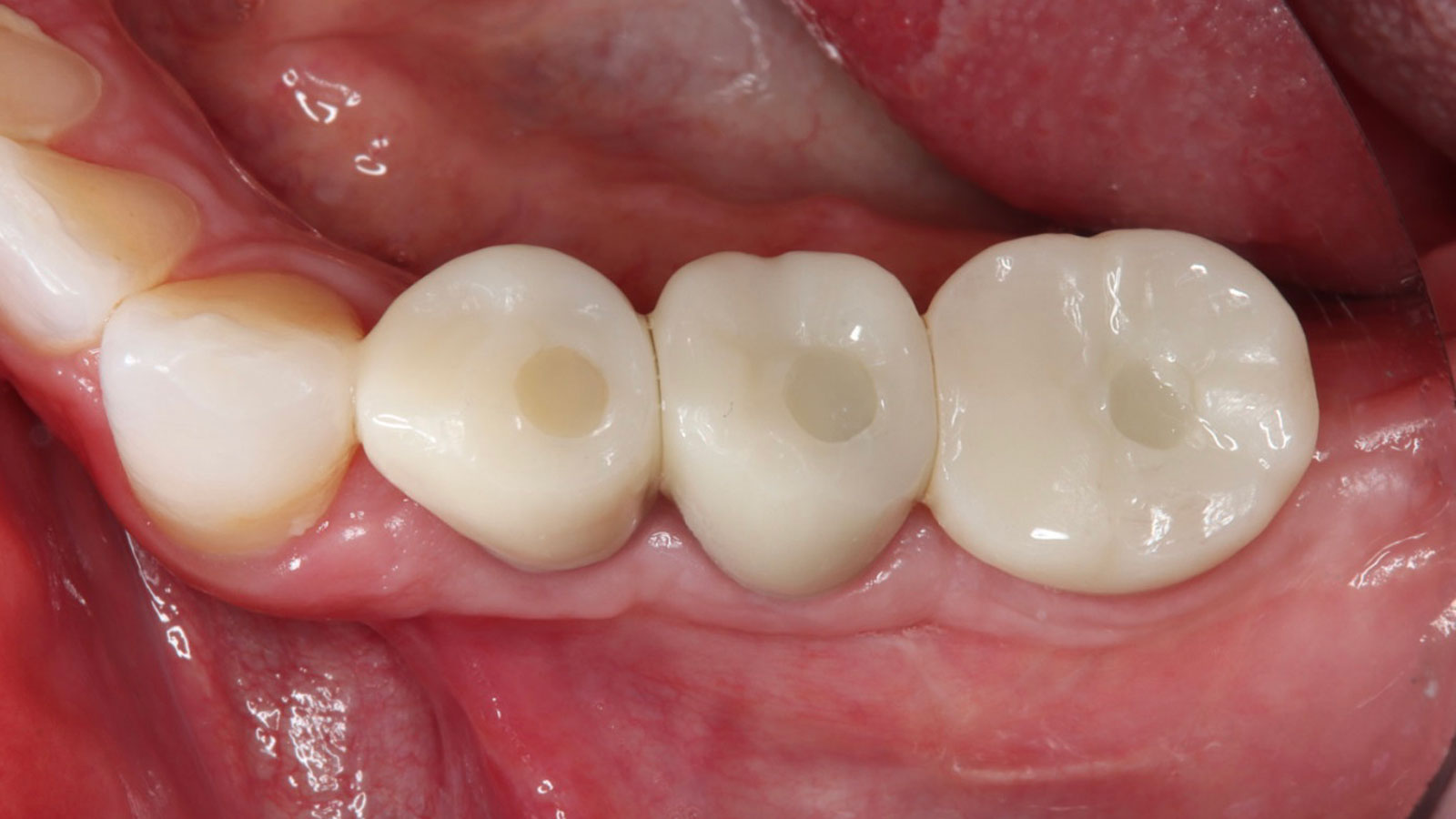
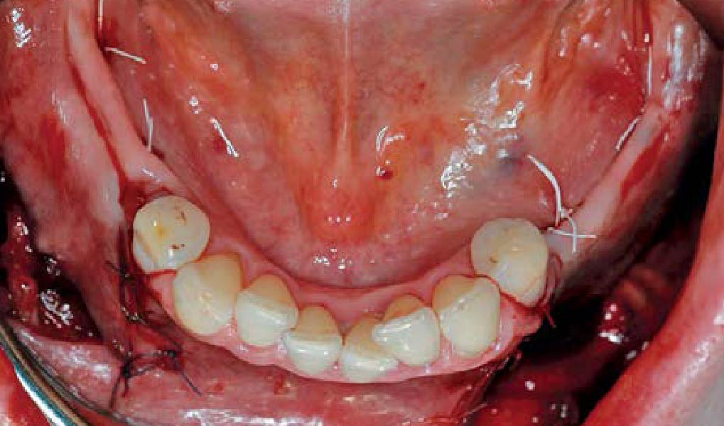
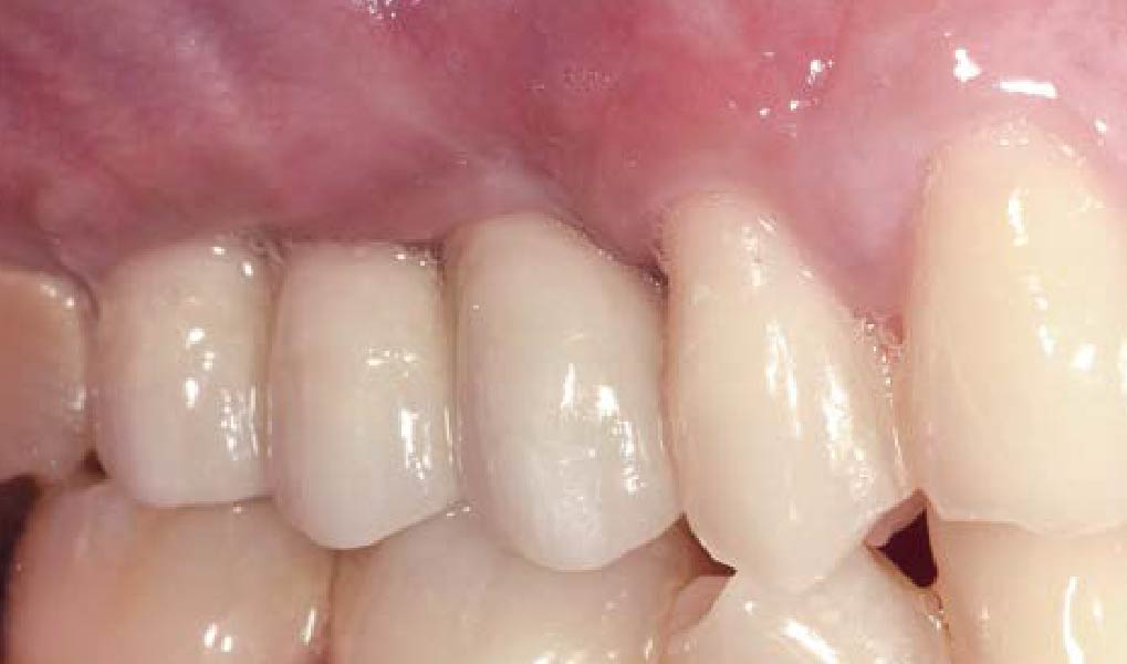
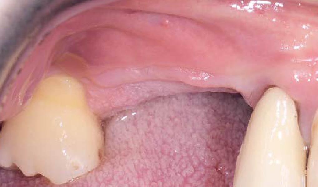
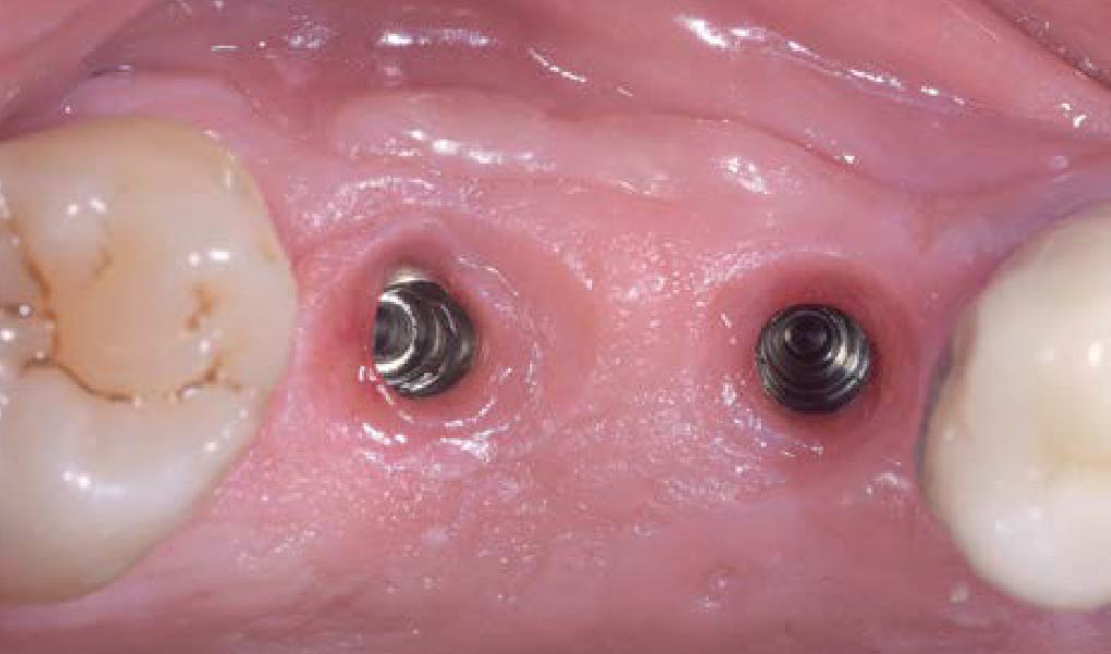
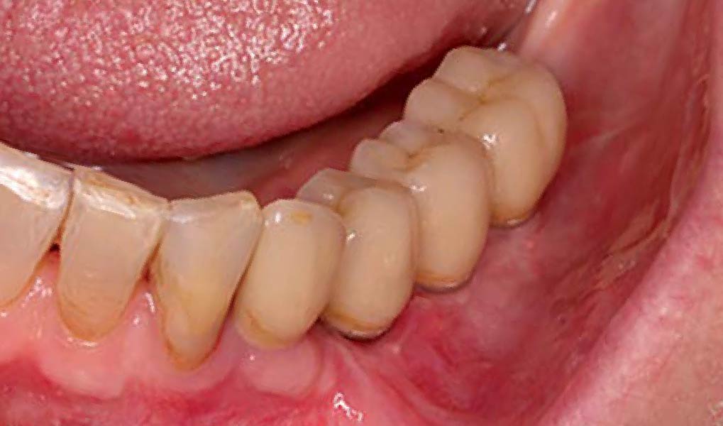
On one hand, the quality of the peri-implant soft tissue was improved by the
soft tissue thickening with Geistlich Fibro‑Gide® and, on the other, by increasing the width of keratinized mucosa with Geistlich Mucograft®. The treatment method chosen resulted in a reduced invasiveness and morbidity by avoiding a donor site for sourcing a transplant.
Arnd Lohmann, MSc
Dr. Arnd Lohmann is a recognized specialist in implantology and periodontology. He earned his dental license in Hamburg in 2002, completed his doctorate in 2003, and has been a partner at a private practice in Bremen since then.
With a Master of Science in Implantology (2007), he specializes in dental implantology and bone augmentation. He is an active speaker at national and international congresses, leads the Bremen study group of the German Society of Oral Implantology (DGOI), and is a member of DGOI, DGZI, and DGI. His practice is equipped with state-of-the-art technology, ensuring high-quality patient care.
BIOBRIEF
Mandibular Ridge Augmentation Using Customized Titanium Mesh
THE SITUATION
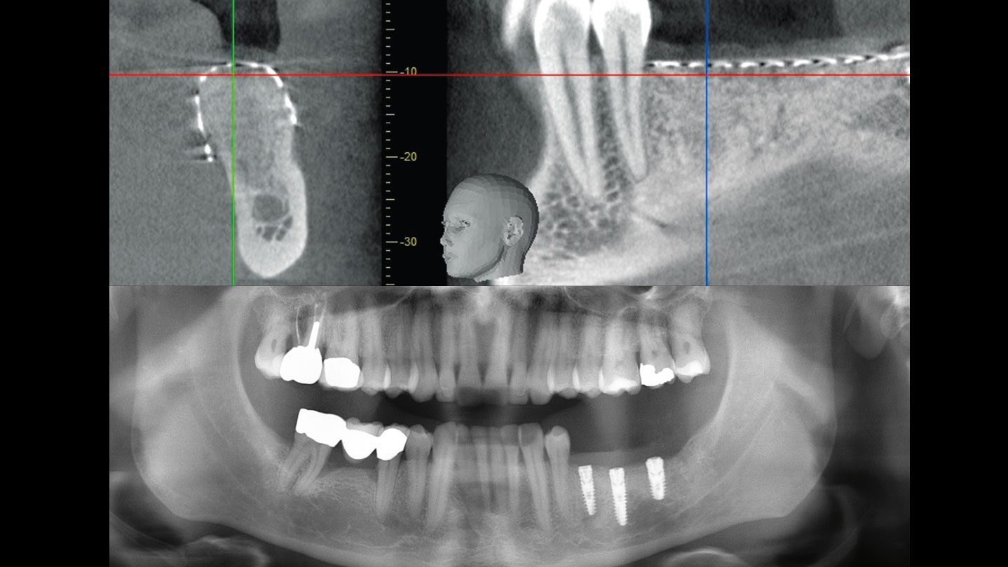
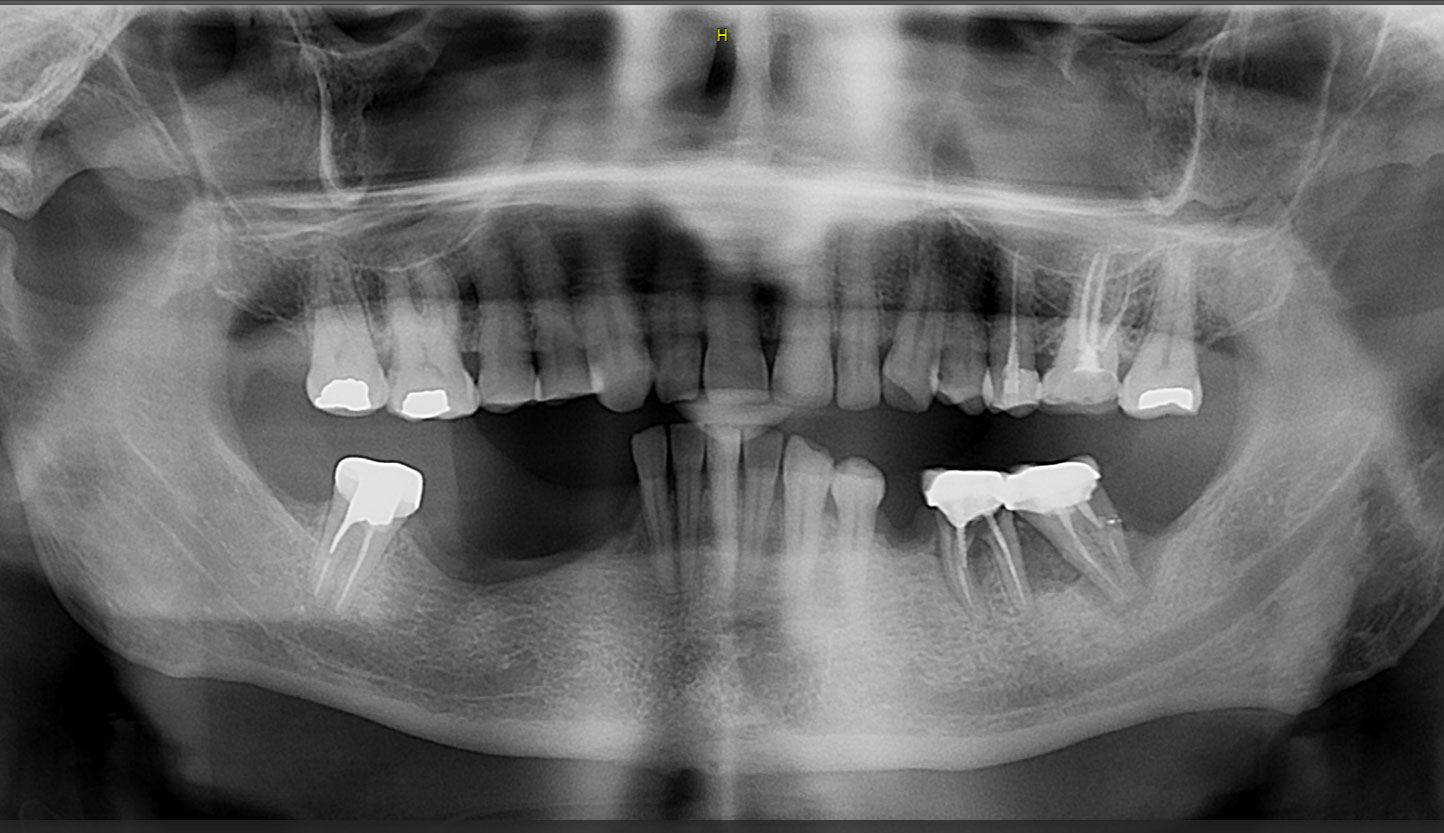
A 60-year-old healthy male presented with a failing lower left bridge. Due to a long history of missing teeth, he had a significantly atrophic mandibular ridge. We decided to use a customized titanium mesh to achieve the necessary vertical and horizontal bone augmentation for dental implant rehabilitation.
THE RISK PROFILE
| Low Risk | Medium Risk | High Risk | |
|---|---|---|---|
| Patient’s health | Intact immune system | Light smoker | Impaired immune system |
| Patient’s esthetic requirements | Low | Medium | High |
| Restorative status of adjacent tooth | Intact | Restored | |
| Width of tooth gap | 1 tooth (≥ 7 mm) | 1 tooth (≤ 7 mm) | 2 teeth or more |
| Bone anatomy of the alveolar ridge | No defect | Horizontal defect | Vertical defect |
THE APPROACH
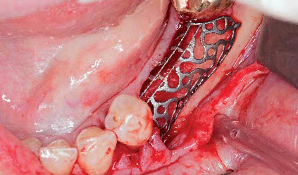
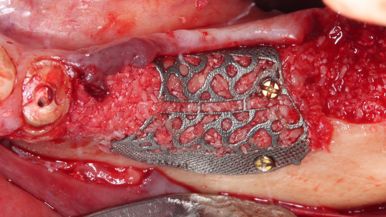
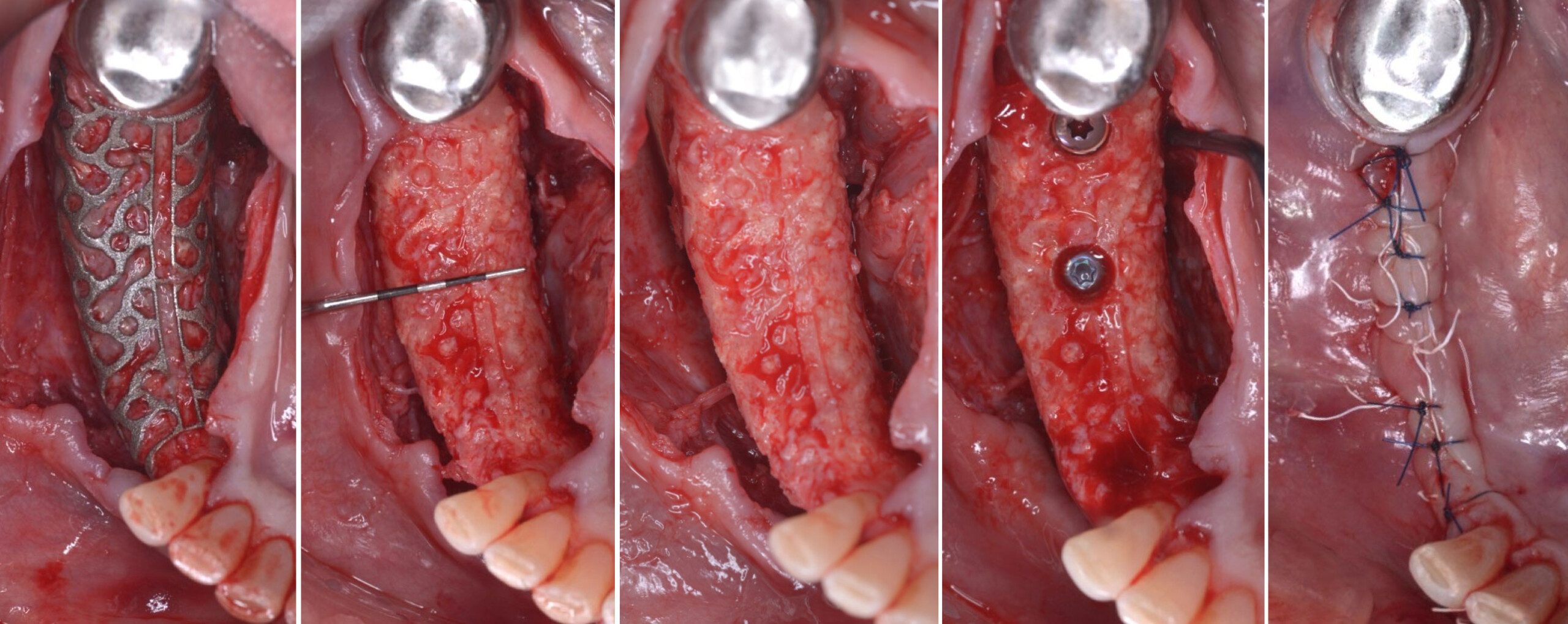
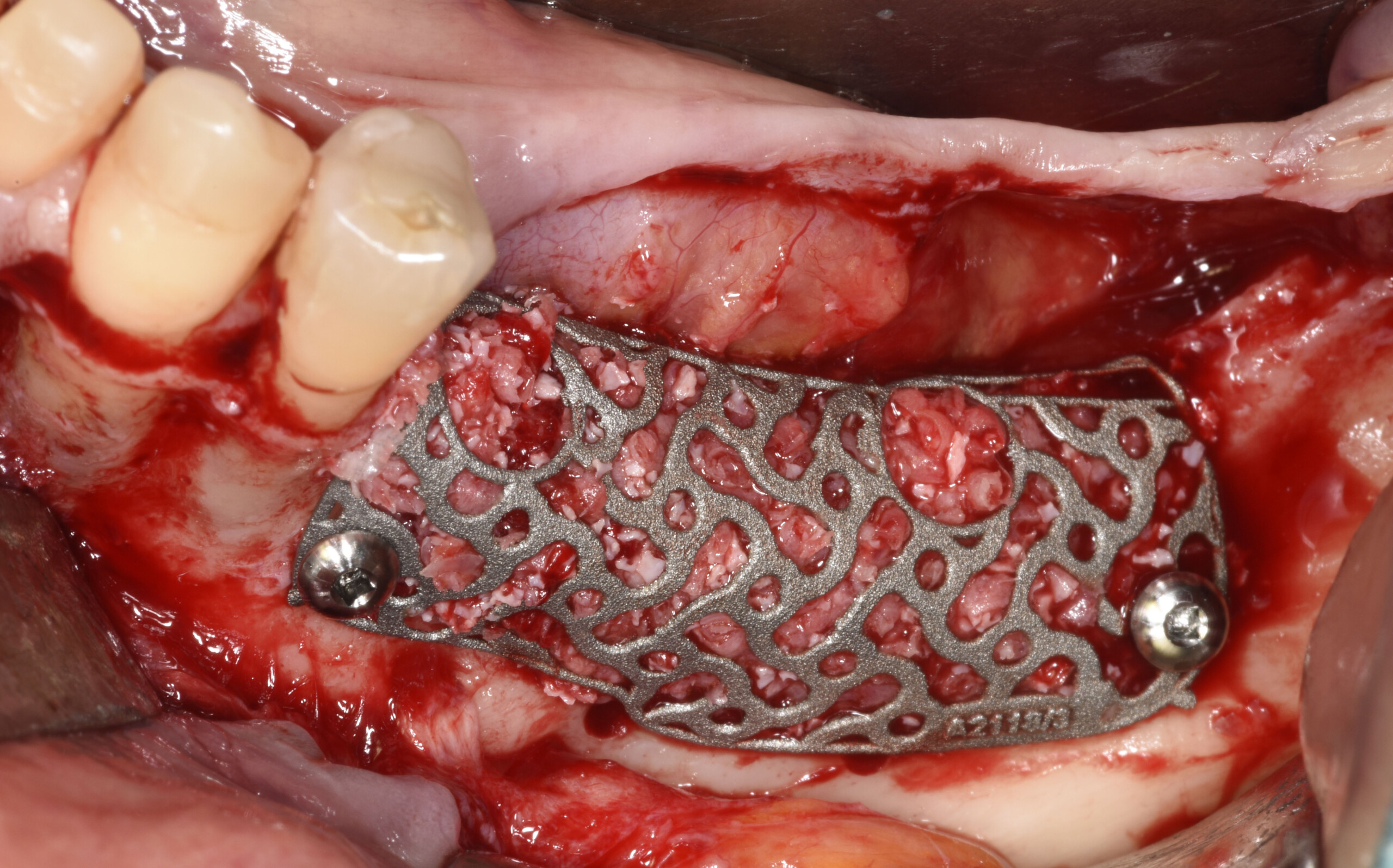
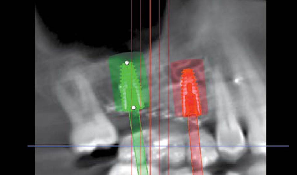
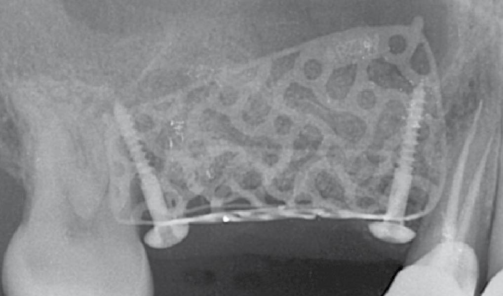
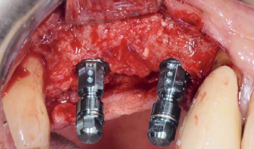
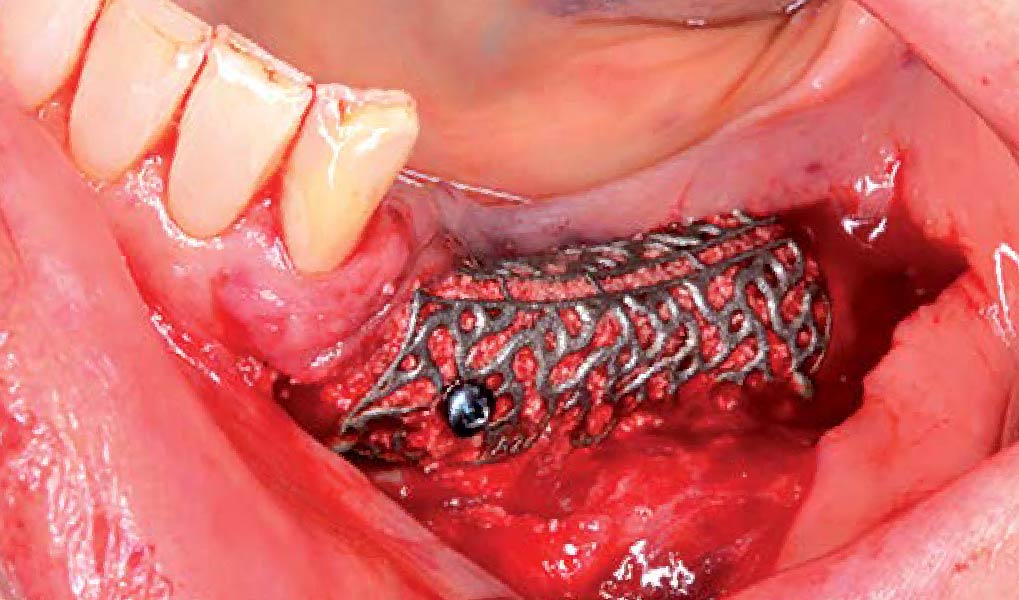
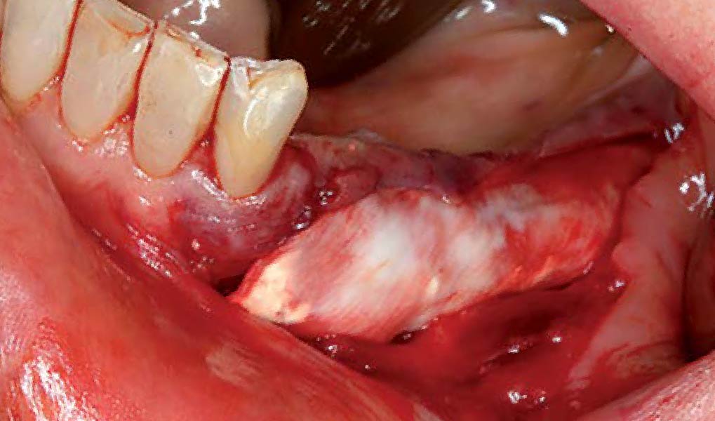
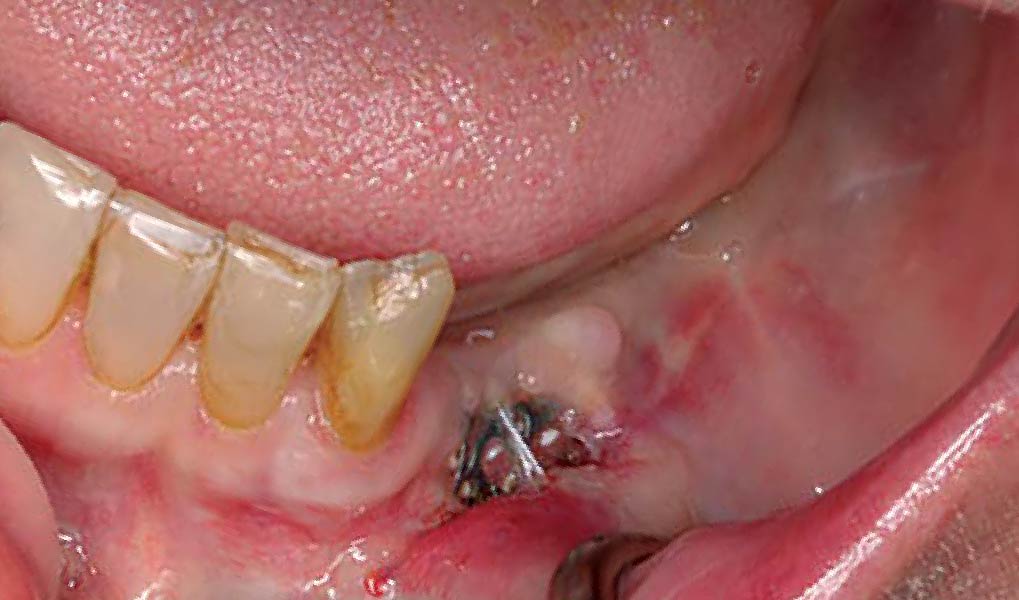
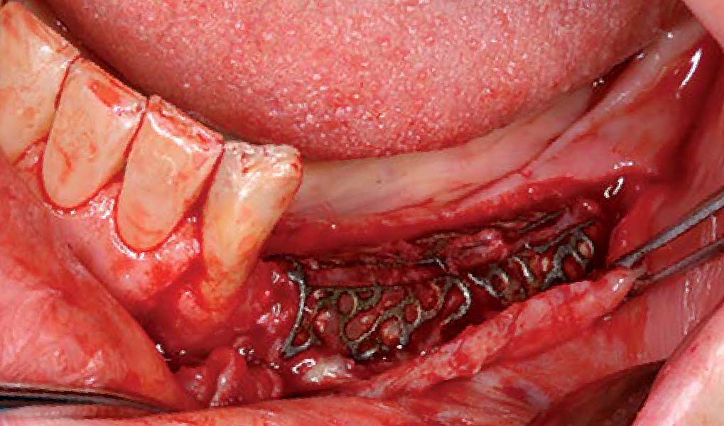
The goal of this procedure was to regenerate sufficient bone to place restoratively driven dental implants. Due to the horizontal and vertical ridge deficiency, we used a customized titanium mesh to predictably achieve this outcome.
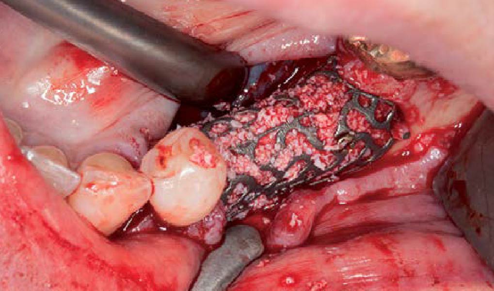
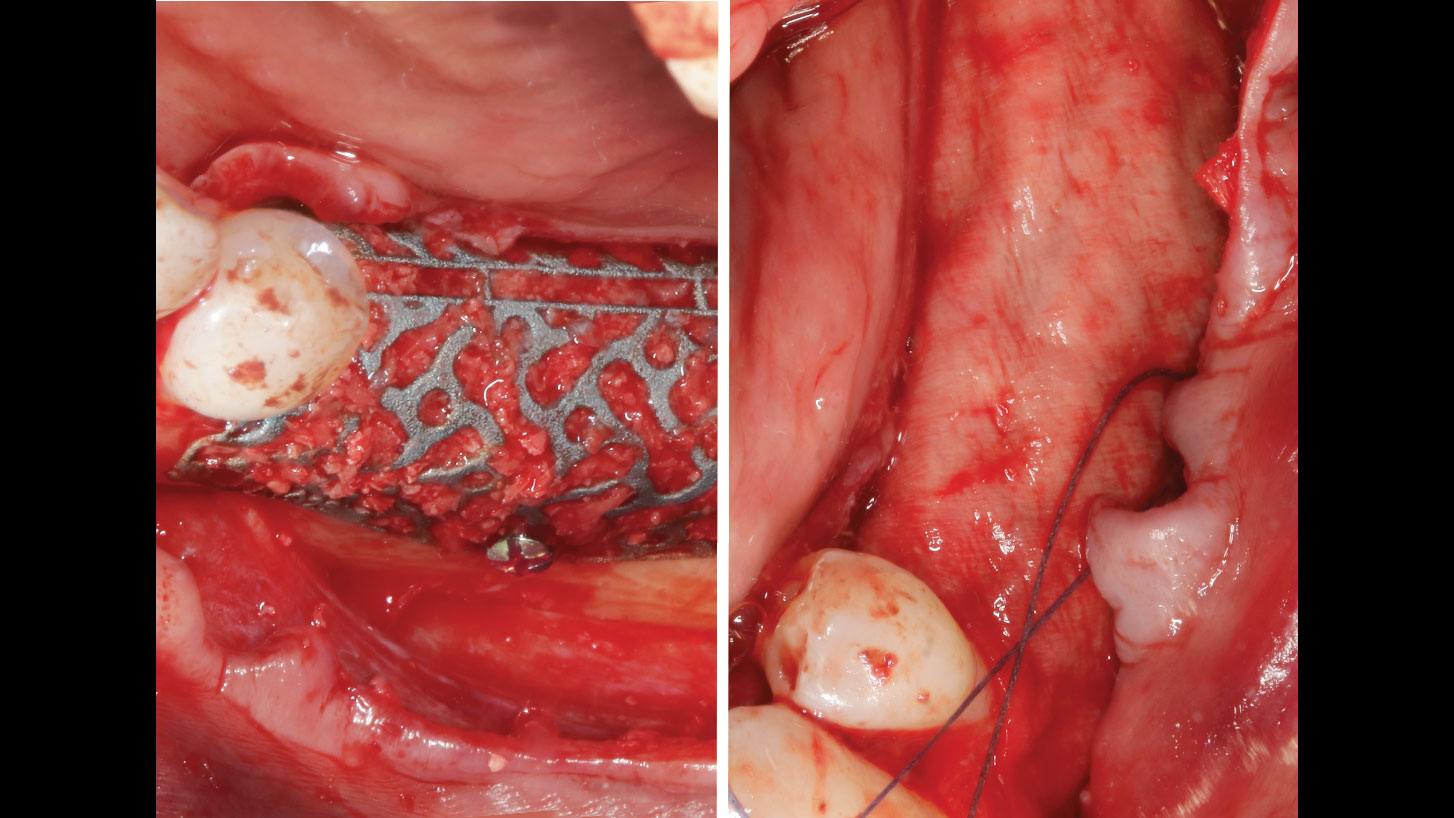
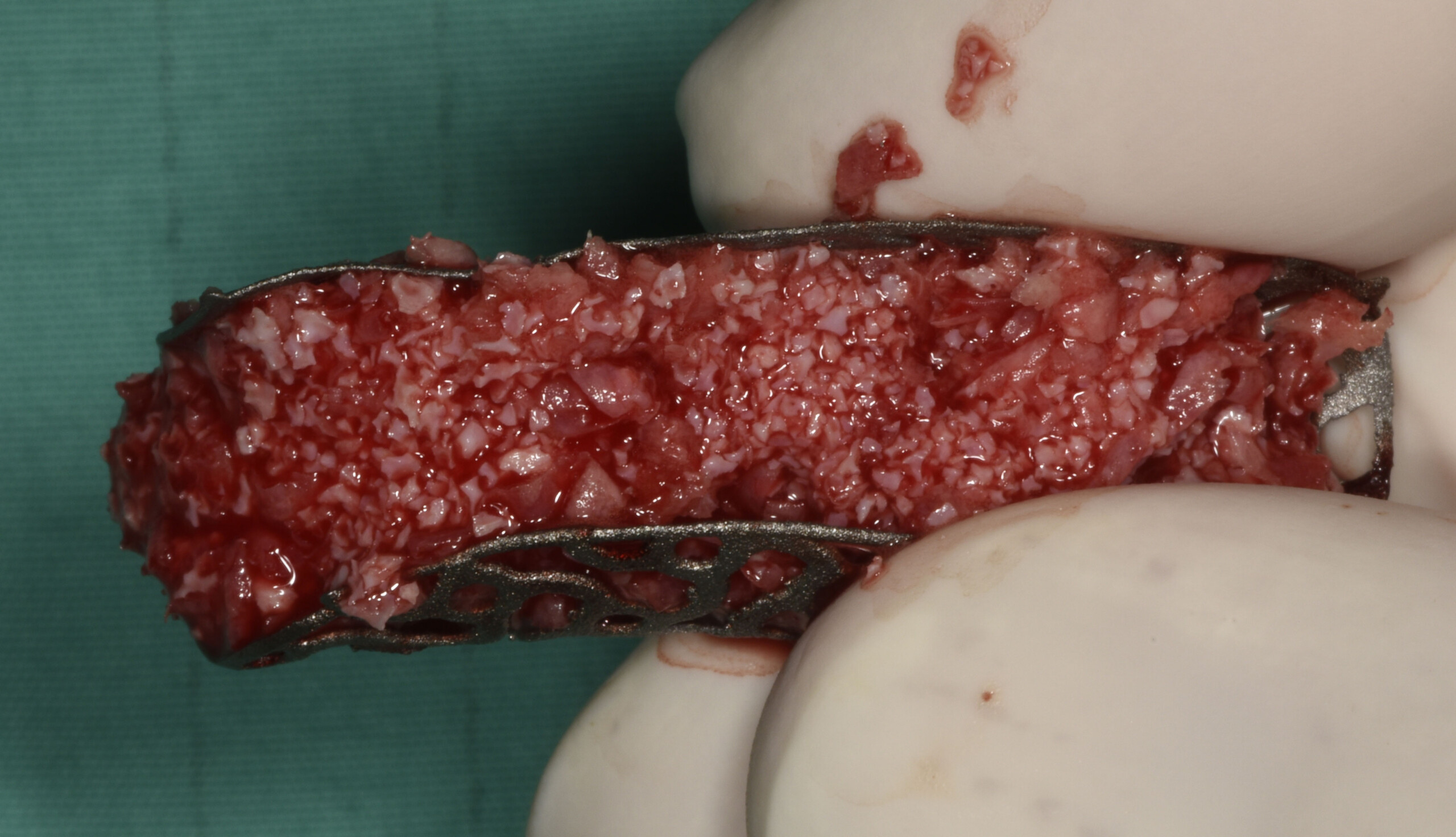
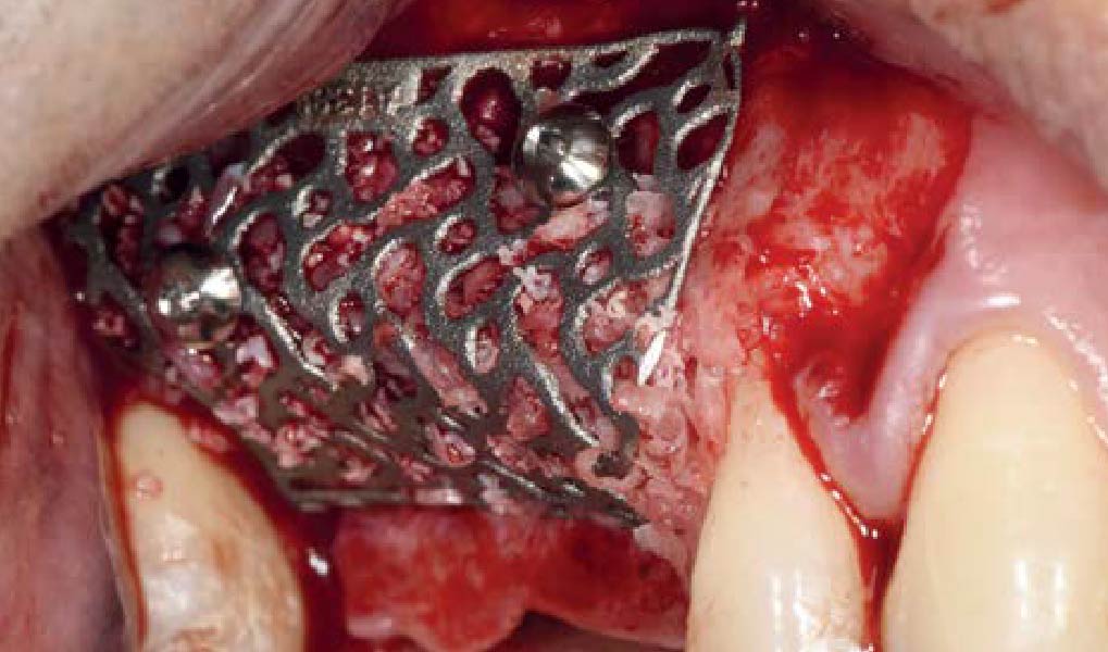
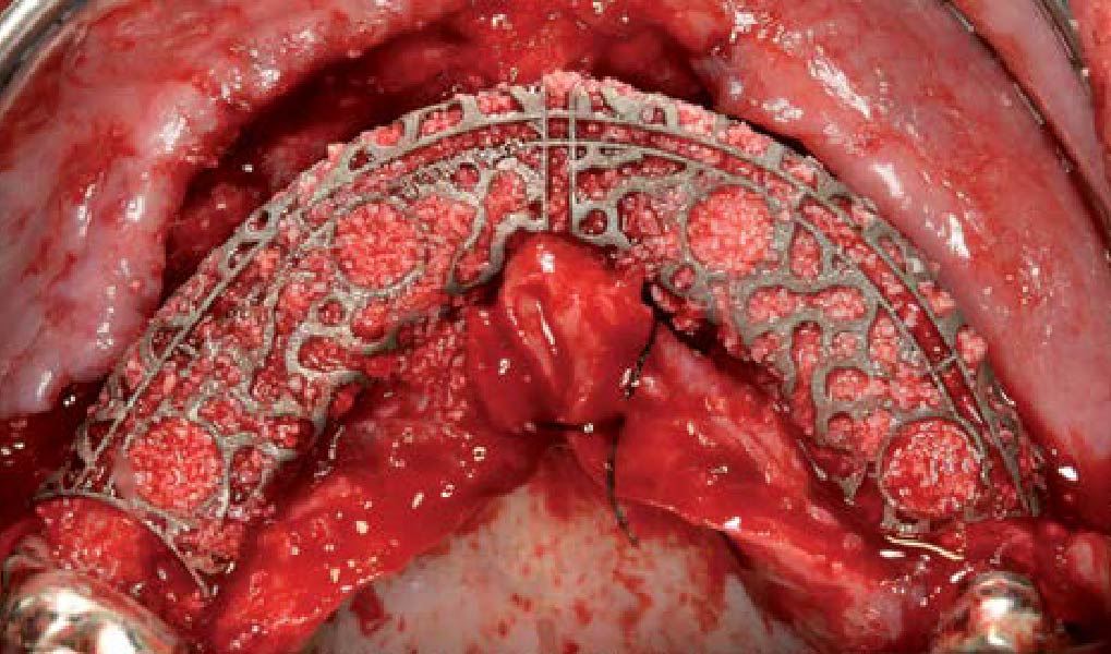
Autogenous bone collected with SafeScraper Twist and Geistlich Bio-Oss® filled the Yxoss CBR® Protect and a Geistlich Bio-Gide® collagen membrane covered the mesh.
.
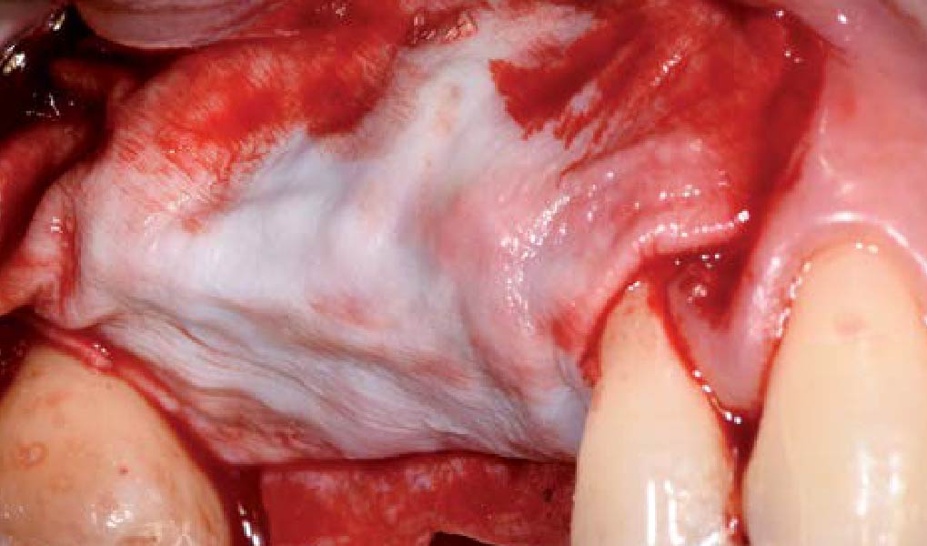
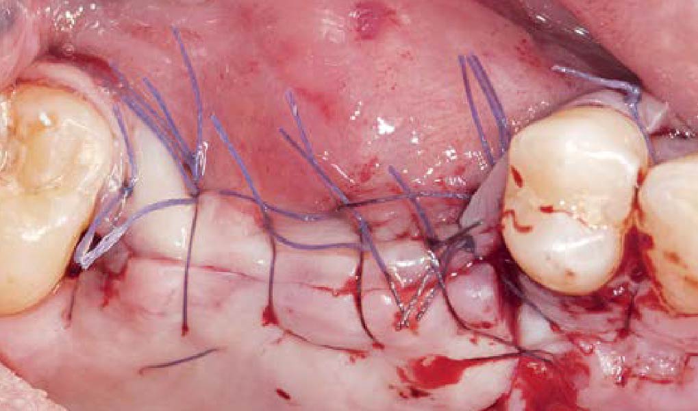
“Success in these cases primarily depends on proper mesh design and careful handling of soft tissue to ensure zero-tension primary closure.”
— Shaun R. Young, DMD
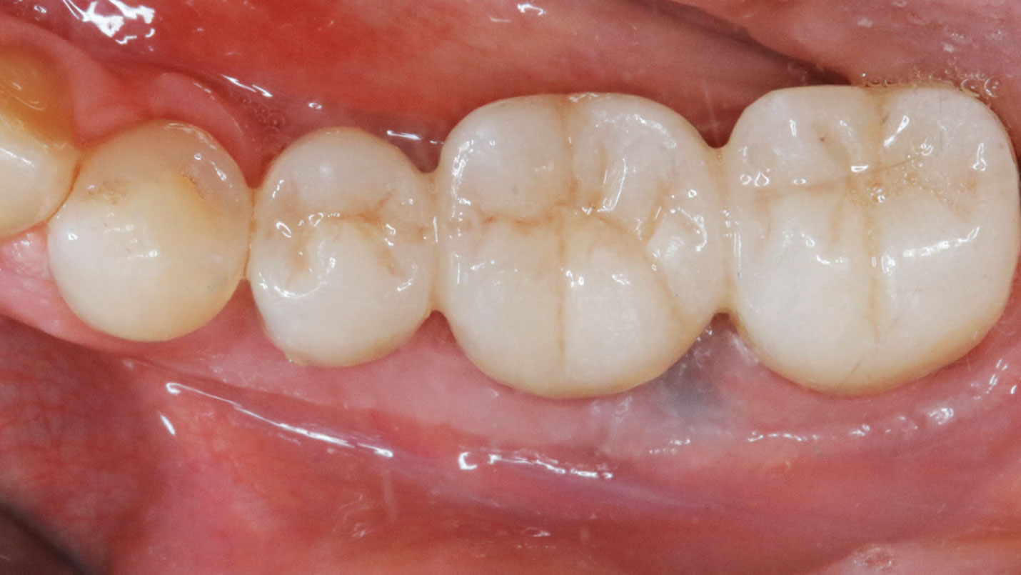
THE OUTCOME
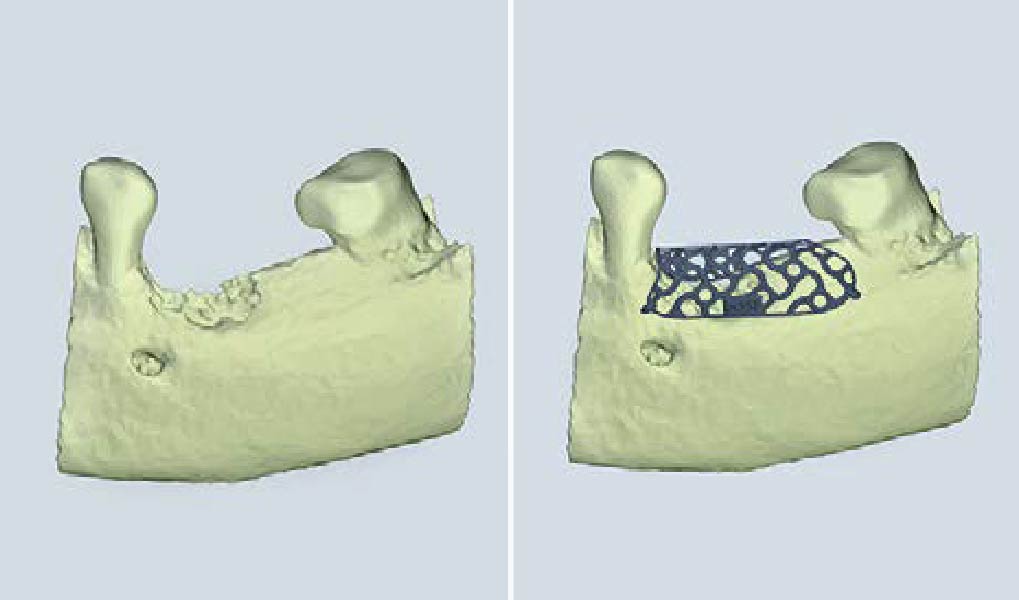
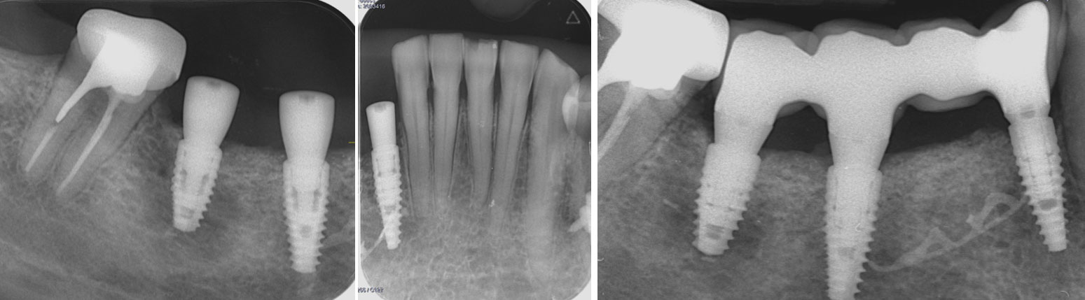
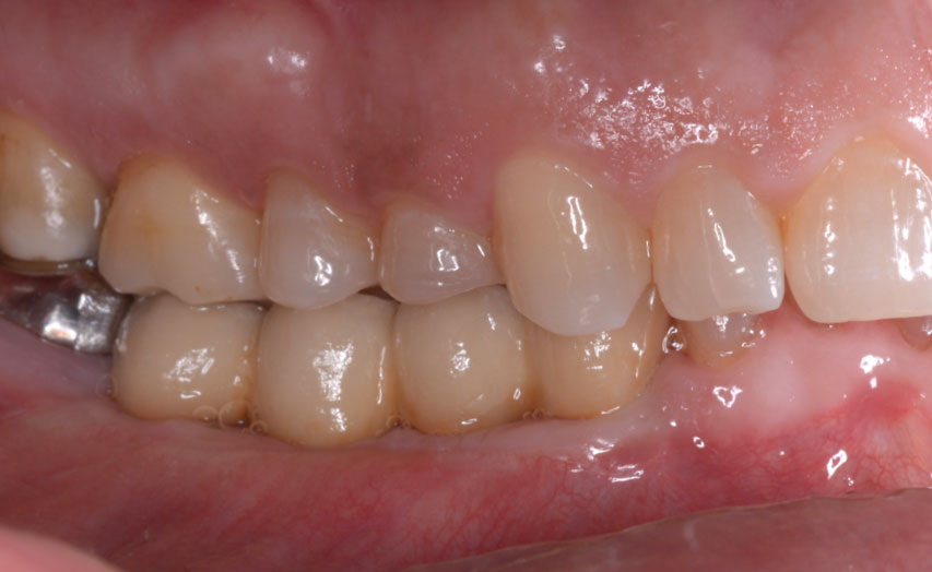
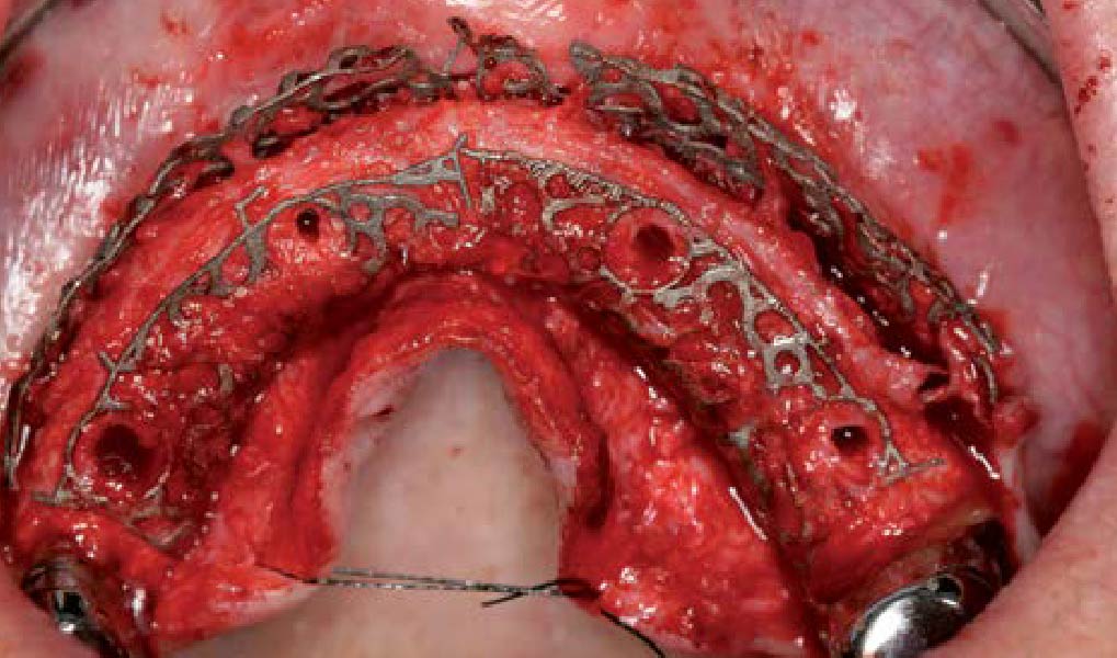
A left mandibular ridge deficiency was corrected using a Yxoss CBR® Protect Customized Bone Regeneration Titanium Mesh, designed from the patient’s CBCT scan.
Shaun R. Young, DMD
Dr. Shaun Young, an Oral and Maxillofacial Surgeon based in Tampa, Florida, specializes in complex ridge augmentation, immediate implants, and All-on-X full arch rehabilitation. He earned his Doctor of Dental Medicine degree from the University of Florida and completed his OMFS residency at Emory University in Atlanta, Georgia, where he served as Administrative Chief Resident. Dr. Young brings his expertise to a full-scope group practice, serving Tampa, Clearwater, and New Port Richey, Florida.
BIOBRIEF
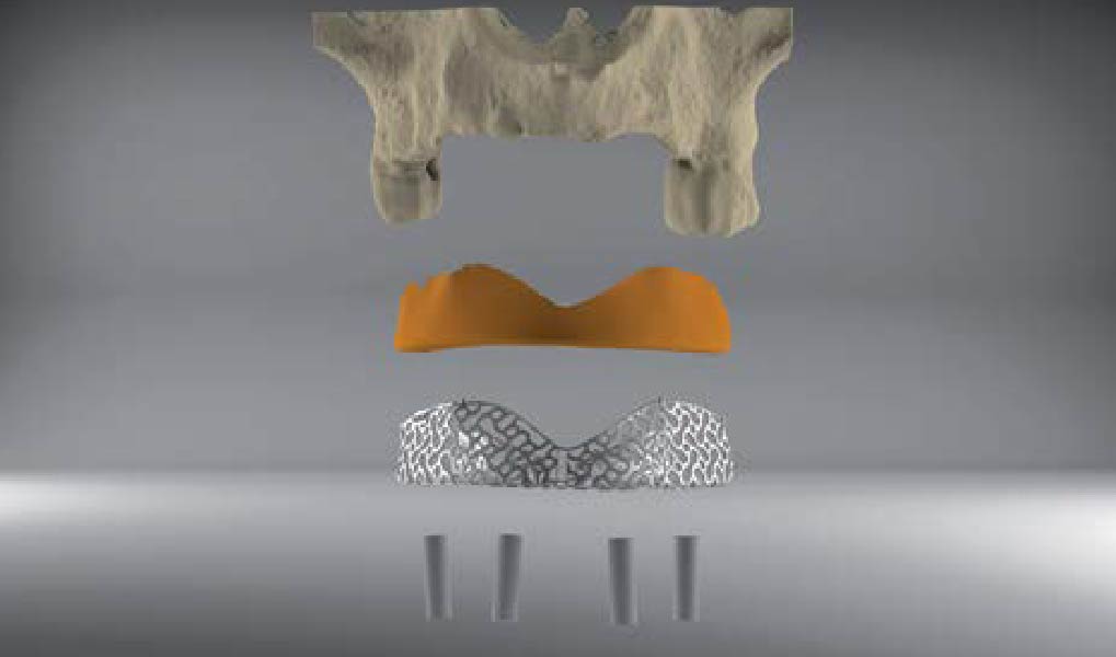
Combined Horizontal and Vertical Regeneration Using a CAD-CAM Titanium Scaffold
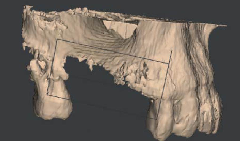
THE SITUATION
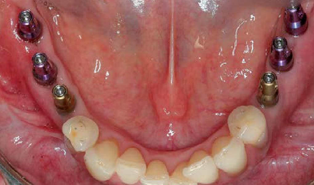
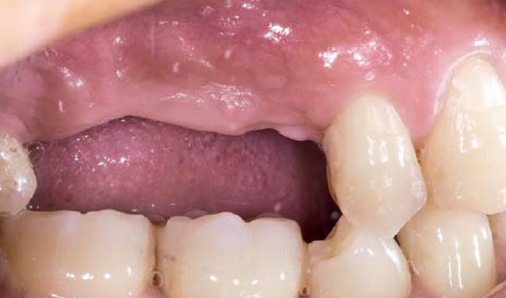
A 54-year-old, systematically healthy male patient (*ASA) came to our attention presenting with partial edentulism in the lower jaw and requiring a fixed and esthetic rehabilitation, refusing any removable solution. The clinical and radiographic evaluation resulted in significant bone atrophy both in the vertical and horizontal components; which makes it impossible to place both conventional implants and short or narrow implants.
*American Society of Anesthesiologists Physical Status Classification System
THE RISK PROFILE
| Low Risk | Medium Risk | High Risk | |
|---|---|---|---|
| Patient’s health | Intact immune system Non-smoker | Light smoker | Impaired immune system |
| Patient’s esthetic requirements | Low | Medium | High |
| Height of smile line | Low | Medium | High |
| Gingival biotype | Thick – “low scalloped” | Medium – “medium scalloped” | Thin – “high scalloped” |
| Shape of dental crowns | Rectangular | Triangular | |
| Infection at implant sight | None | Chronic | Acute |
| Bone height at adjacent tooth site | ≤ 5 mm from contact point | 5.5 – 6.5 mm from contact point | ≥ 7 mm from contact point |
| Restorative status of adjacent tooth | Intact | Restored | |
| Width of tooth gap | 1 tooth (≥ 7 mm) | 1 tooth (≤ 7 mm) | 2 teeth or more |
| Soft-tissue anatomy | Intact | Compromised | |
| Bone anatomy of the alveolar ridge | No defect | Horizontal defect | Vertical defect |
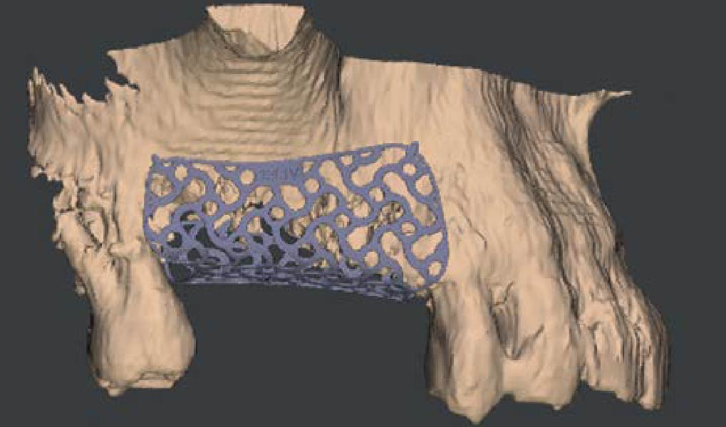
THE APPROACH
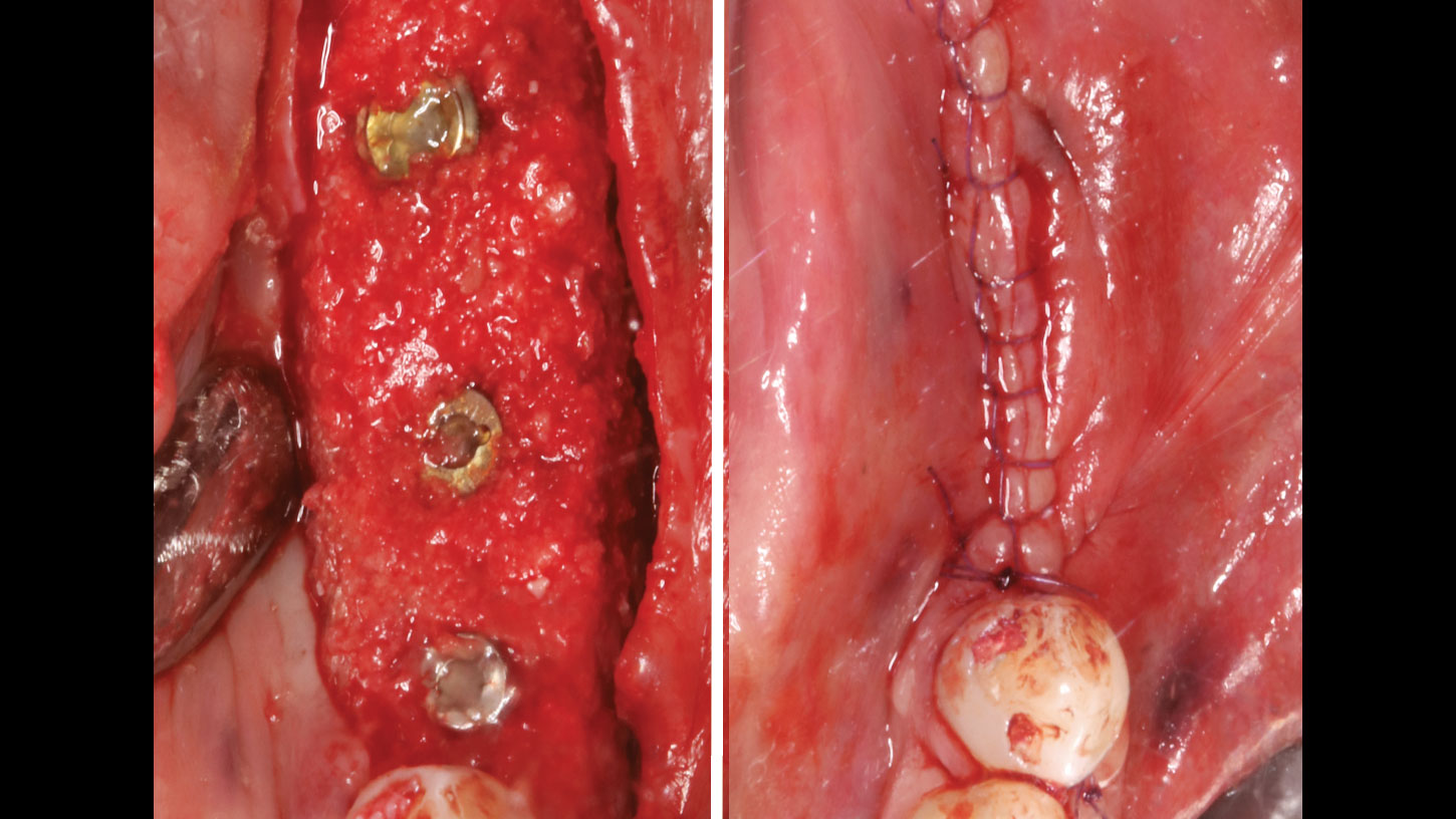
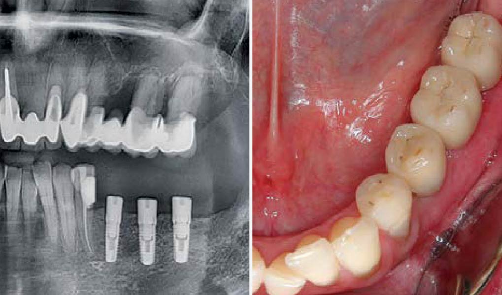
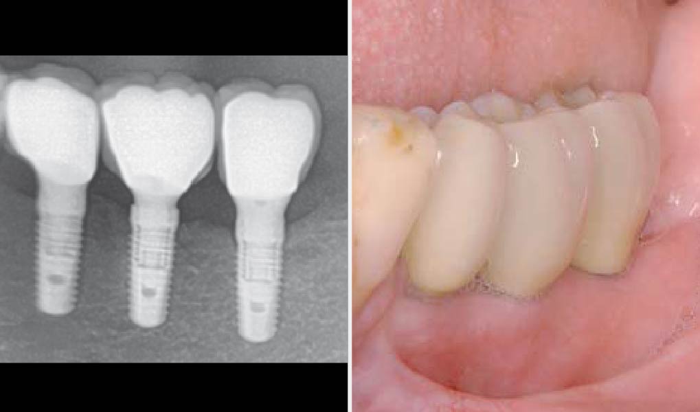
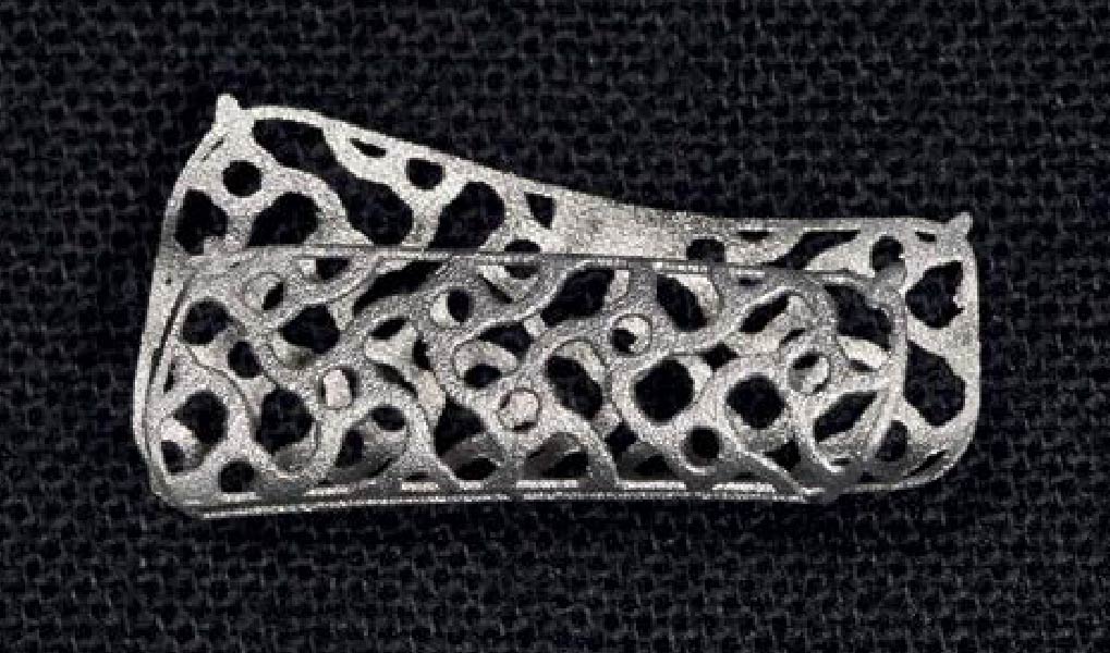
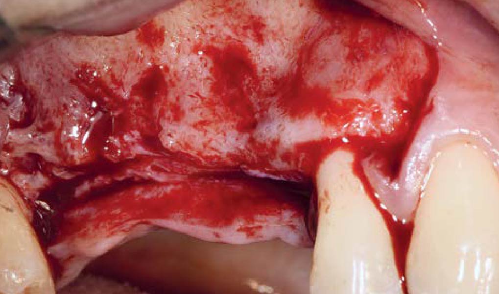
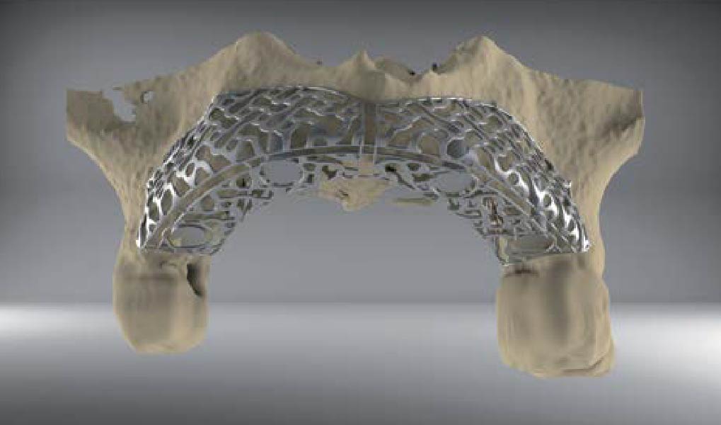
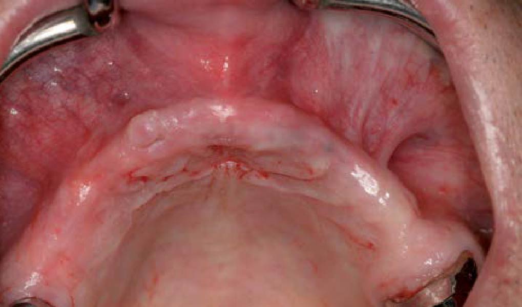
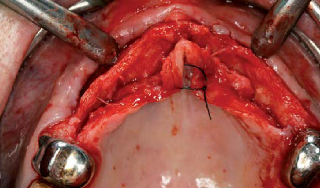
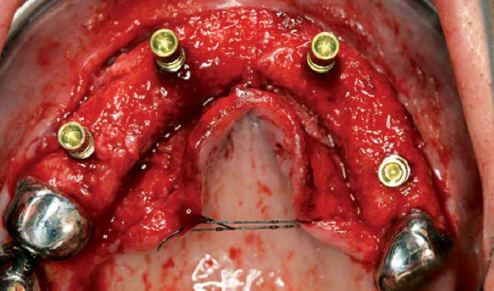
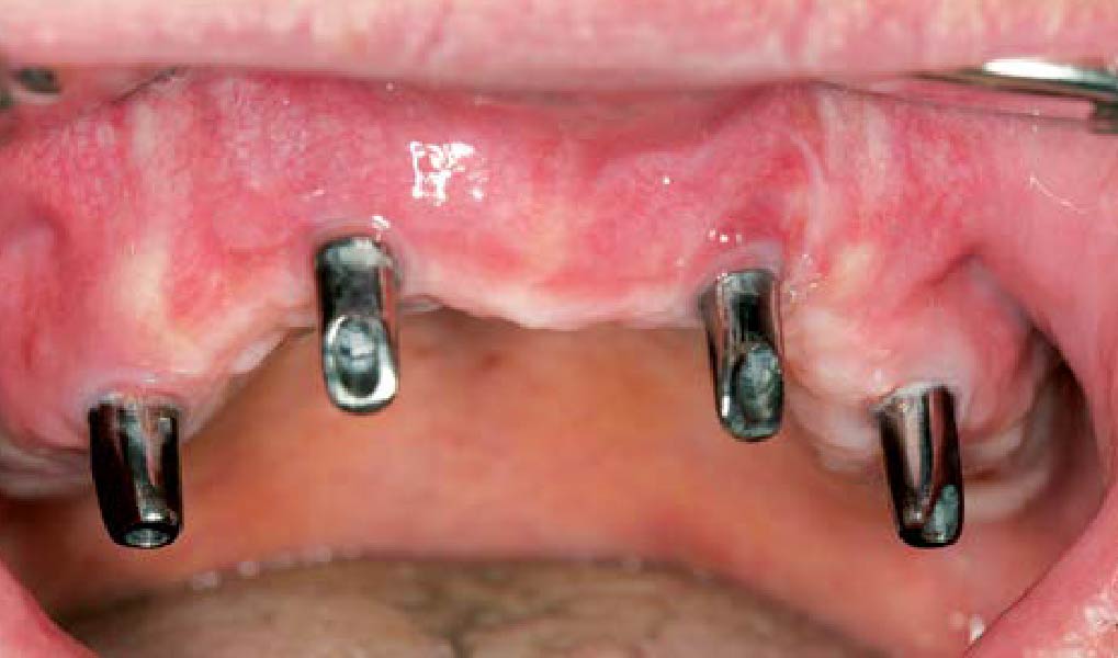
Solving the case was developed in two steps: first bone reconstruction to restore the ideal anatomy, second positioning of the prosthetically guided implants. An individualized regeneration technique was chosen using a CAD-CAM titanium scaffold (Yxoss CBR®) in conjunction with a mix of 60% autogenous bone and 40% Geistlich Bio-Oss®, covered by Geistlich Bio-Gide®. At 9 months, the titanium scaffold was easily removed and 3 prosthetically guided implants were placed, completely surrounded by bone. At 12 months, a free gingival graft was performed to re-establish the missing amount of keratinized mucosa. Finally, at 16 months, the final rehabilitation was carried out with a fixed prosthesis on implants.
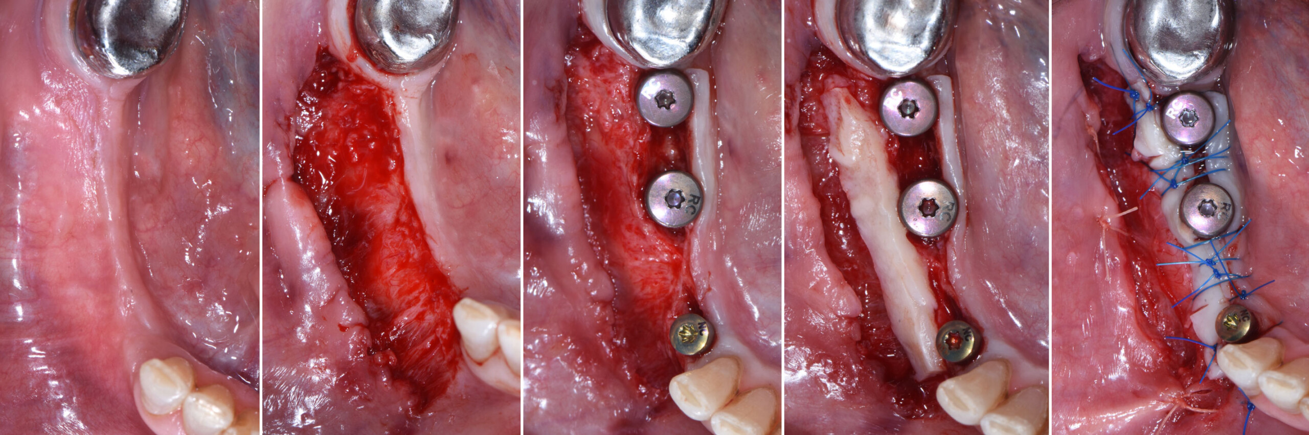

“Combined horizontal and vertical bone augmentation utilizing a CAD CAM titanium scaffold can be achieved with less surgical time and less complications.”
THE OUTCOME
The final resolution of the case was very satisfactory. There were no complications during all the procedures performed. The Yxoss CBR® allowed for easier reconstructive surgery and a significant reduction in surgical times, thanks to the precise dimensions of the scaffold. This resulted in a favorable post- operative situation for the patient and complications were prevented.
Dr. Gian Maria Ragucci
Universitat Internacional de Catalunya (UIC), Barcelona Dental degree at Universidad Europea de Madrid 2015
International Master in oral surgery at UIC, Barcelona 2018
PhD student at UIC, Barcelona 2018
EAO Certification program in implant dentistry 2018
EAO European prize in implant dentistry 2019
Prof. Federico Hernández-Alfaro
Full professor & Chairman, Department of Oral and Maxillofacial Surgery, UIC, Barcelona
Institute of Maxillofacial Surgery, Teknon Medical Center, Barcelona
BIOBRIEF
3D Bone Augmentation Using Customized Titanium Mesh in Conjunction with Autogenous Bone and Bovine Bone Material Granules
THE SITUATION
A 75-year-old systemically healthy female came to our attention presenting with absent mandibular second bicuspids and molars and requiring a fixed rehabilitation supported by implants as she refused a removable solution. The clinical and radiographic evaluation showed a relevant vertical and horizontal bone atrophy of such an extent that short or narrow implants were not considered a reliable option. The patient smoked <10 cigarettes per day.
THE RISK PROFILE
| Low Risk | Medium Risk | High Risk | |
|---|---|---|---|
| Patient’s health | Intact immune system | Light smoker | Impaired immune system |
| Patient’s esthetic requirements | Low | Medium | High |
| Height of smile line | Low | Medium | High |
| Gingival biotype | Thick – “low scalloped” | Medium – “medium scalloped” | Thin – “high scalloped” |
| Shape of dental crowns | Rectangular | Triangular | |
| Infection at implant sight | None | Chronic | Acute |
| Bone height at adjacent tooth site | ≤ 5 mm from contact point | 5.5 – 6.5 mm from contact point | ≥ 7 mm from contact point |
| Restorative status of adjacent tooth | Intact | Restored | |
| Width of tooth gap | 1 tooth (≥ 7 mm) | 1 tooth (≤ 7 mm) | 2 teeth or more |
| Soft-tissue anatomy | Intact | Compromised | |
| Bone anatomy of the alveolar ridge | No defect | Horizontal defect | Vertical defect |
THE APPROACH
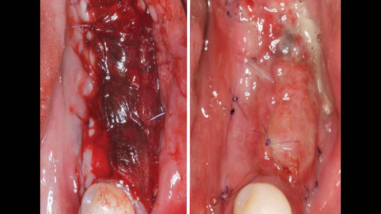
The main goal was to obtain a horizontal and vertical reconstruction of the deficient alveolar bone in order to allow safe and prosthetically-guided implant placement. Reconstruction was obtained by means of a customized titanium mesh, Yxoss CBR®, in combination with a mixture of autologous bone chips harvested from the mandibular ramus and bovine bone mineral, Geistlich Bio-Oss®.
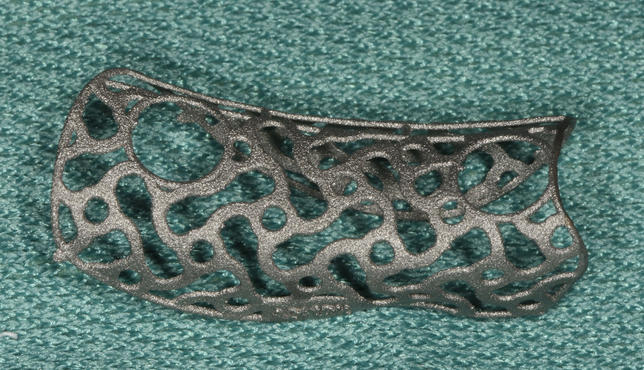
The 3-dimensional reproduction of the left edentulous area permits the production of a precise and customized Ti-mesh.
THE OUTCOME
Post-operative recovery of this patient was uneventful, no complications such as dehiscence or late exposure of the customized mesh, with complete correction of the initial defect. The Yxoss CBR® allowed an easy and faster reconstruction thanks to the precision of the prefabricated mesh filled with autologous chips, Geistlich Bio-Oss® and Geistlich Bio-Gide®.

Matteo Chiapasco, D.D.S., M.D.
Graduated in Medicine and specialized in Maxillofacial Surgery at the University of Milan, Italy. Professor, Unit of Oral Surgery, University of Milan; Associate Professor, Loma Linda University, Los Angeles, California, USA.
Grazia Tommasato, D.D.S., M.S.C.
Graduated in Dentistry in 2013, specialized in Oral Surgery at the University of Milan magna cum laude. PhD student and a medical consultant of the Clinical Unit of Oral Surgery (“G. Vogel” Clinic, Milan).

WEBINAR

CLINICAL CASE

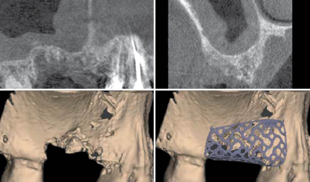
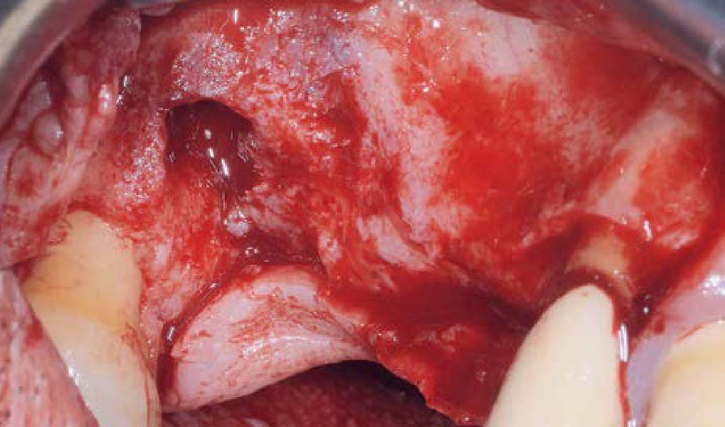
CLINICAL SITUATION:
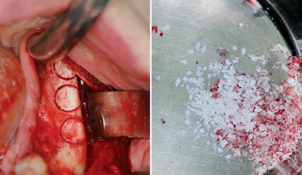
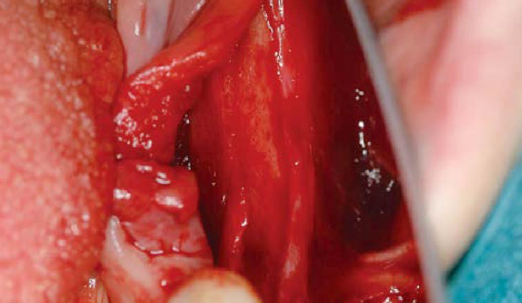
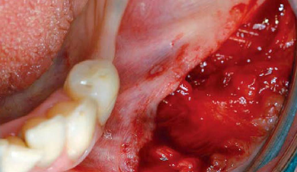
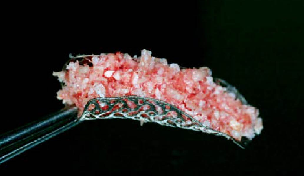
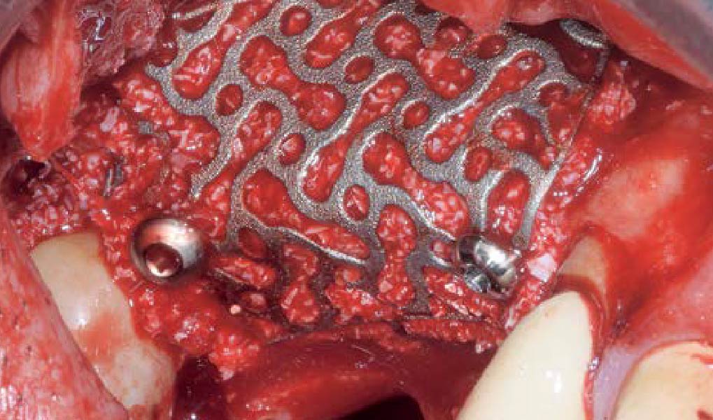
A 60-year-old female presented to the periodontics clinic at UTHSA for implant placement at sites #18 and #19. Upon clinical and radiographic examinations, the lower left edentulous ridge was diagnosed as a Siebert class III due to the presence of bucco-lingual and apico-coronal tissue defects. The treatment proposed included soft tissue grafting for increase of keratinized tissue followed by ridge augmentation using Yxoss CBR®mesh and a mix of autograft, vallos™ fibers, and platelet-rich plasma (PRP)
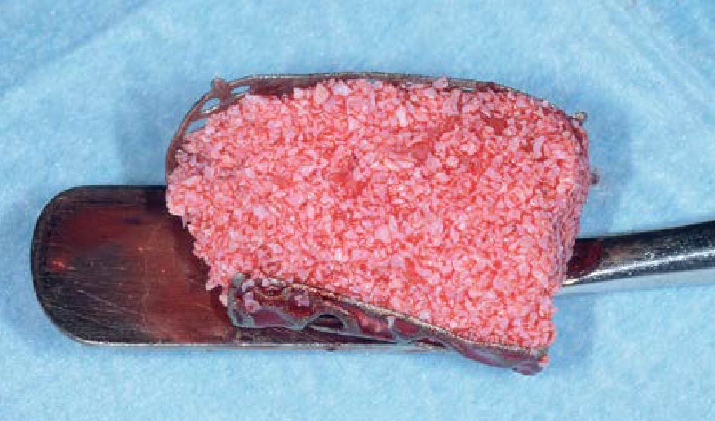
OUTCOME:
The vallos™ fibers combined with autogenous bone and the PRP created a stable fibrin bone graft that could be easily molded and contained within the mesh. Hydration with PRP was progressive until the graft reached the desired consistency. Wound healing following ridge augmentation was uneventful. There were no signs of infection or membrane exposure at the site. Mesh removal and implant placement is planned at 6-months following ridge augmentation.
CLINICAL CASE
CLINICAL CHALLENGE:
- The planning of the patient’s case takes local and general patient-specific risk factors into consideration according to the principles of backward planning for implant positioning.
AIM/APPROACH:
- Highlights step-by-step the important procedures to regenerate the bone (horizontal and vertical) with the 3-D printing technology, Yxoss CBR®.
CLINICAL CASE
CLINICAL CASE
CLINICAL CASE

CLINICAL CASE
CLINICAL CASE