CLINICAL CASE

THE OUTCOME

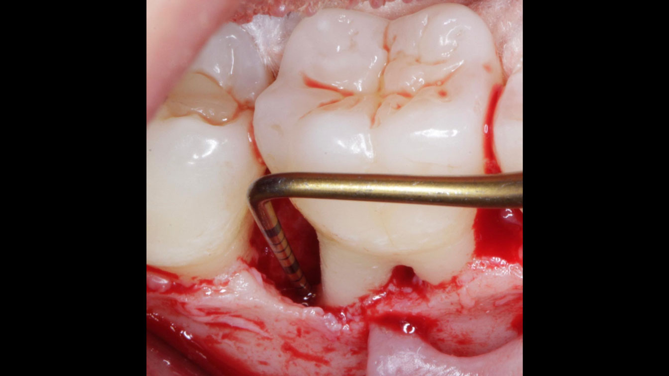
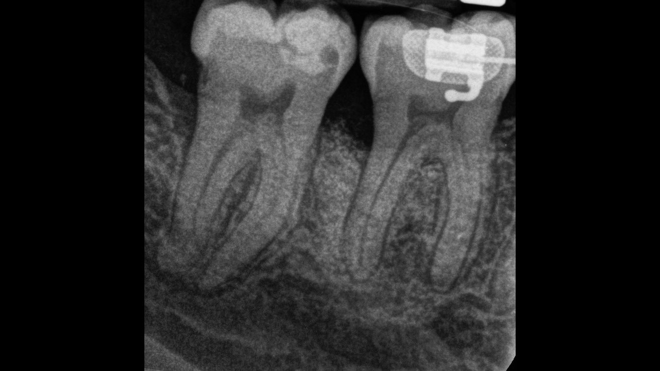
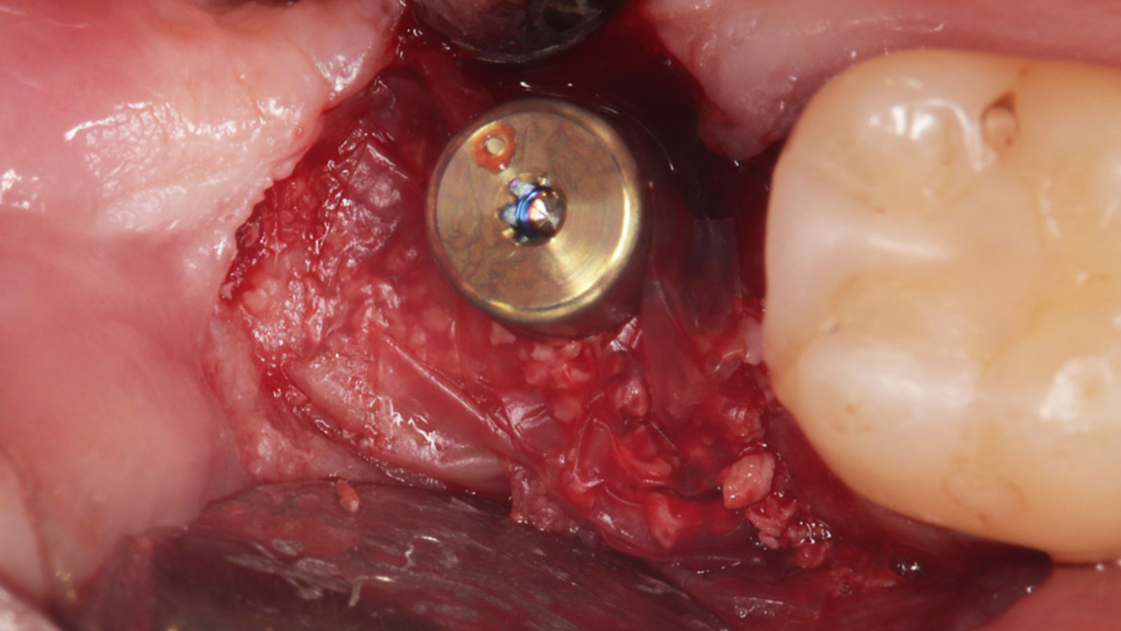
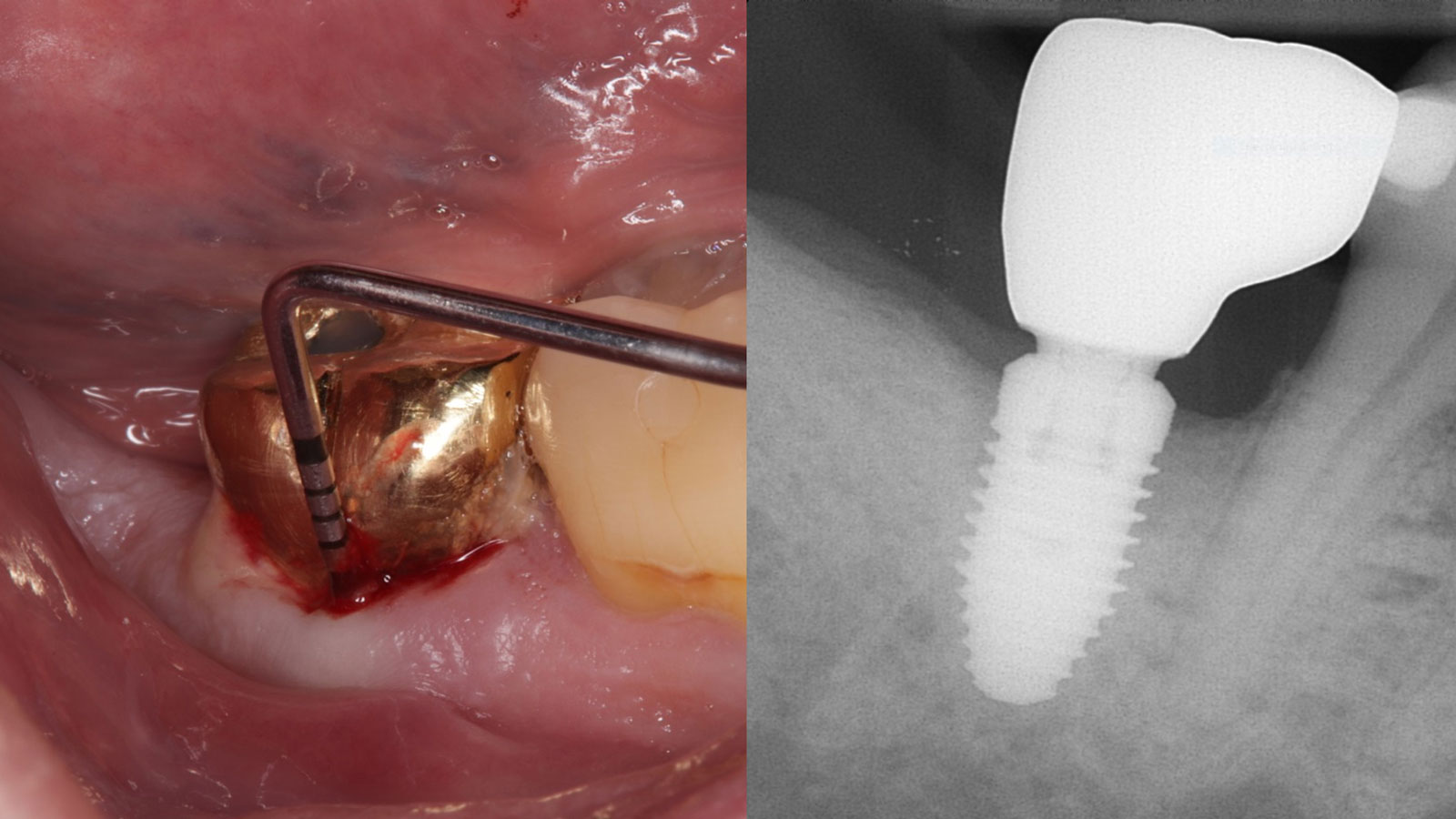
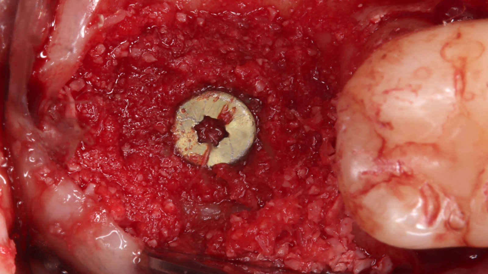
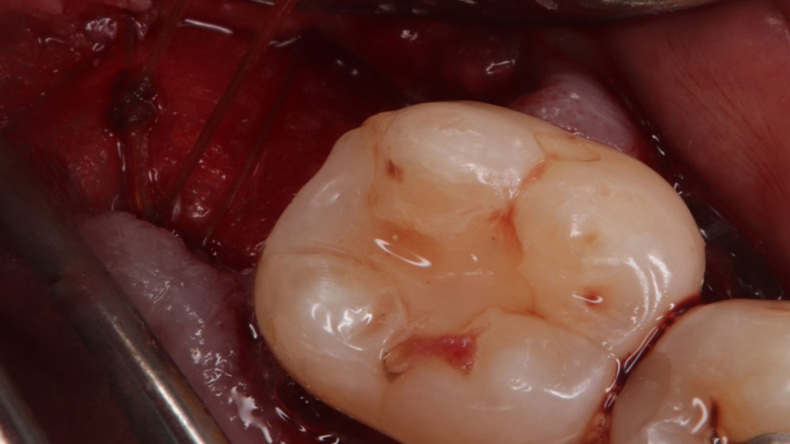
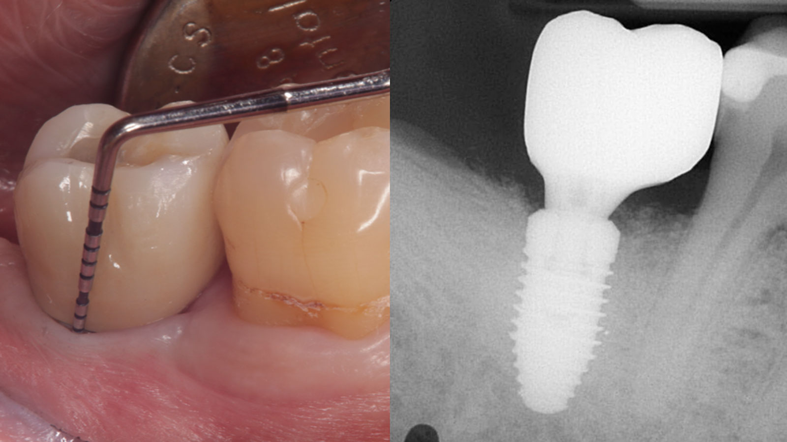
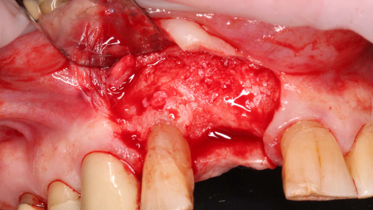
At 6 months, healing was complete with a bone fill of 10x10x6.5 mm. Tooth #7, affected by the combined defect, was fully regenerated. The adjacent ridge defect was also restored, enabling the placement of a regular-sized implant without the need for additional grafting.

CLINICAL CASE

THE APPROACH

Considering the patient’s age, condition, and good compliance, a regenerative approach was chosen to preserve all teeth. Due to the extent and severity of the defect, a bone graft, biologics, and a membrane were utilized. The goal was to restore lost attachment and stabilize the teeth through splinted therapy.

THE OUTCOME

At the 7-month follow-up, probing depths decreased from 7 mm (MB, DB) to 3 mm, with radiographic bone fill indicating a 90% success. Attachment gain of 3 mm was achieved, improving from 7 mm to 4 mm. The patient reported satisfactory outcomes, and splinting effectively maintained tooth stability with no mobility. 

CLINICAL CASE

THE APPROACH

Following flap elevation and thorough defect debridement, a xenograft bone substitute mixed with PDGF (GEM 21S®) was used to fill the defect. The graft was left uncovered without a membrane.

THE OUTCOME

The 1-year follow-up radiograph demonstrated near-complete bone fill in a previously deep defect. Clinically, shallow probing depths and healthy tissue were also observed at the 1-year follow-up.

CLINICAL CASE

THE APPROACH

The crown was biologically shaped, and the root surface was detoxified using Ellman burs. Following flap elevation and thorough defect debridement, an allograft mixed with GEM 21S® was used to fill the defect. The graft was then covered with Geistlich Bio-Gide®.

THE OUTCOME

At the 10-month follow-up, radiographs revealed evidence of bone fill, and clinically, the interdental tissue showed signs of maturation. At the 9-year follow-up, clinical photos demonstrated long-term stability, with maintained bone levels, minimal interproximal recession, and lack of facial recession. Radiographic analysis further confirmed the sustained stability of the bone.

BIOBRIEF

Prosthetic-Surgical Approach to Regenerative Treatment for Peri-Implantitis

Andrea Ravidà, DDS, MS, PhD
Anu Viswanathan DDS, MDS

THE SITUATION

A 68-year-old male patient, who received an implant in tooth position #31 about 8 years prior, presented for an examination. He reports bleeding during brushing around the implant and some discomfort. Clinically, there was severe vertical bone loss, profuse bleeding on probing, and deep probing depths, but no pain. The condition was diagnosed as peri-implantitis according to the 2018 classification.

THE RISK PROFILE

Low RiskMedium RiskHigh Risk
Patient’s healthIntact immune systemLight smokerImpaired immune system 
Patient’s esthetic requirementsLowMediumHigh
Height of smile lineLowMediumHigh
Gingival biotypeThick – “low scalloped”Medium – “medium scalloped”Thin – “high scalloped”
Infection at implant sightNoneChronicAcute
Restorative status of adjacent toothIntactRestored
Soft-tissue anatomyIntactCompromised
Bone anatomy of the alveolar ridgeNo defectHorizontal defectVertical defect

Additional Risk Factors: The patient exhibited bleeding on probing and deep pocket depths. He also reported occasional marijuana use and was inconsistent with periodontal maintenance and oral hygiene visits.

THE APPROACH

The treatment goals were to eliminate peri-implant infection, regenerate lost hard and soft tissues, and ensure long-term implant stability. A closed regenerative approach was utilized, including crown removal, thorough implant decontamination with Perioflow®, an airpolishing technology, application of the correct bone grafting materials (Geistlich Bio-Oss®, vallos® and GEM 21S®), enclosed healing, and fabrication of a new crown to facilitate hygiene.

Clinical presentation of tooth #31 showing radiographic evidence of bone loss, profuse bleeding on probing (BOP), deep probing depths, and suppuration, indicative of peri-implantitis with a Class I-infraosseous (c) circumferential-type defect, as described in the study by Monje et al. (2019) Clin Implant Relat Res, 21(4)635-643.
Crown removal by the prosthodontist, followed by placement of a healing abutment for non-surgical therapy using PerioFlow®. After therapy, a cover screw was placed, and the tissue was allowed to heal over the implant for eight weeks.
Surgical treatment initiated with a midcrestal incision and full-thickness flap elevation. Granulation tissue was removed using a surgical curette, revealing a deep infrabony defect.
Implant thoroughly decontaminated using Perioflow® with erythritol powder to ensure a clean surface for regeneration.
rhPDGF-BB was used to hydrate bone grafting materials (vallos® Demineralized Cortical Granules and Geistlich Bio-Oss®), which were first hydrated with sterile water before rhPDGF-BB was added. The materials were mixed in a 1:1 ratio and allowed to sit for 10 minutes before being applied to the deep infrabony defect to promote regeneration.
Flap closed primarily with 5-0 PTFE horizontal mattress and single interrupted suture for secure closure.
Collagen membrane stabilized with 5-0 chromic gut sutures using the lasso technique.
After 5 months of healing, significant bone gain is evident. Geistlich Bio-Oss® was placed on the buccal site to enhance thickness, covered with an amnion-chorion membrane. A healing abutment was placed at this stage.
Two-year follow-up shows disease resolution with shallow probing depths, no bleeding or suppuration, and complete bone gain. A new crown was fabricated with an increased final abutment height (>2mm), contributing to optimal maintenance and long-term stability based on evidence supporting its role in promoting long-term success. A second surgery may be necessary to gain additional tissue thickness or cover residual thread exposure to achieve the desired long-term results.

“The implant presented with significant bone loss, deep probing depths, and bleeding on probing, placing it at risk of failure and requiring intervention to preserve function and longevity.”

— Andrea Ravidà, DDS, MS, PhD

THE OUTCOME

At the two-year follow-up, clinical and radiographic assessments showed disease resolution, complete bone gain, and stable peri-implant tissues. Probing depths were within healthy ranges, and no bleeding on probing was observed, confirming the long-term success of the treatment.

Enclosed healing, meticulous implant decontamination, appropriate selection of bone grafting materials, and customized crown design, combined with patient compliance and regular maintenance, contributed to disease resolution and complete bone regeneration.”

Andrea Ravidà, DDS, MS, PhD

The air polishing device with erythritol powder ensured thorough implant decontamination, while the bone grafting materials combined with rhPDGF-BB provided essential biologic support for regeneration and improved peri-implantitis treatment outcomes.”

Andrea Ravidà, DDS, MS, PhD

Andrea Ravidà, DDS, MS, PhD

Dr. Andrea Ravidà is the Director of the Graduate Periodontics Program in the department of Periodontics at the University of Pittsburgh. He conducts clinical research focusing on peri-implantitis, periodontitis and short implants. He has published more than 70 peer-reviewed articles and conference abstracts/presentations related to periodontics and implant therapy. He is section editor of the International Journal of Oral Implantology and the Journal of Translational Medicine.

Anu Viswanathan DDS, MDS

Dr. Anu Viswanathan is a Diplomate of the American Board of Periodontology and Implant Dentistry. She earned her Doctor of Dental Surgery degree from the University of Colorado School of Dental Medicine in 2019. Dr. Viswanathan completed a Certificate in Periodontics and earned a Master of Dental Science at the University of Pittsburgh School of Dental Medicine. She also obtained a Certificate in IV Sedation. Dr. Viswanathan is currently in private practice in Shoreline, Connecticut.

Sorry, you do not have permission to view this content.

Sorry, you do not have permission to view this content.

Sorry, you do not have permission to view this content.

Sorry, you do not have permission to view this content.

Sorry, you do not have permission to view this content.

Sorry, you do not have permission to view this content.

Sorry, you do not have permission to view this content.

Sorry, you do not have permission to view this content.

Sorry, you do not have permission to view this content.

Sorry, you do not have permission to view this content.