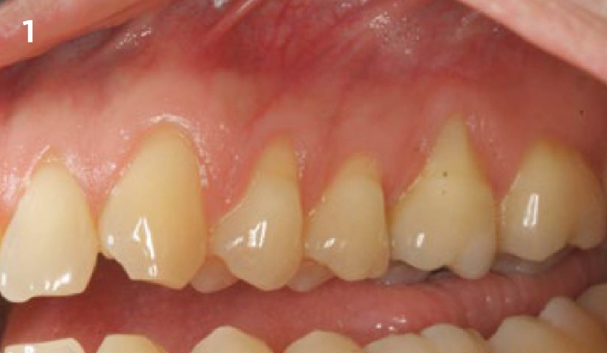
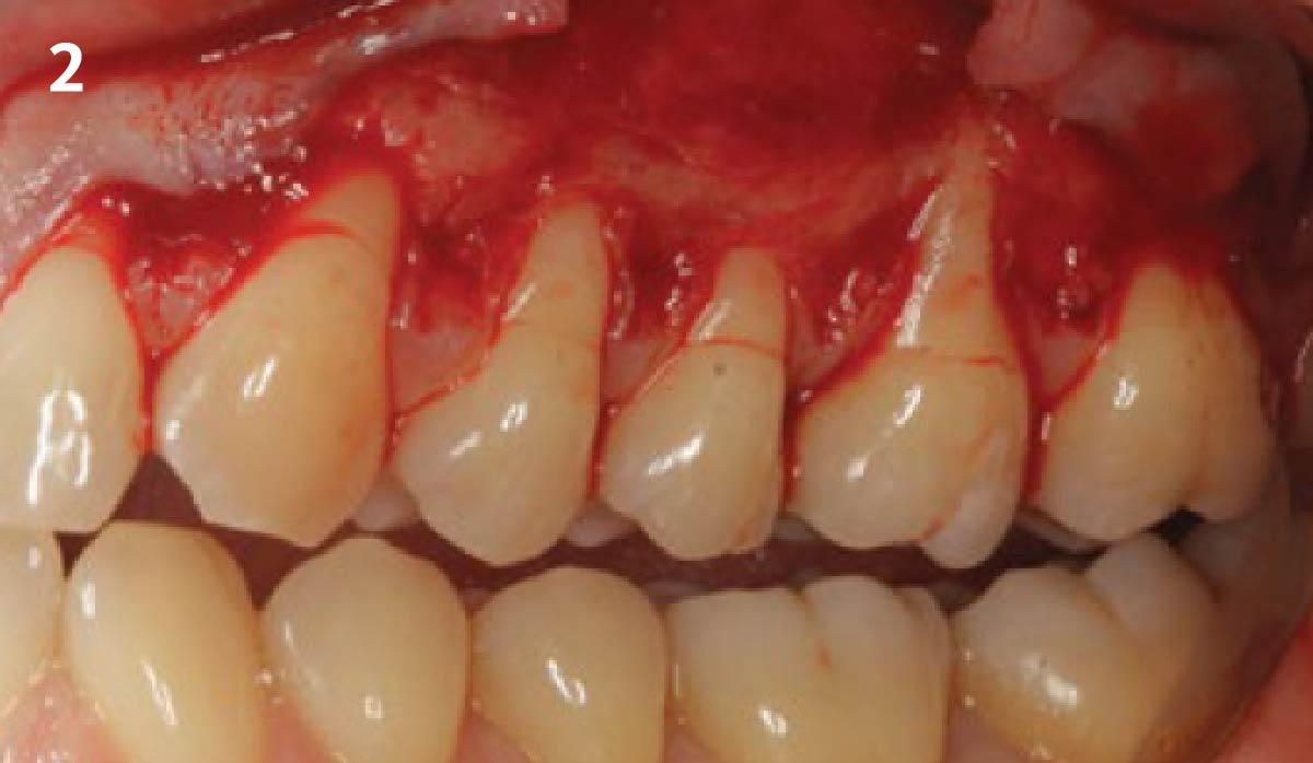
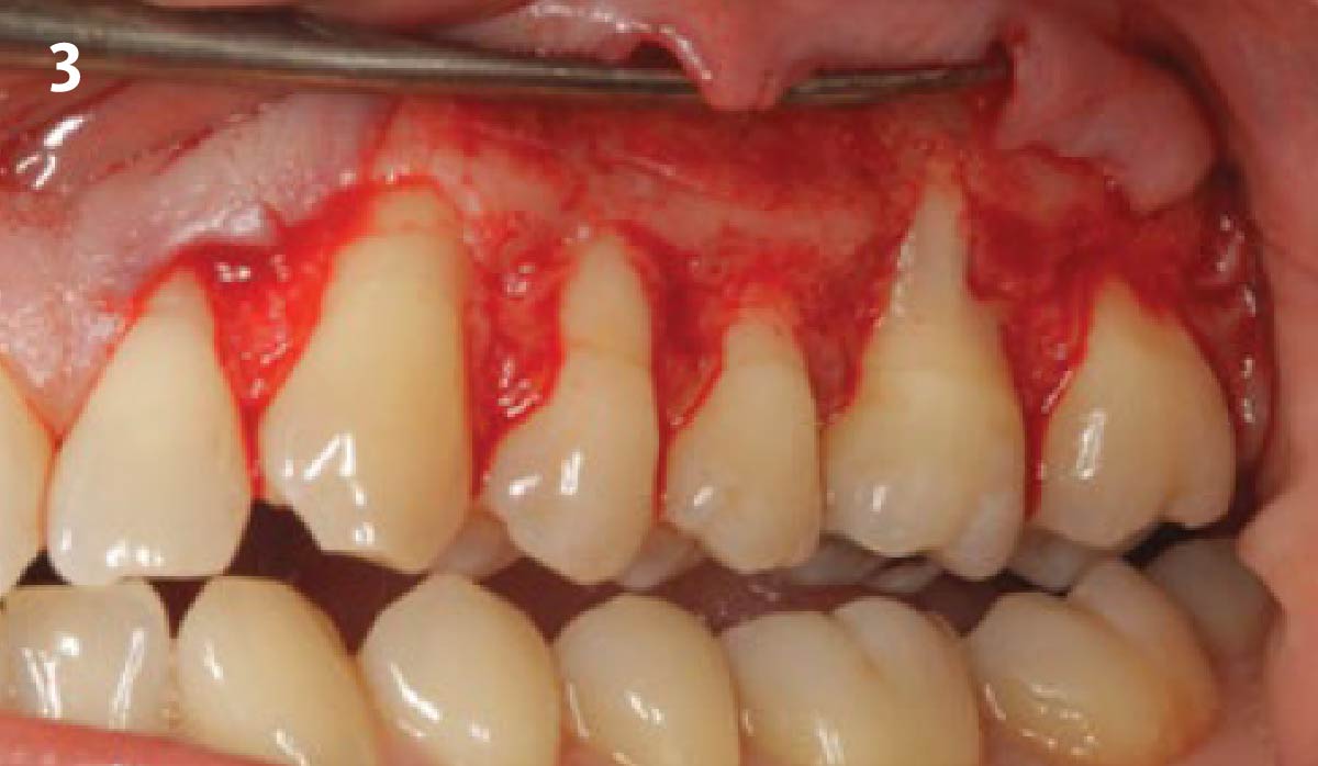
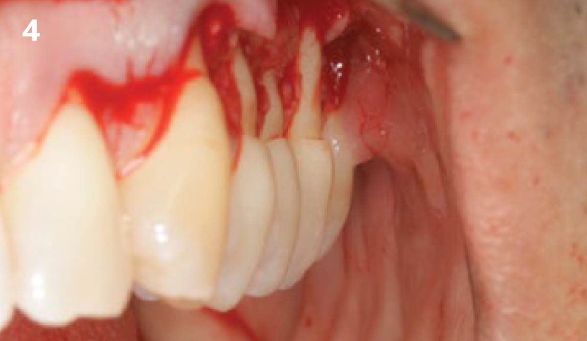
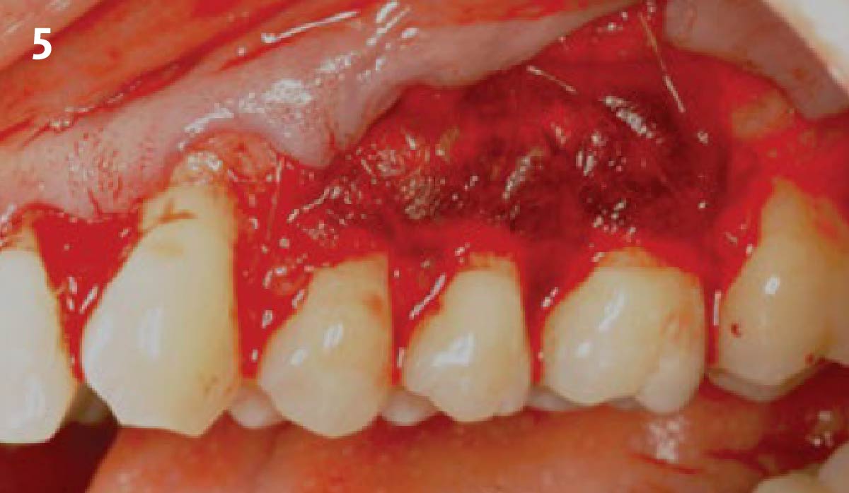
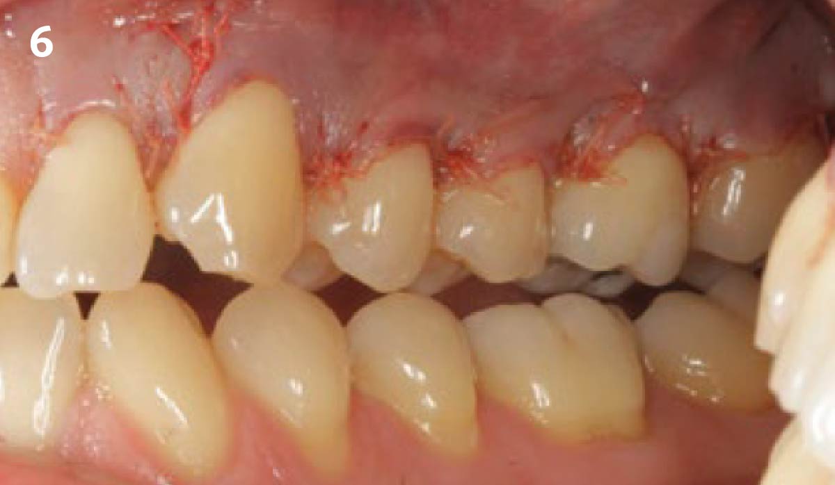
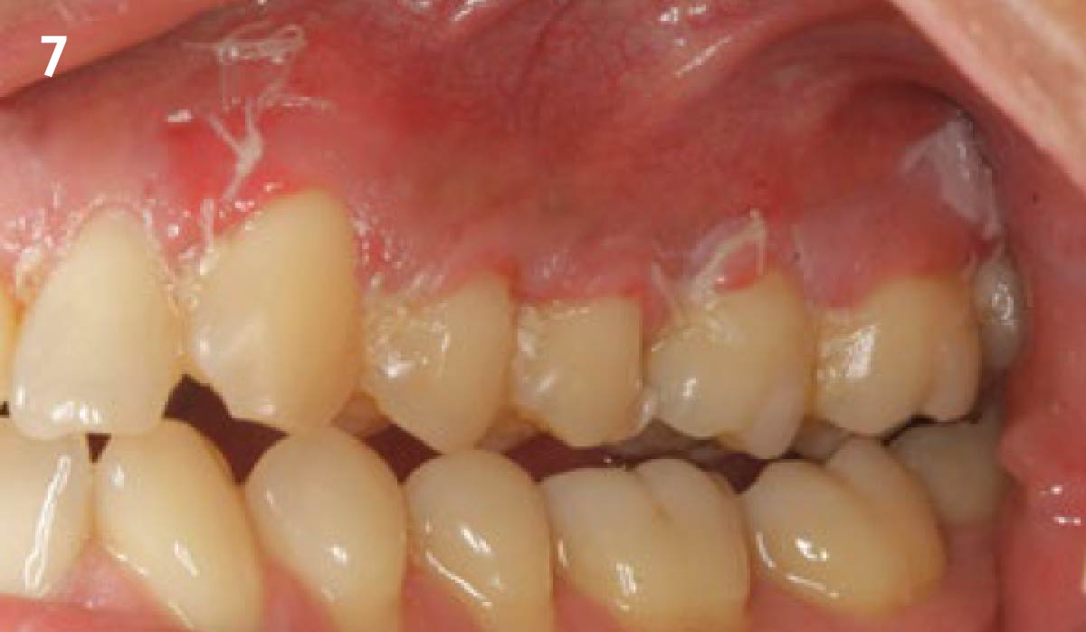
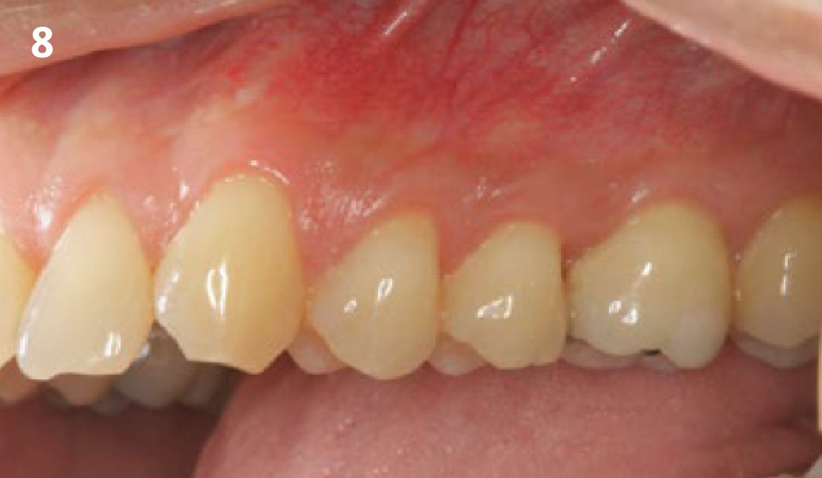
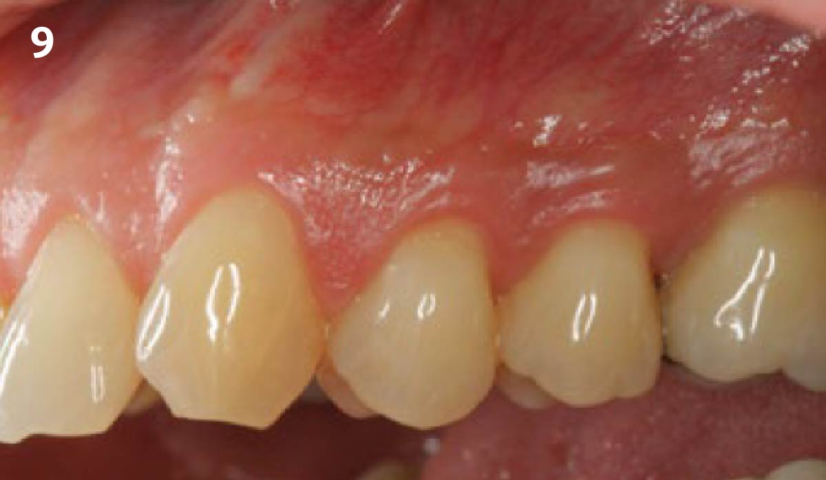
CLINICAL CASE Pre-operative view of recession defects that ranges from 2-5 mm. There is minimal keratinized tissue on #14. Pocket depths are within limits with no loss of interproximal tissue. A 15c blade is used to make sulcular incisions with scooping incisions at the level of the CEJ. A combination flap consisting of full-thickness coronally followed by a partial-thickness dissection apically is reflected. The papillary tissue is de-epithelialized with a football diamond bur on a rotary hand piece. This exposes a vascular bed for the graft and intended coronal positioning of the flap. Side-view of the recession defects. It is clearly visible how deep the recession defects are. Geistlich Mucograft® is trimmed and positioned to extend beyond the root surfaces. A combination of simple interrupted sutures at its coronal edge and mattress sutures extending over the entire graft are used to adapt the graft to the recipient site. The flap is then advanced and coronally positioned with horizontal mattress sutures to release tension and simple interrupted sutures to approximate the flap edges to the de-epithelialized papillas. Follow-up after 1 week: note that the flap margins appear stable. Erythema and edema evident with maturation of the tissue beginning. Follow-up after 3 months: maturation of the tissue evident with complete root coverage. An increase in the zone of keratinized tissue by #14 is also visible. Follow-up after 1 year: stability of the graft is evident, complete coverage and a healthy and maintainable gingival situation have been achieved.