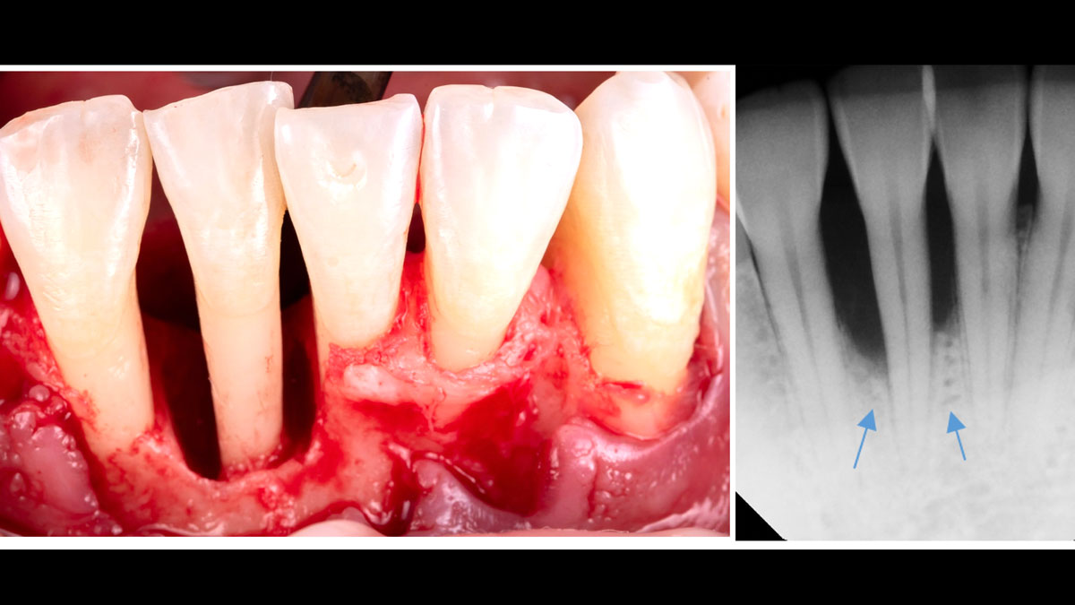
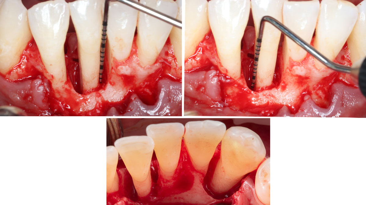
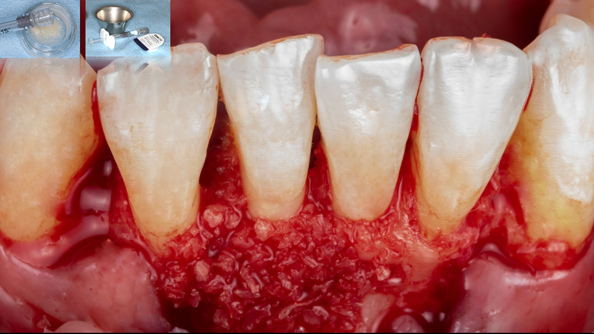
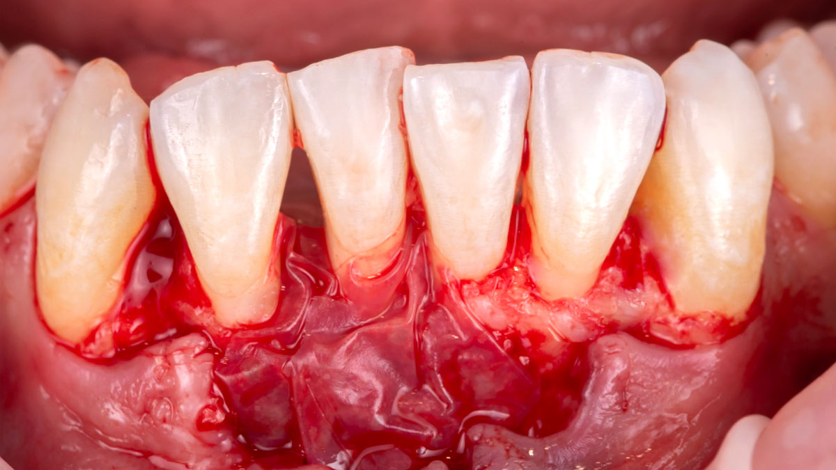
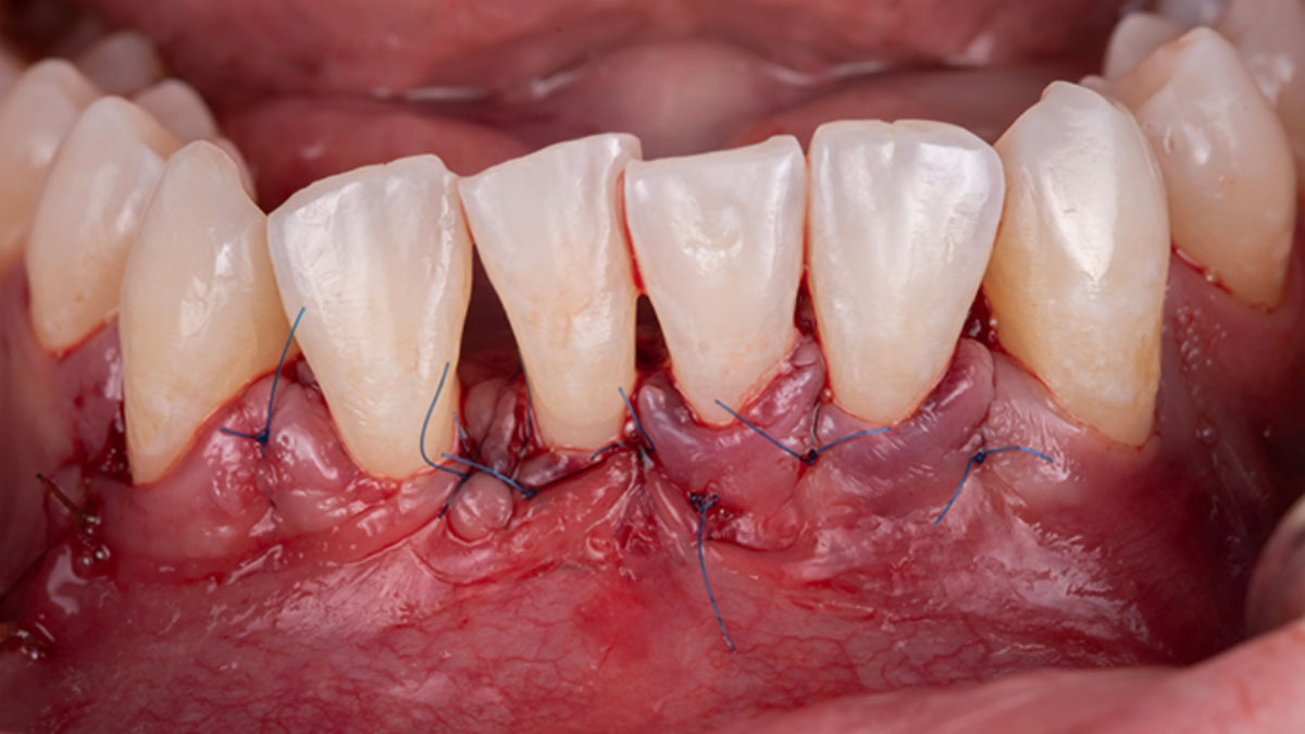
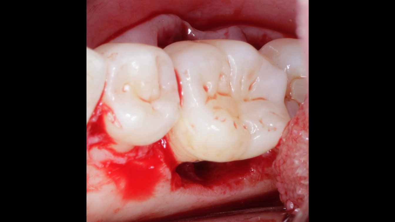
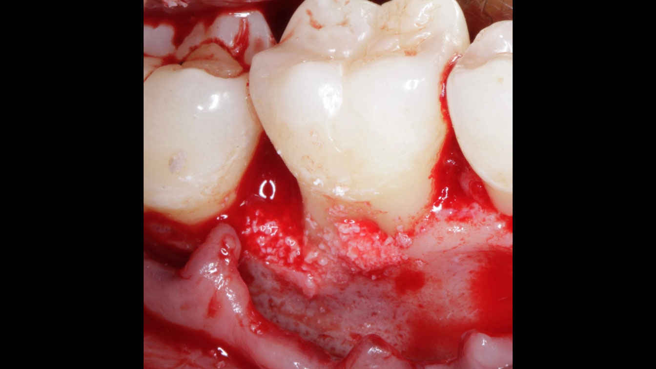
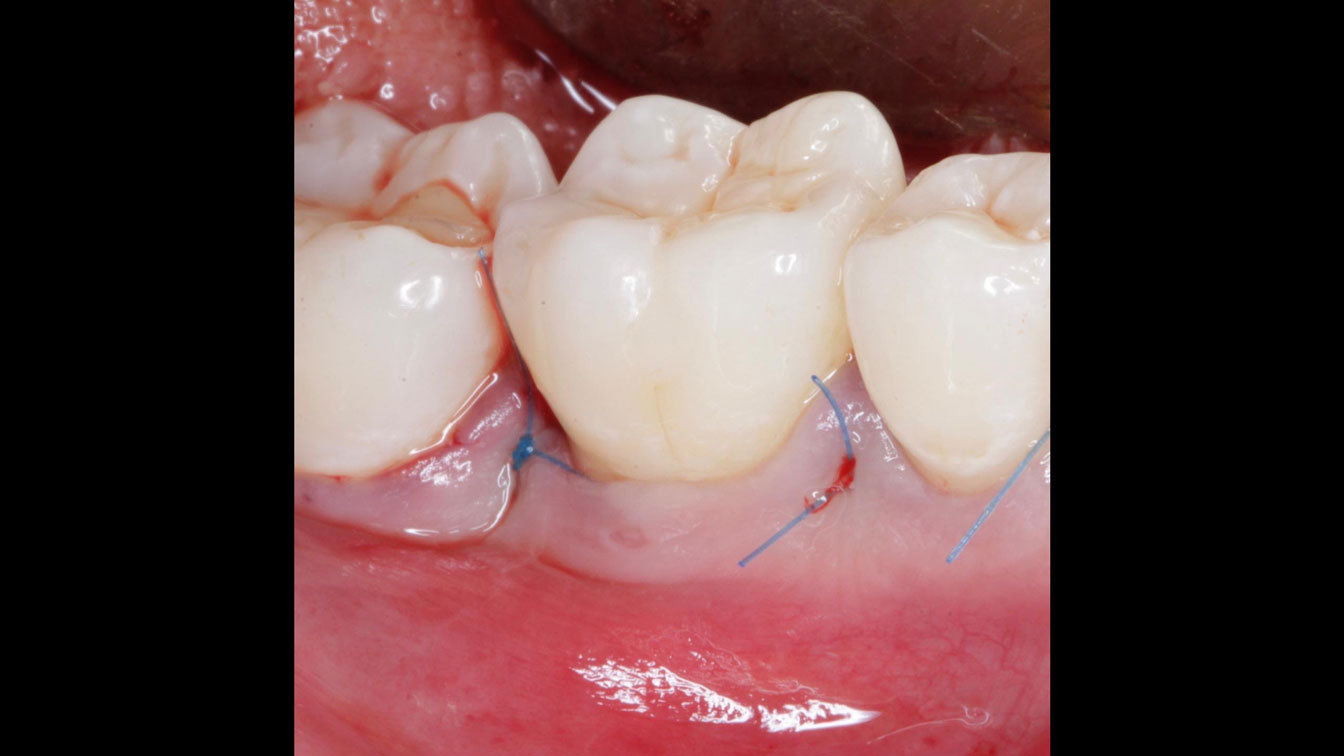
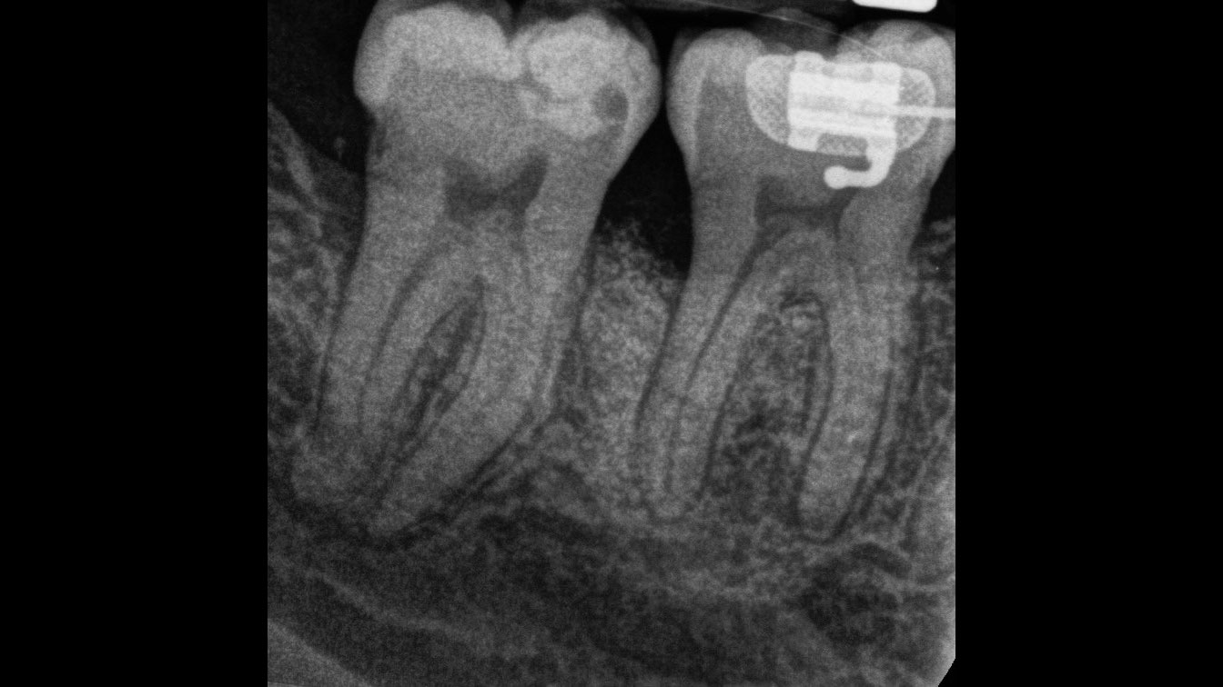
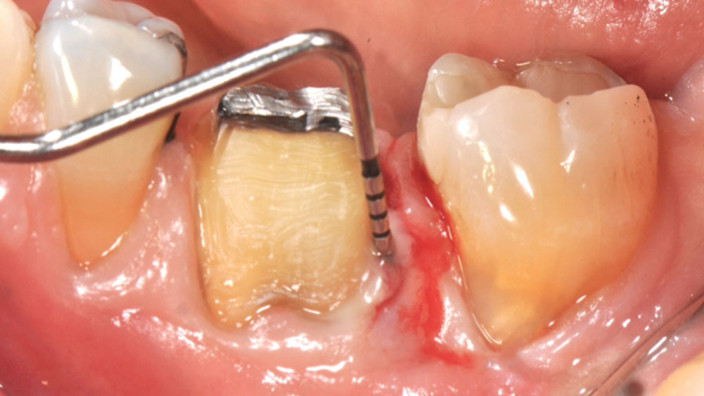
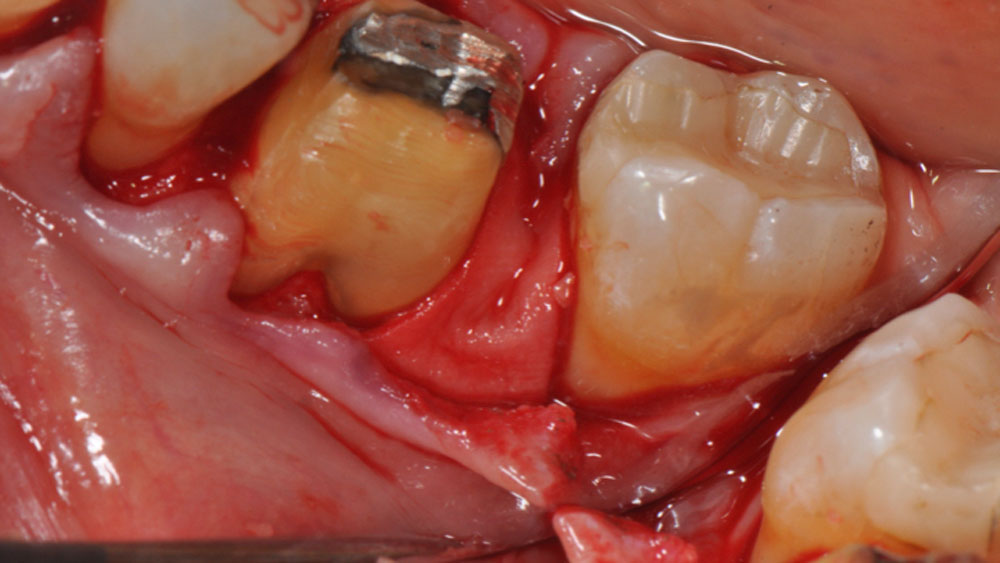
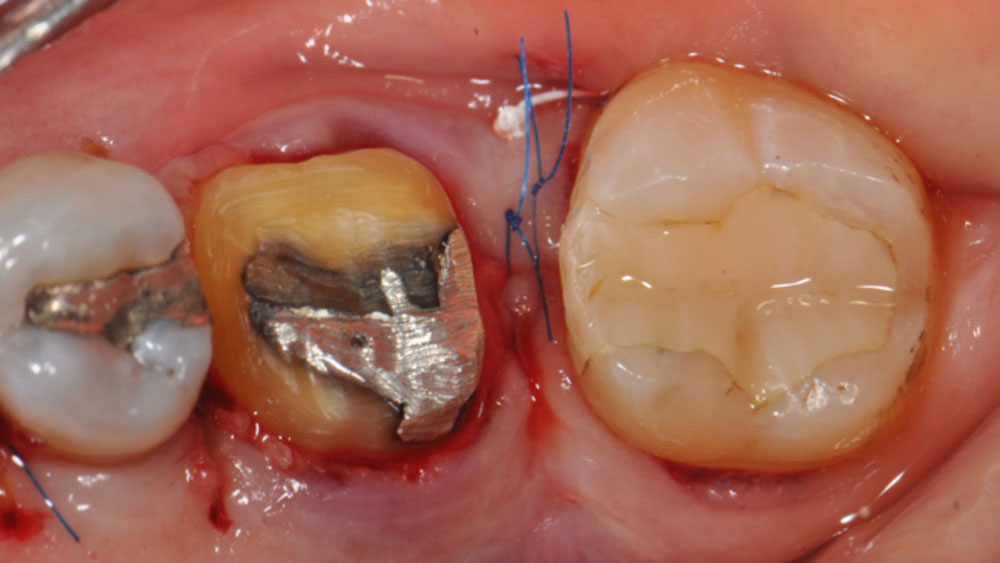
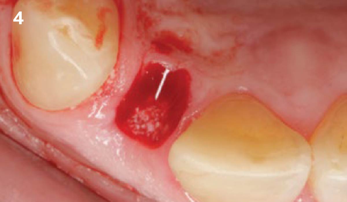
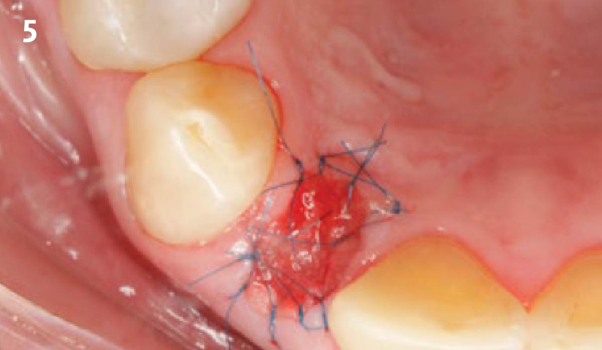
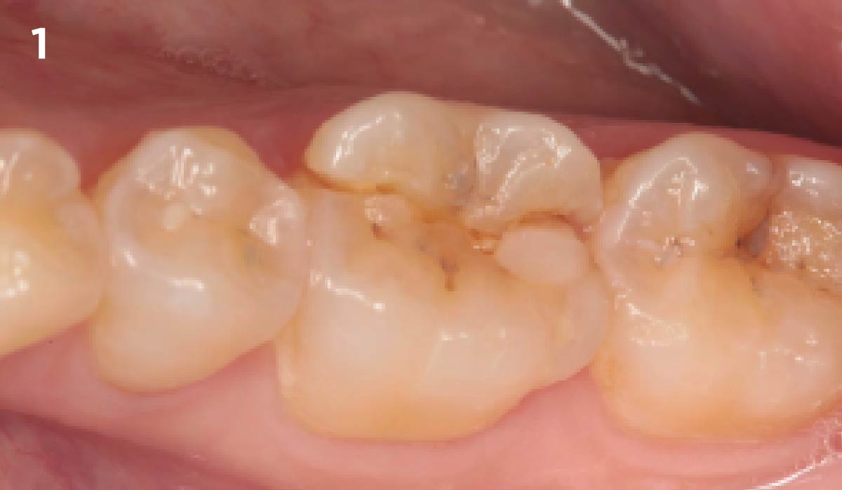
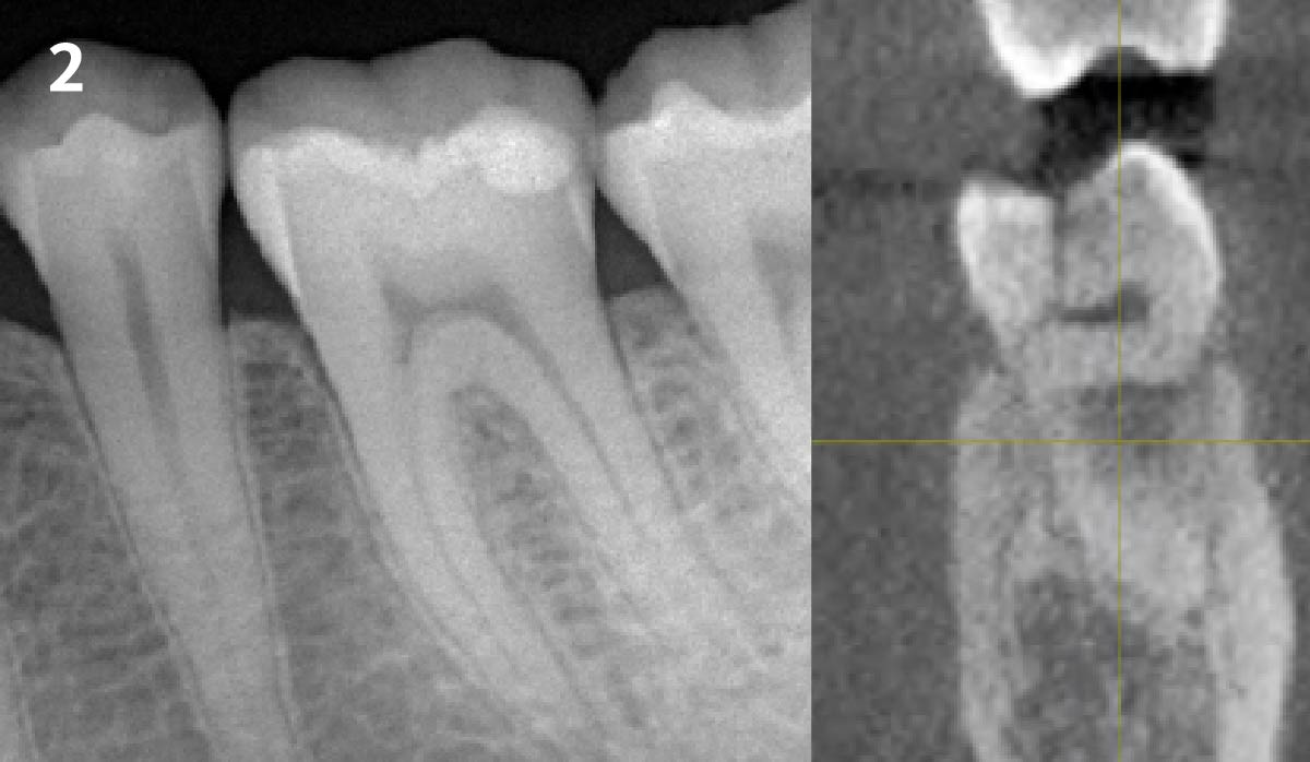
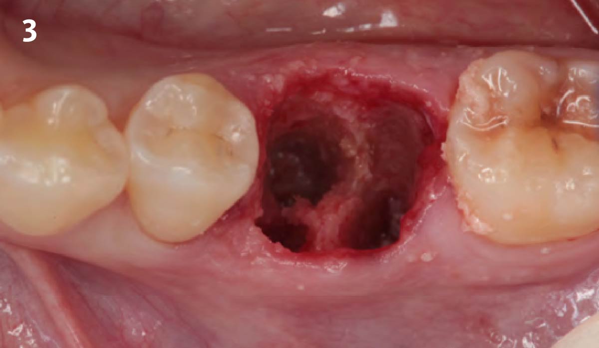
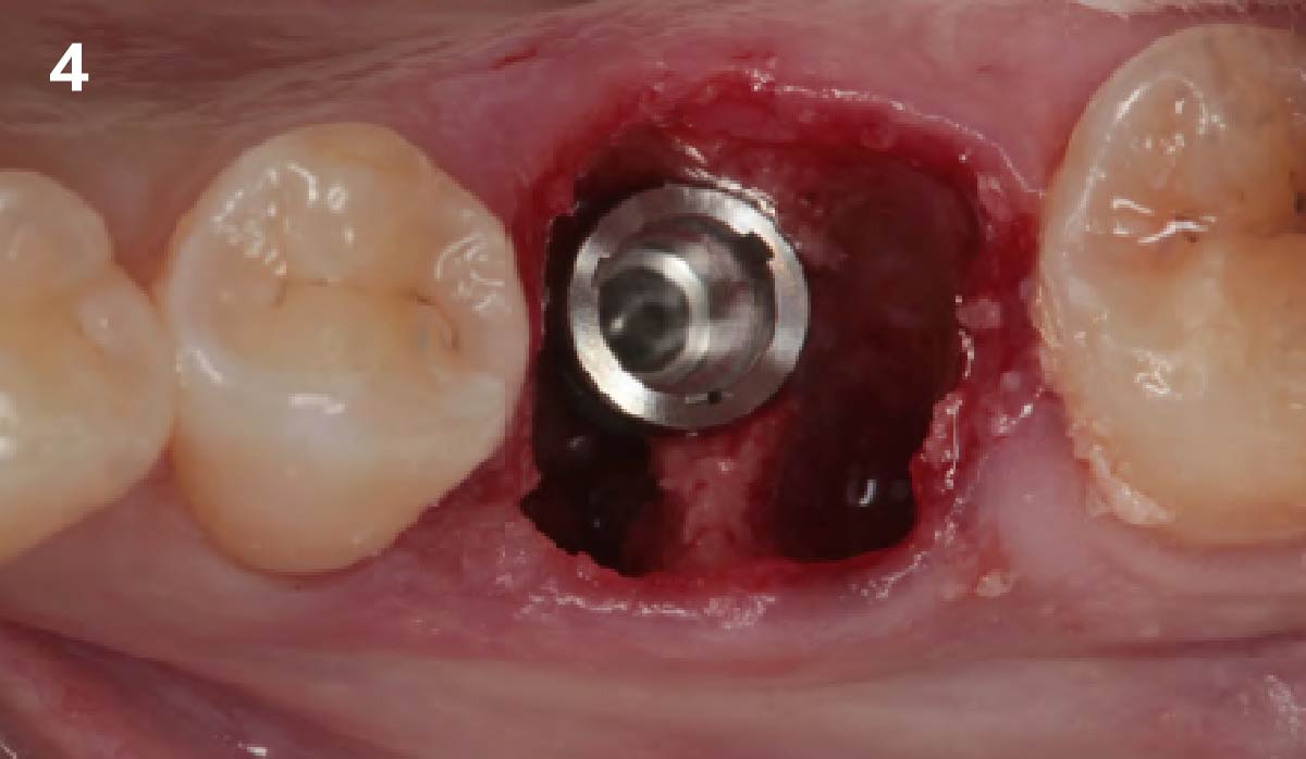
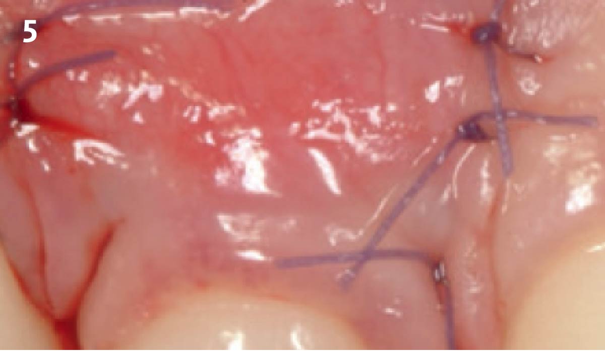
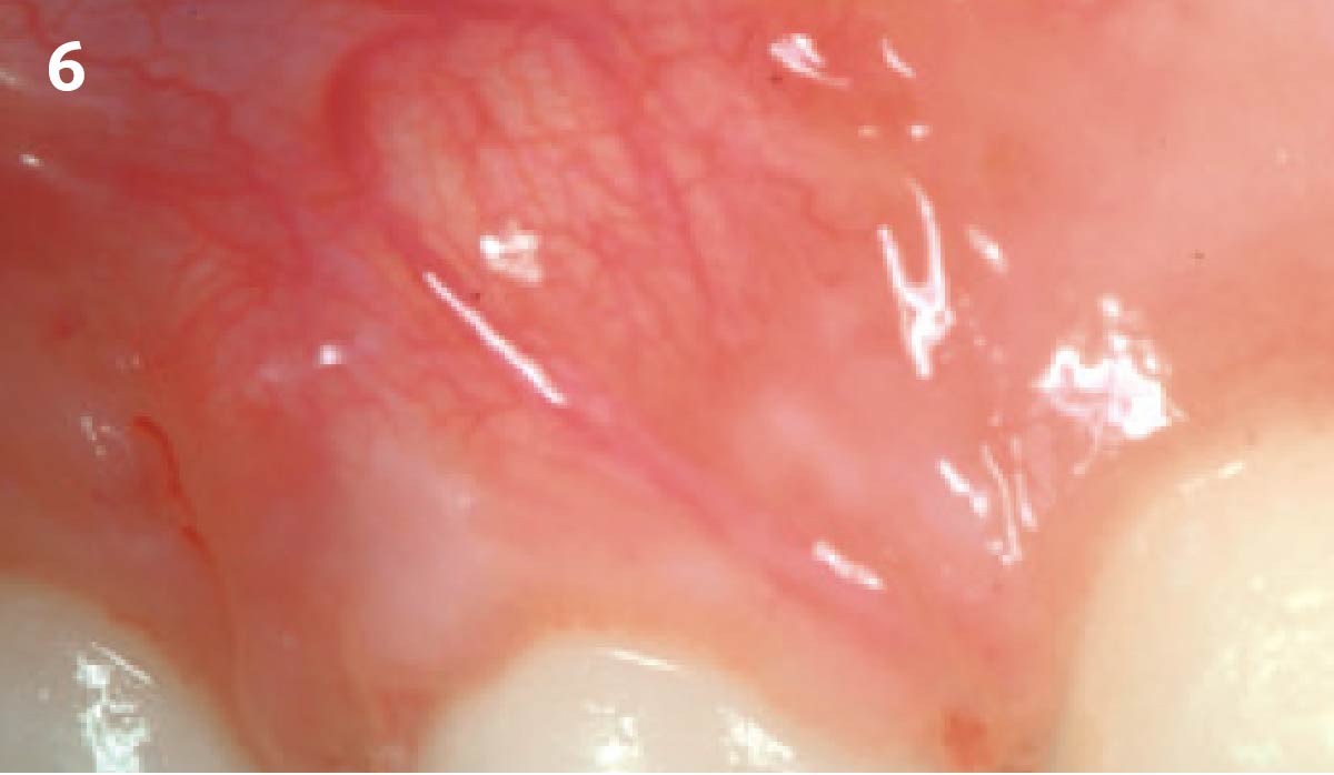
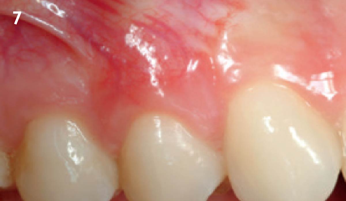
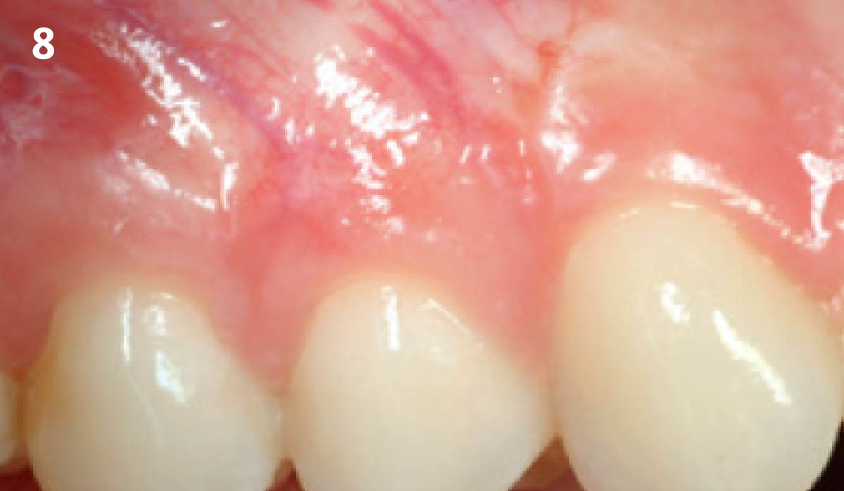
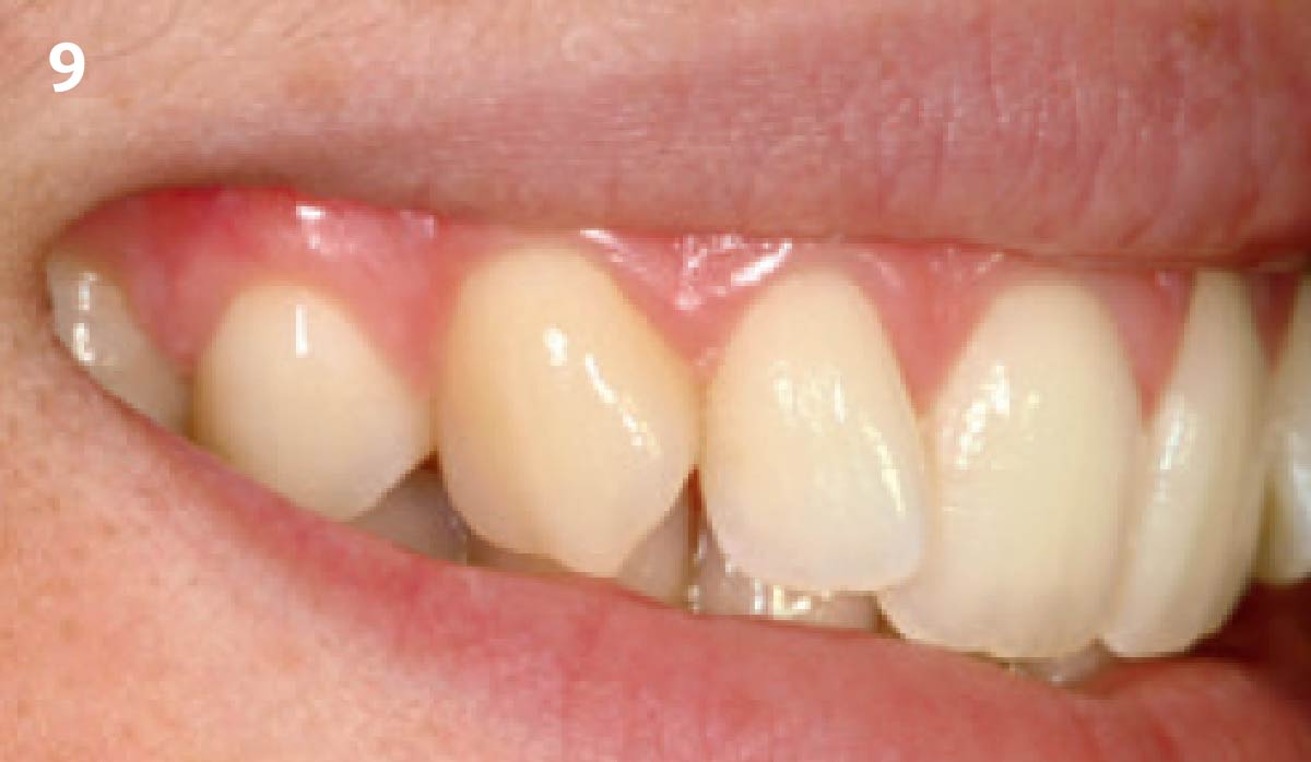
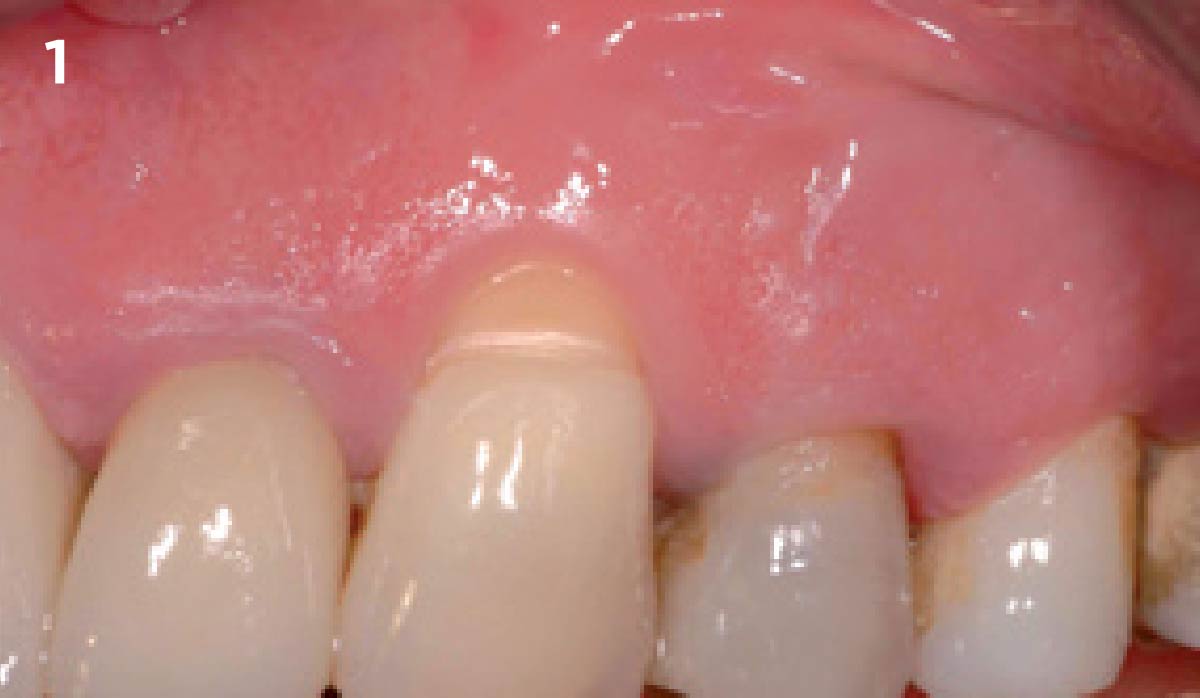
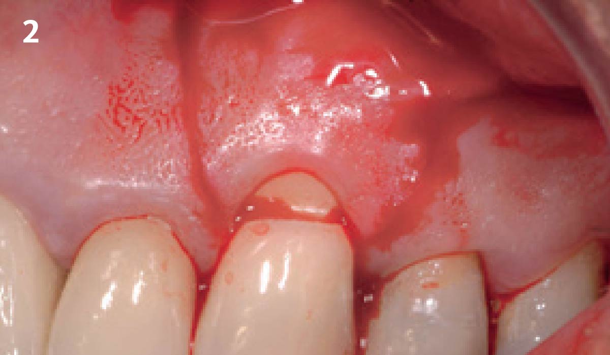
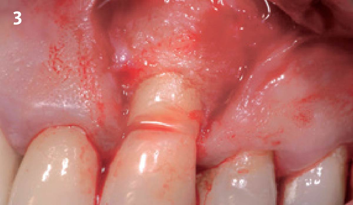
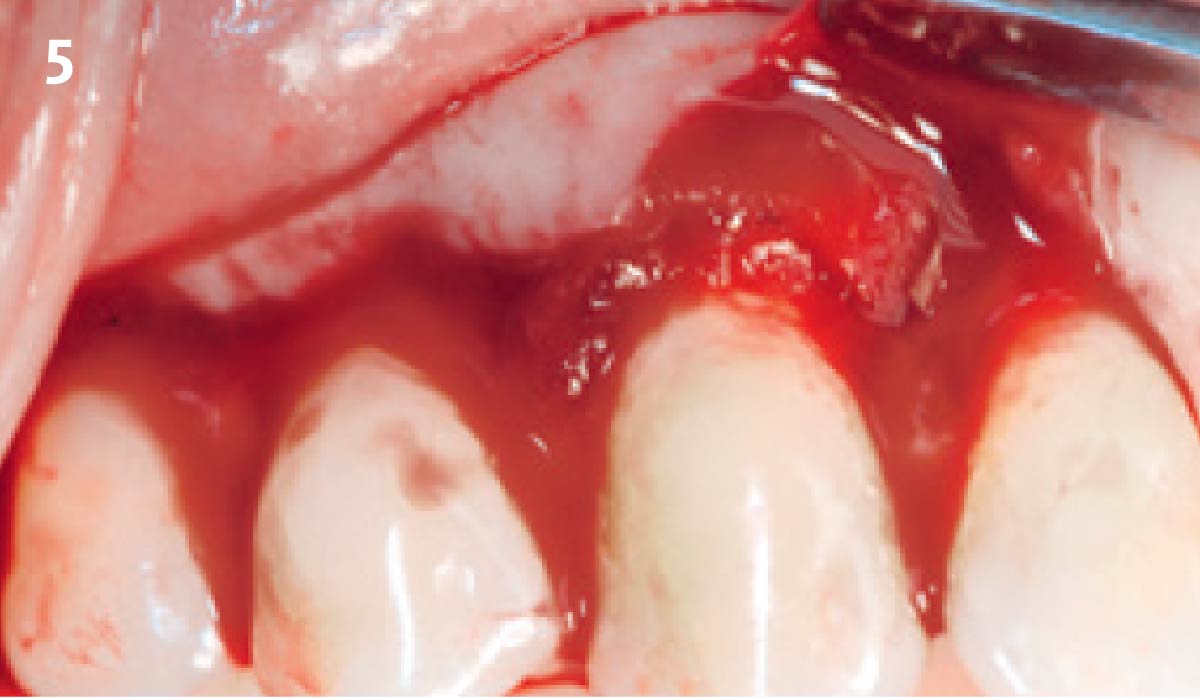
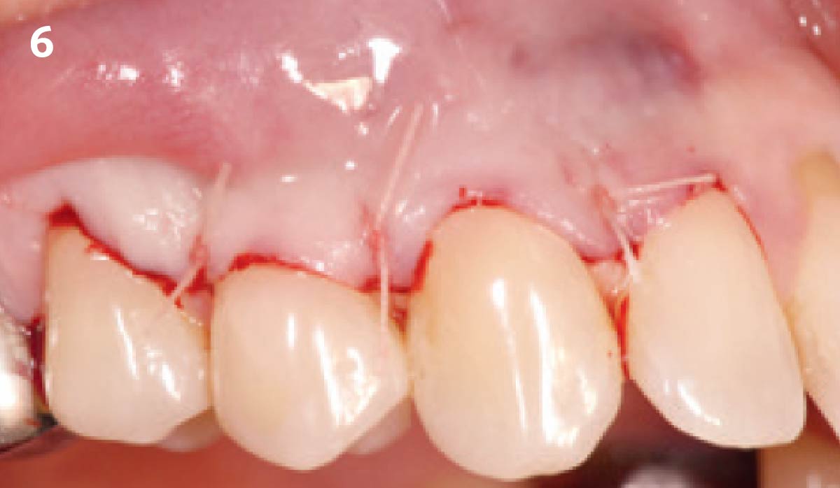
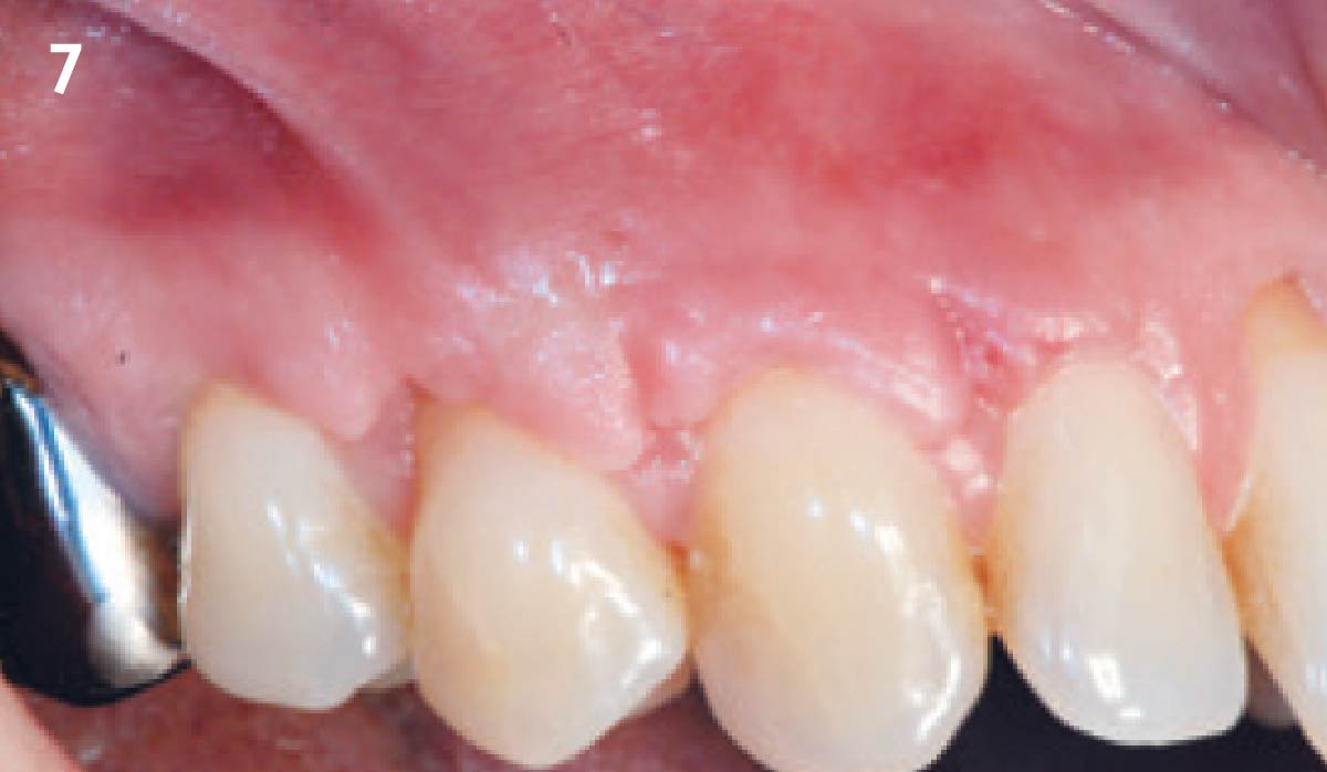
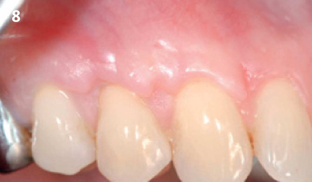
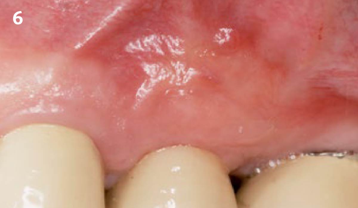
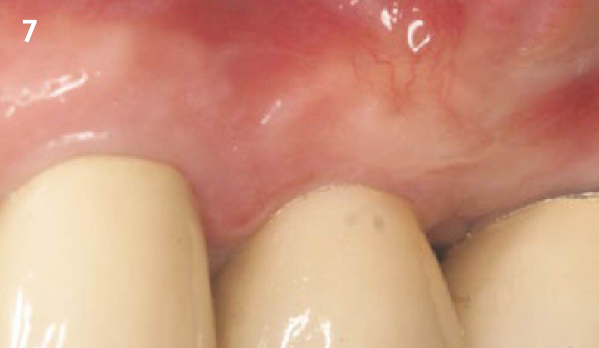
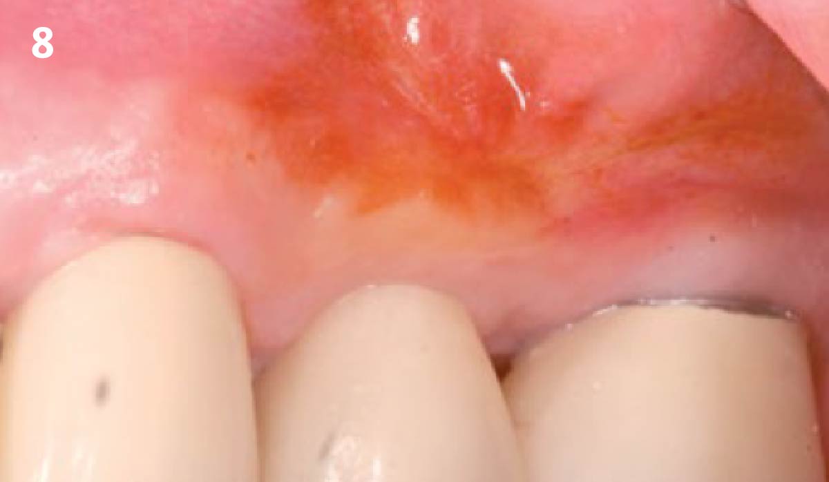
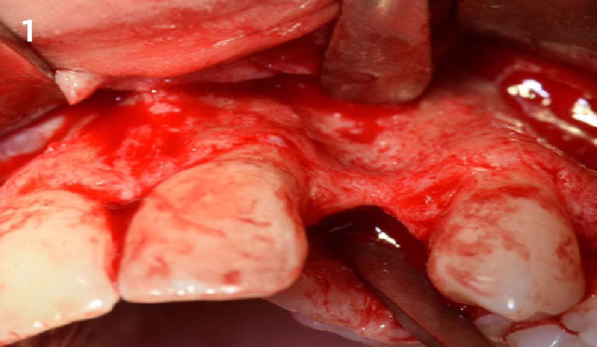
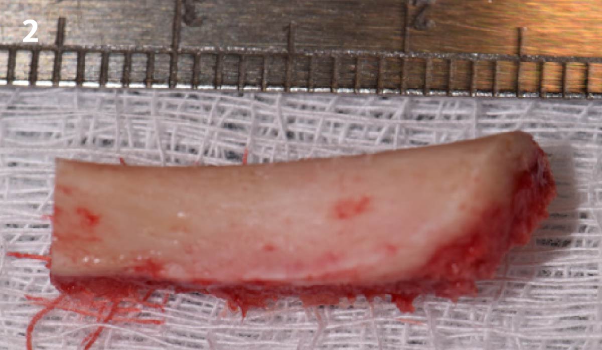
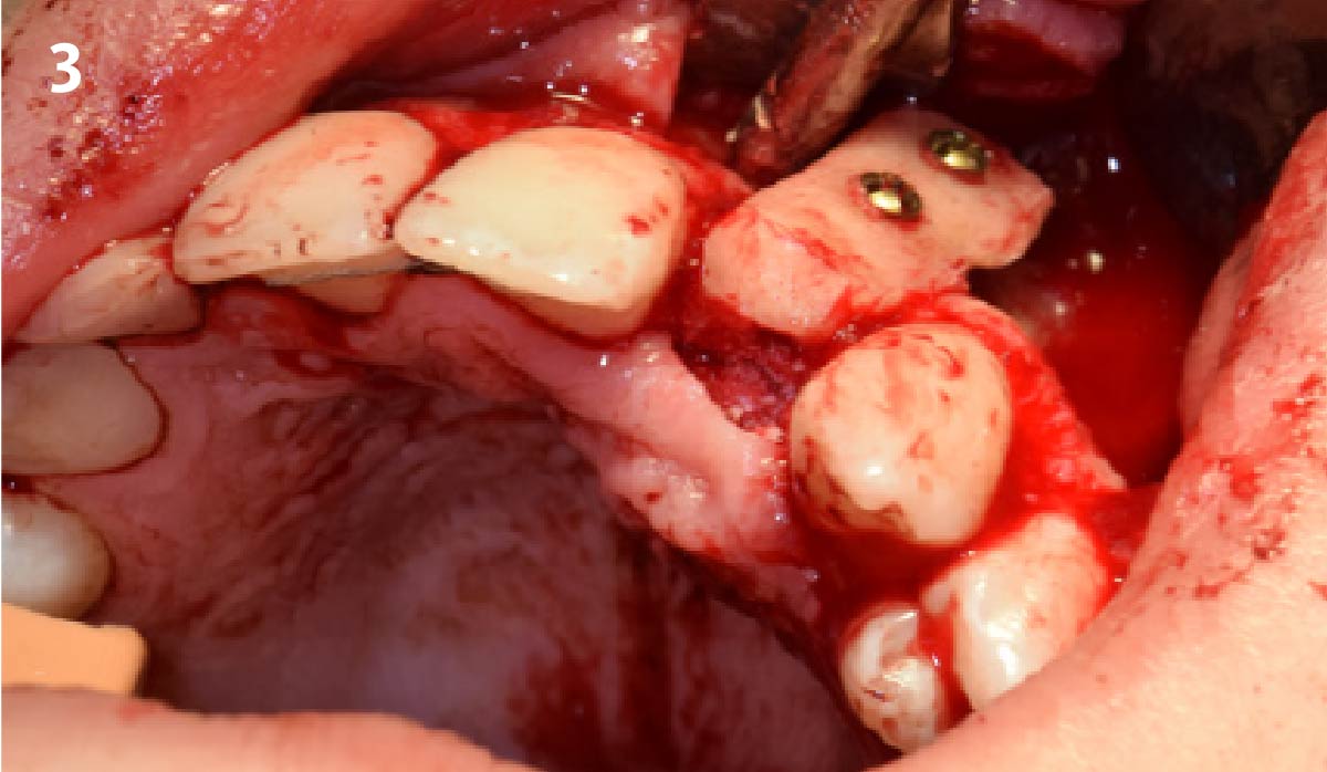
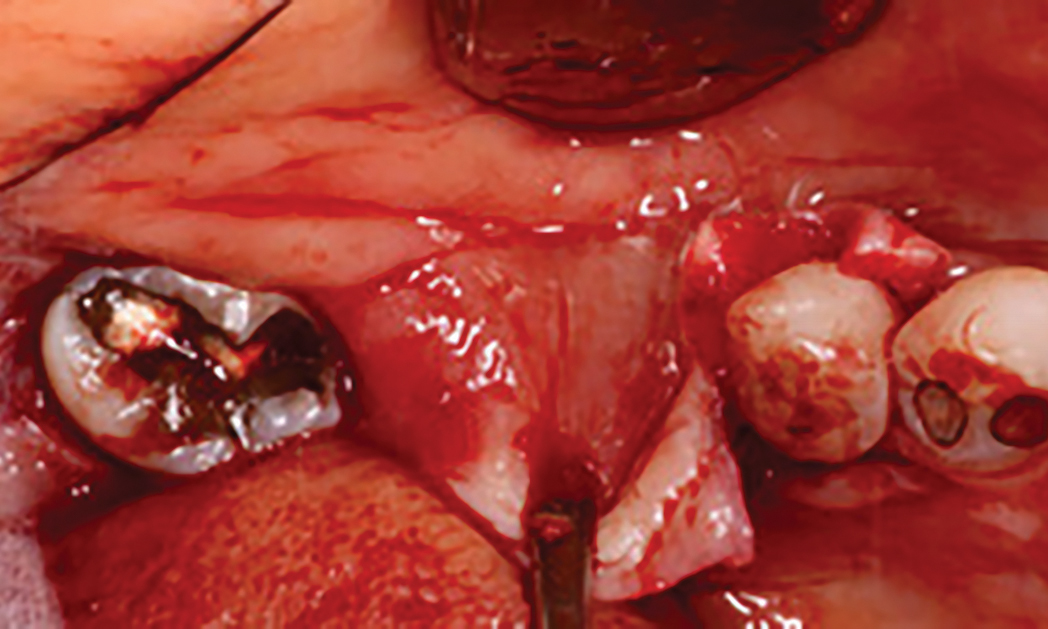
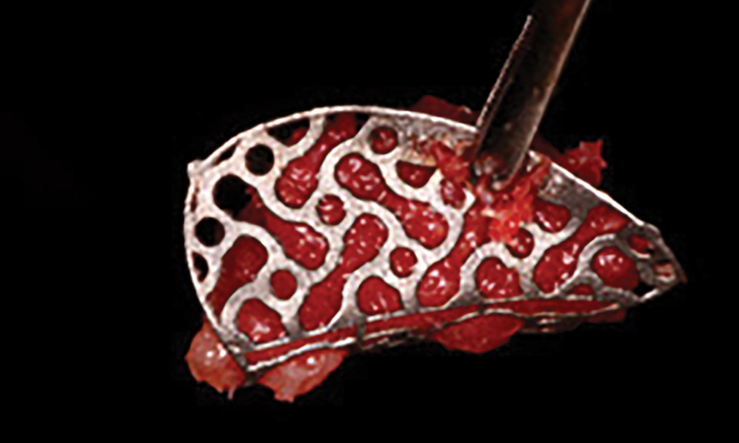
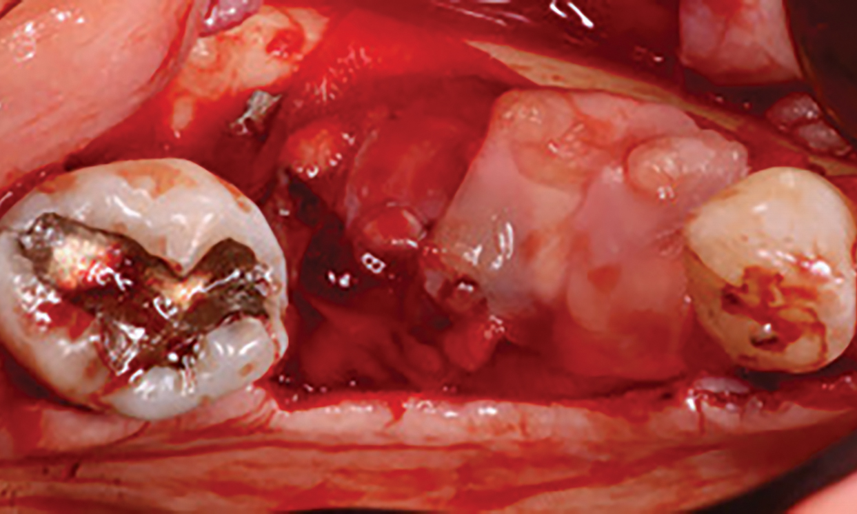
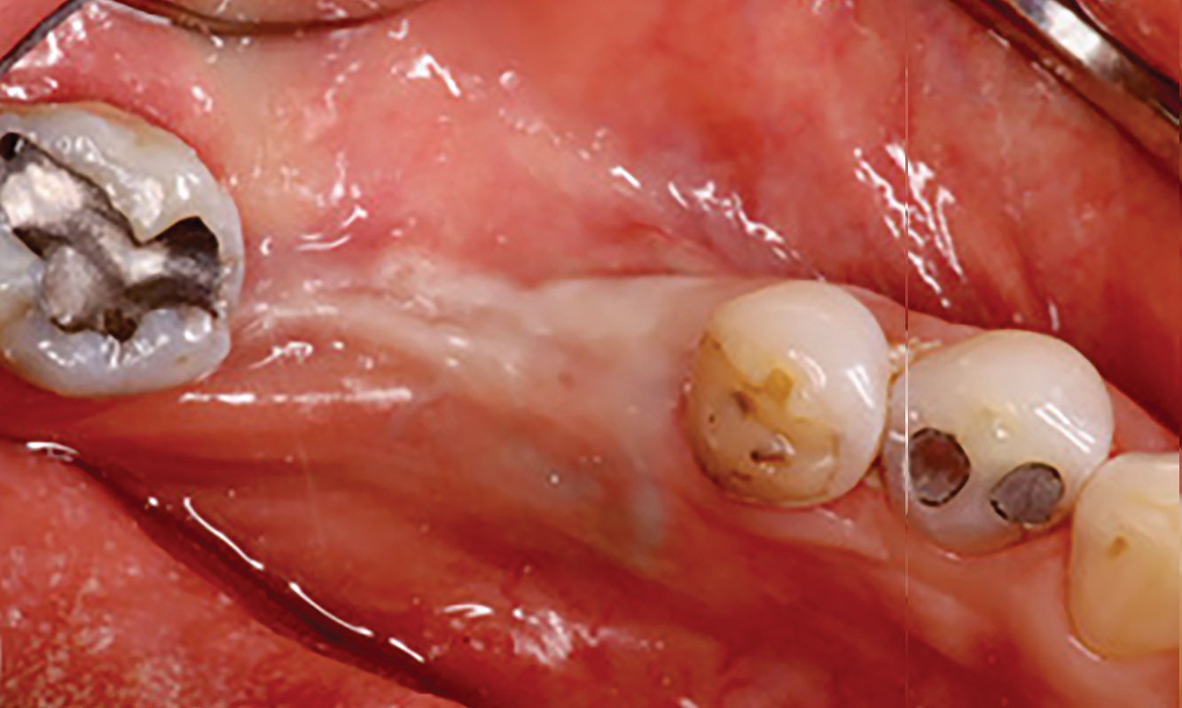
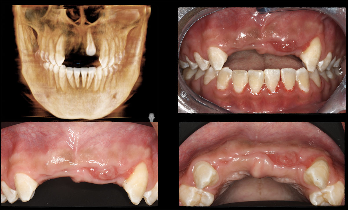
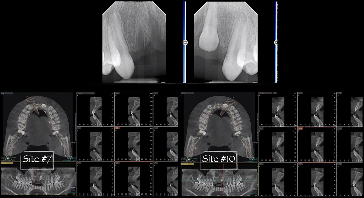
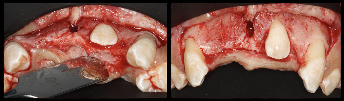
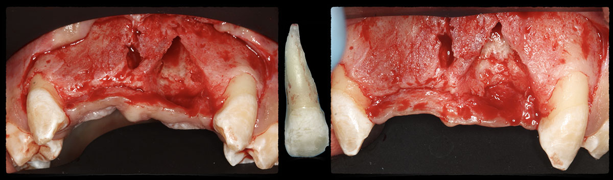
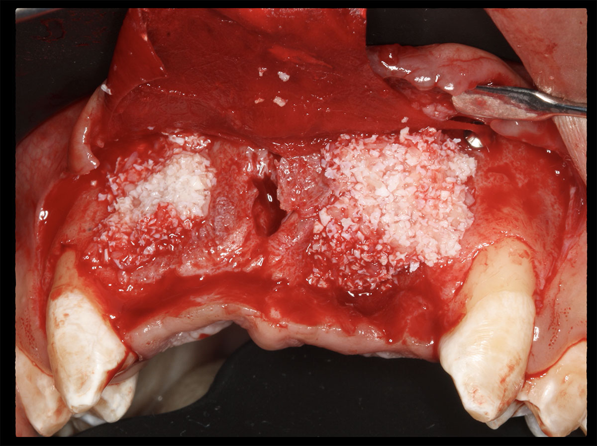
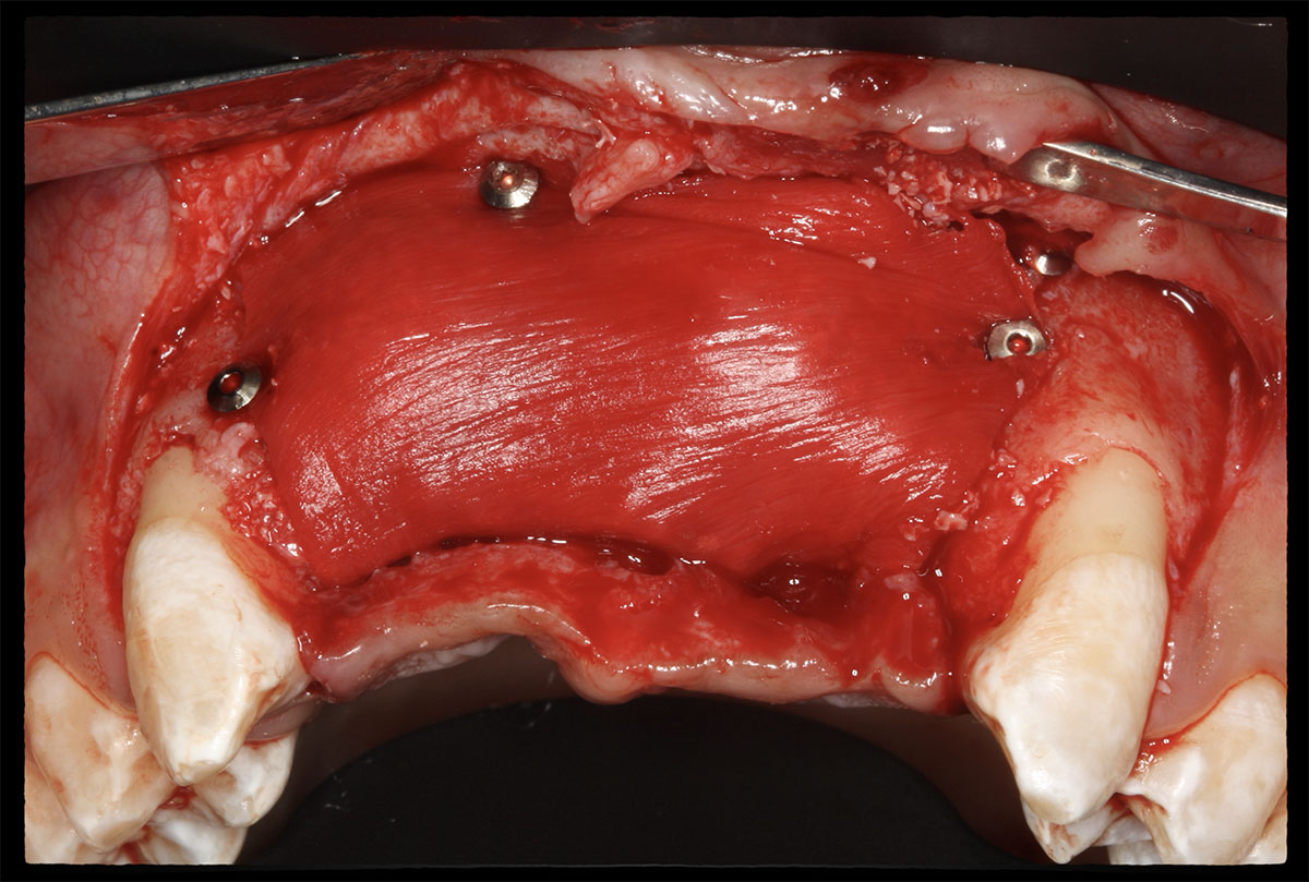
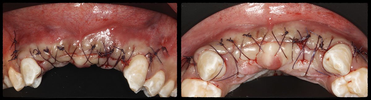
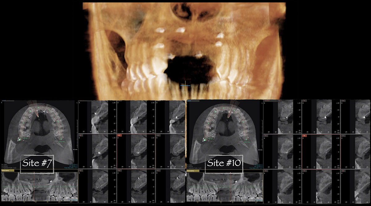
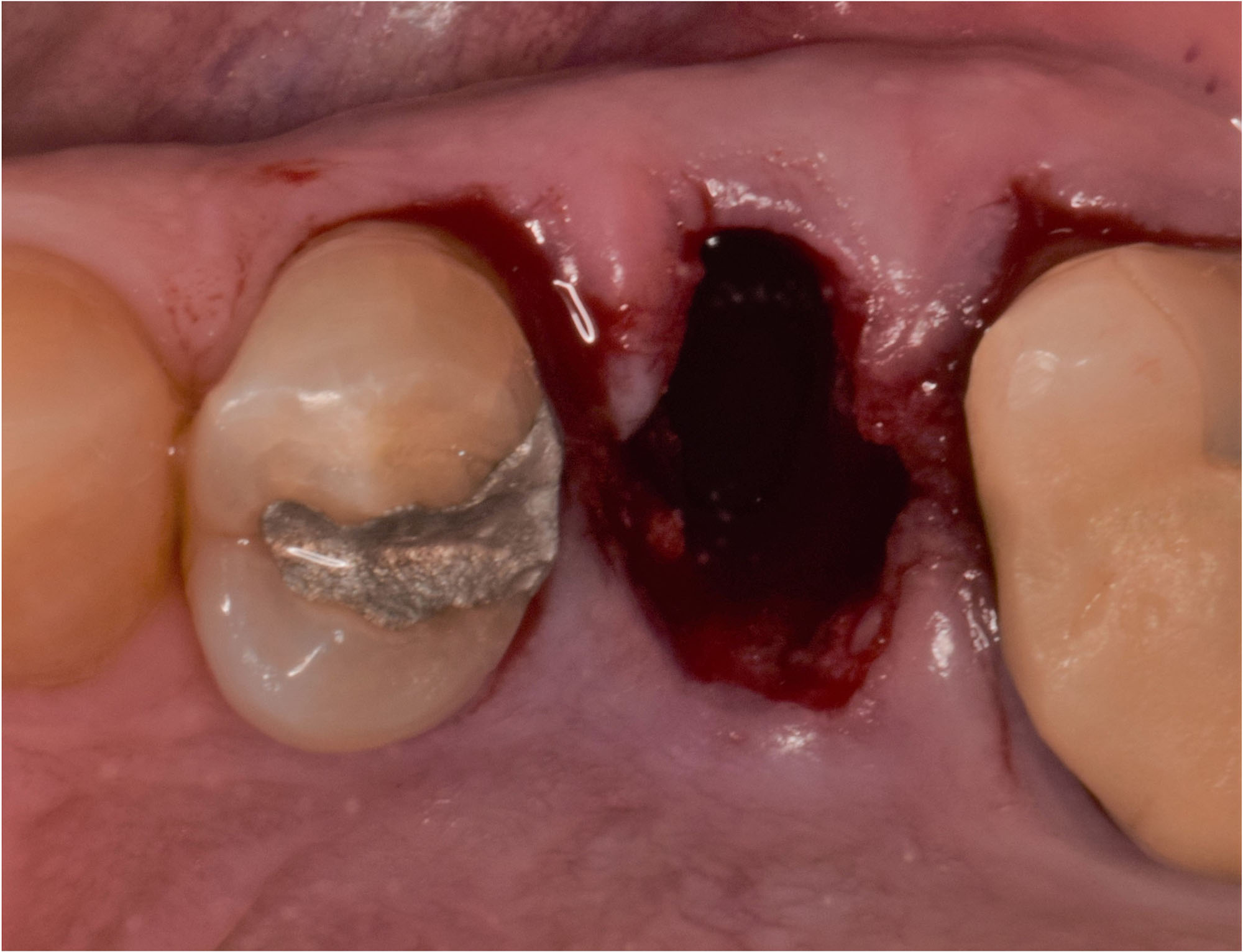
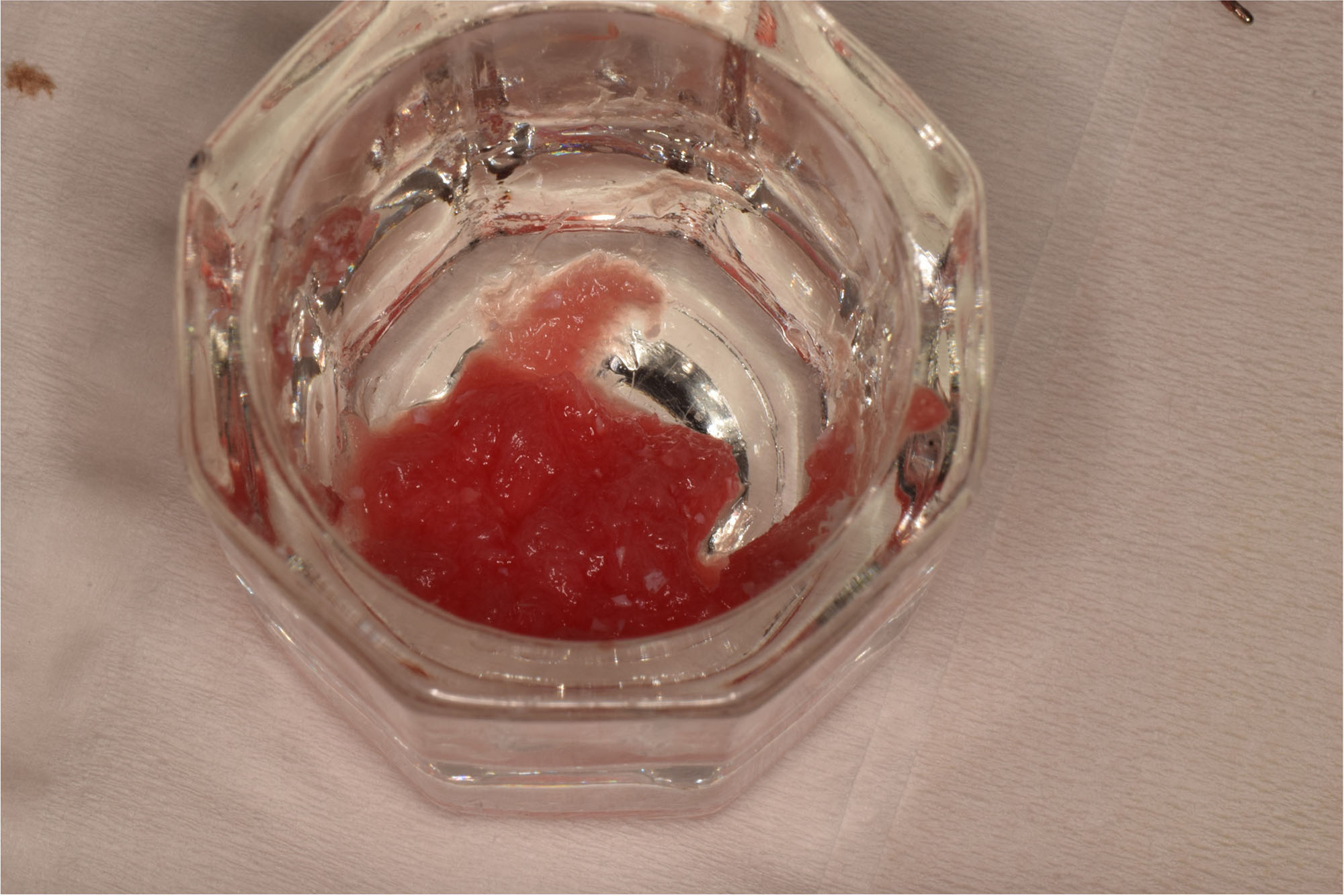
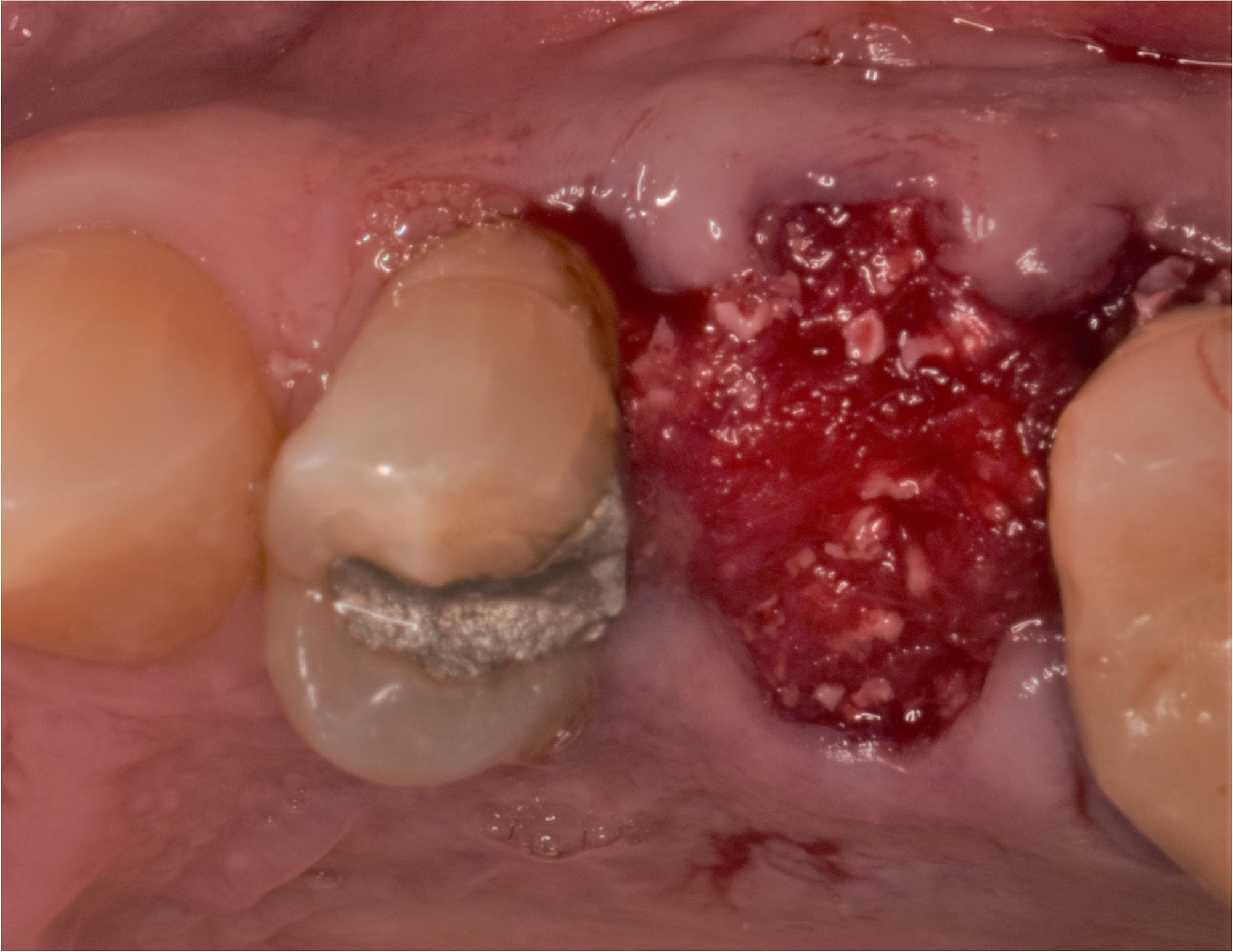
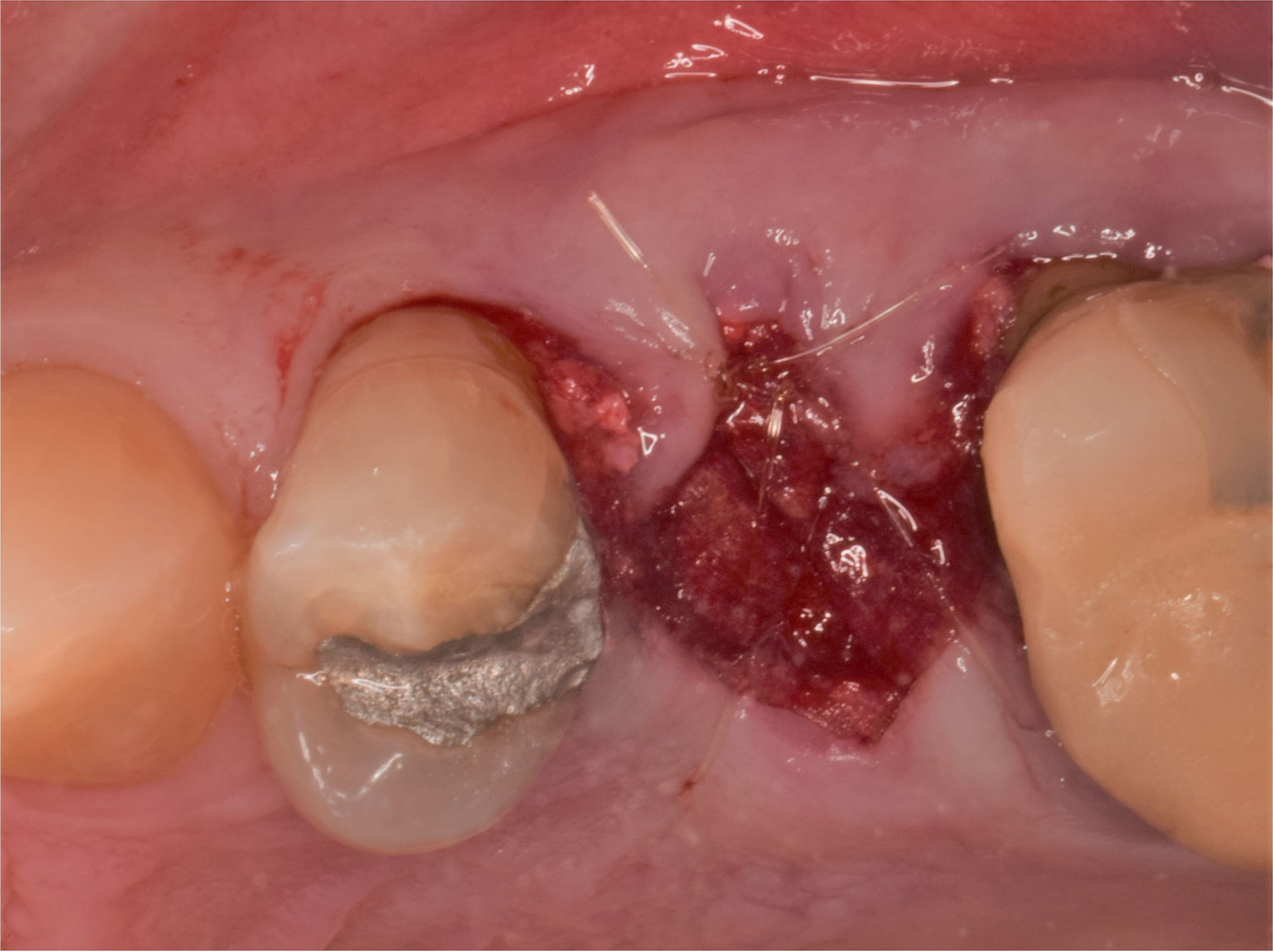
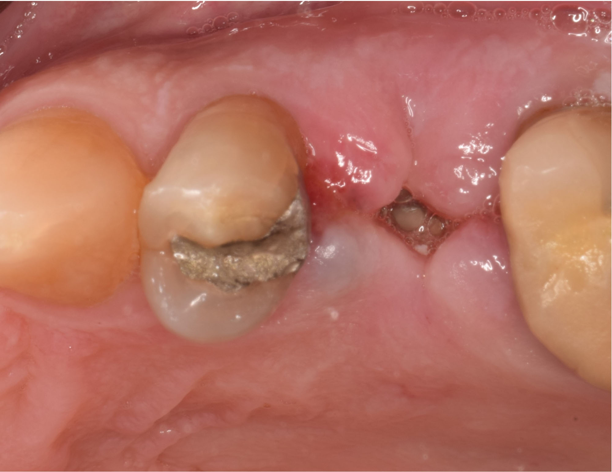
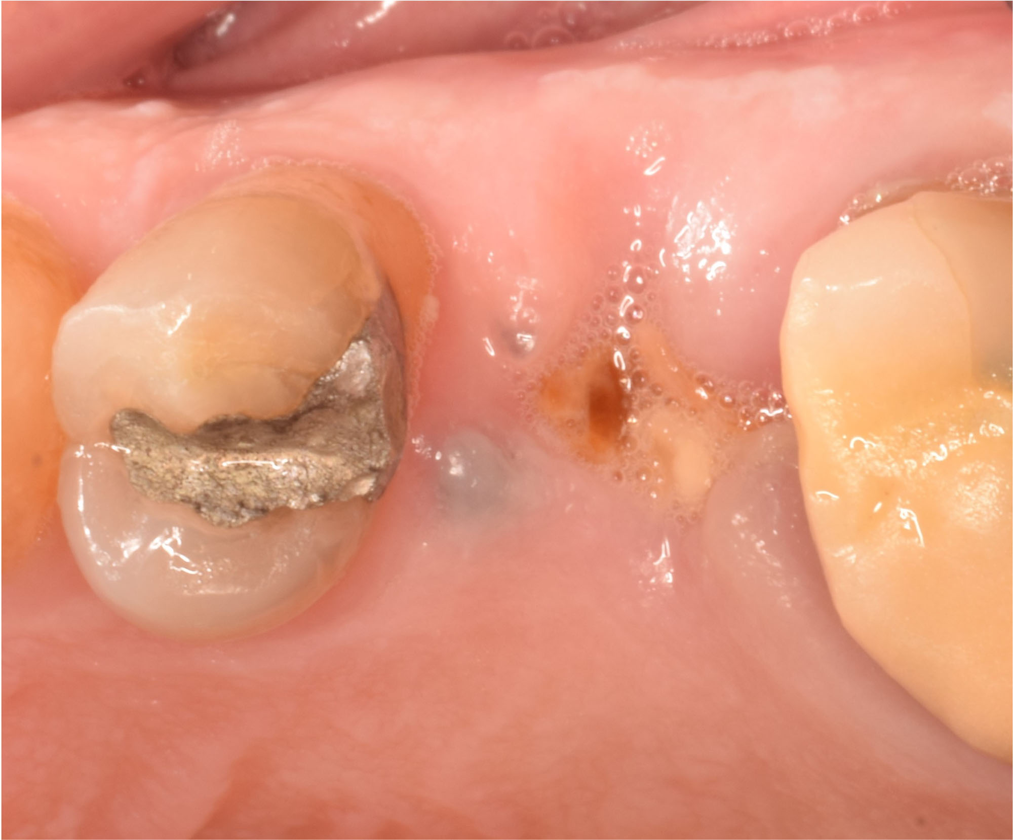
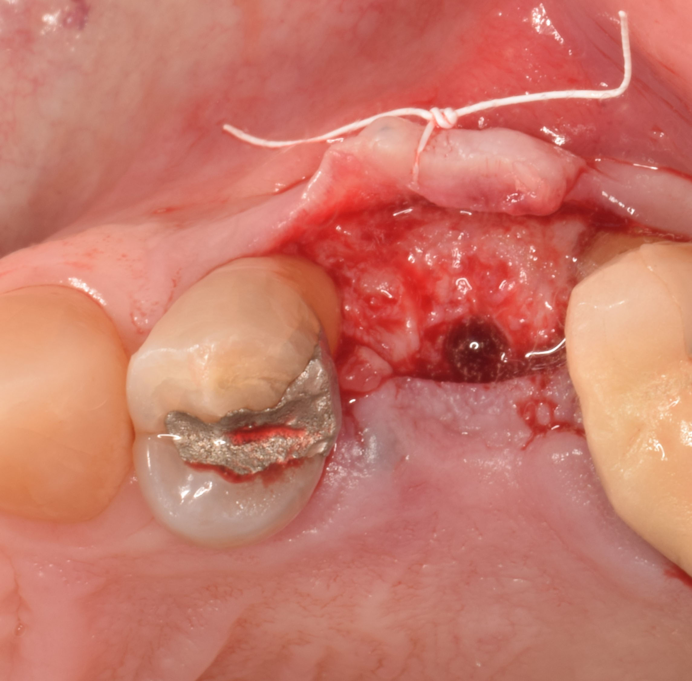
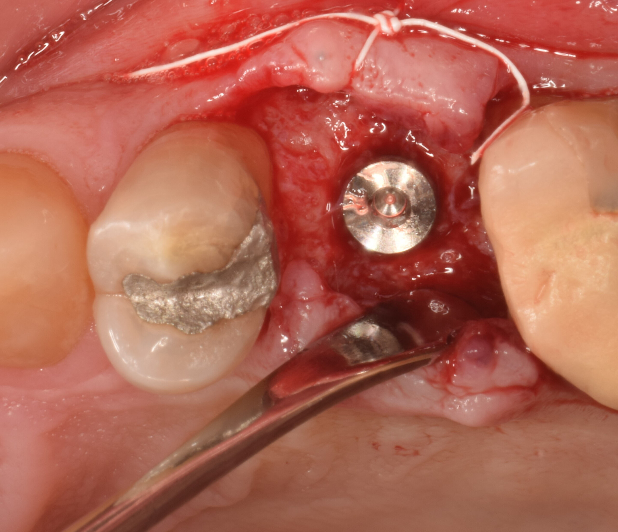
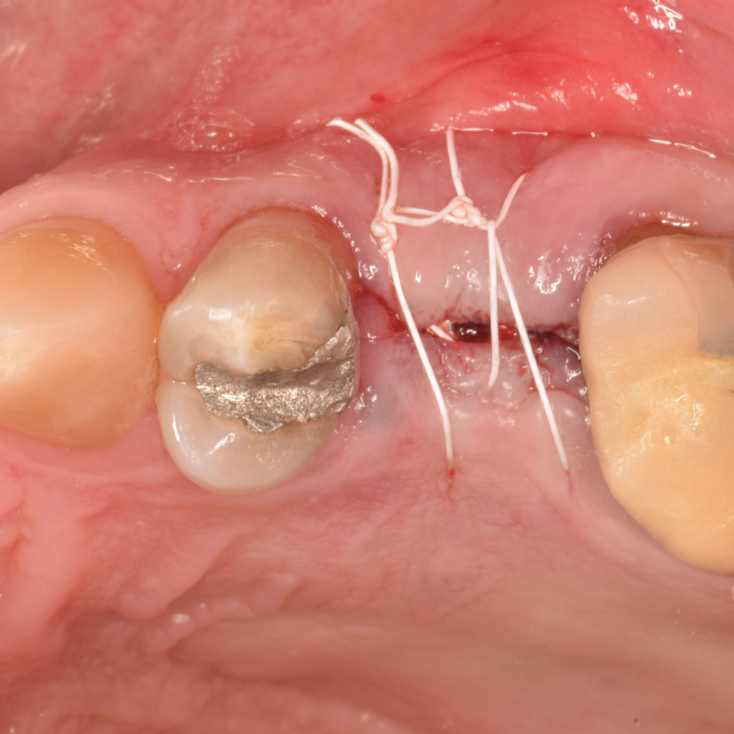
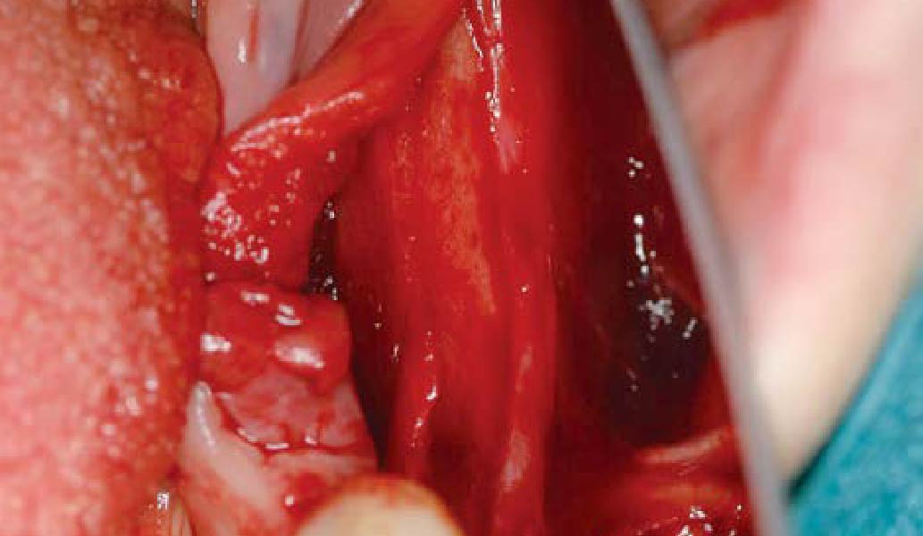
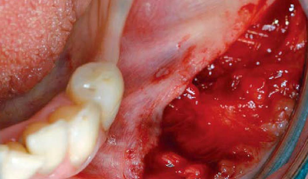
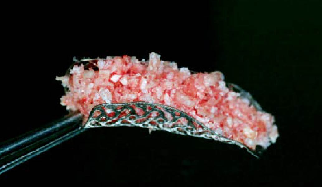
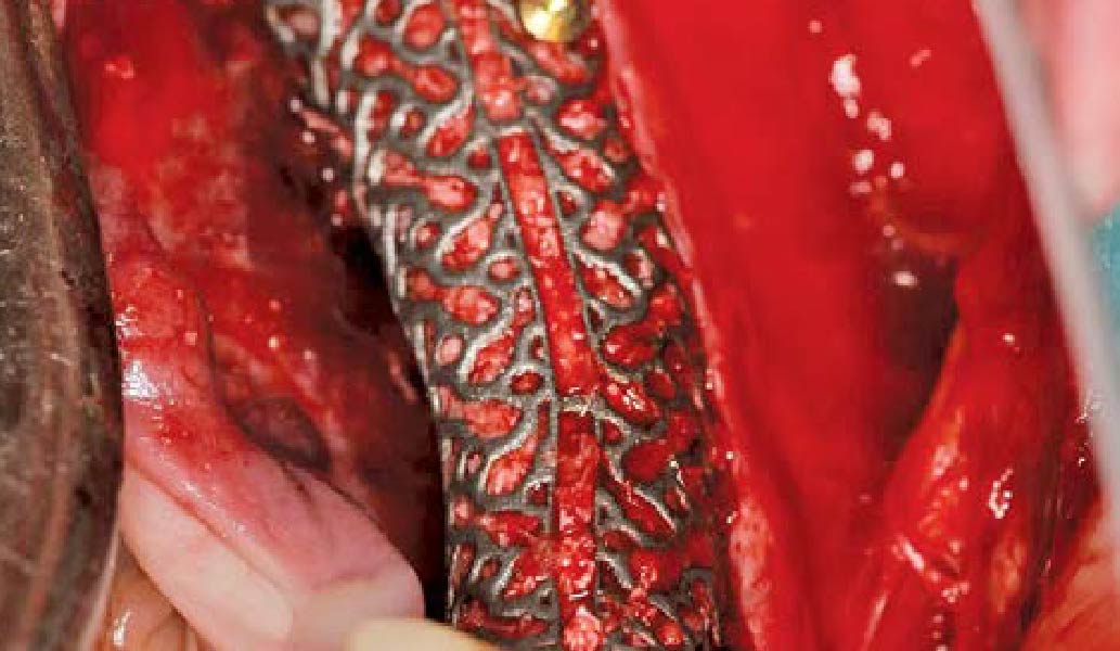
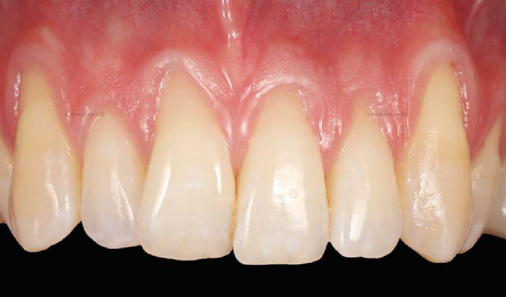
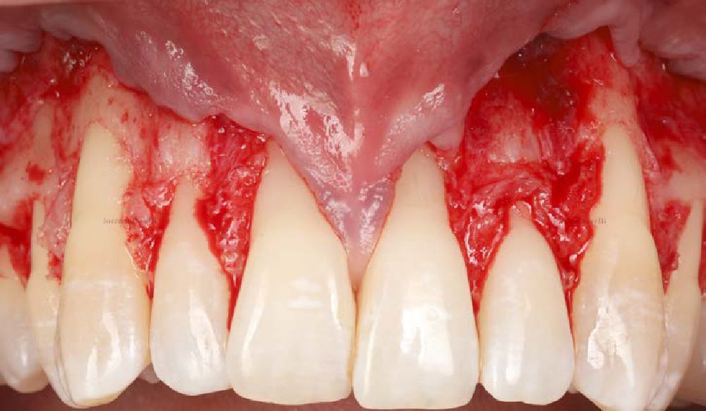
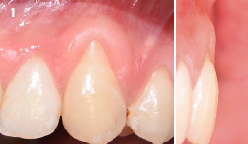
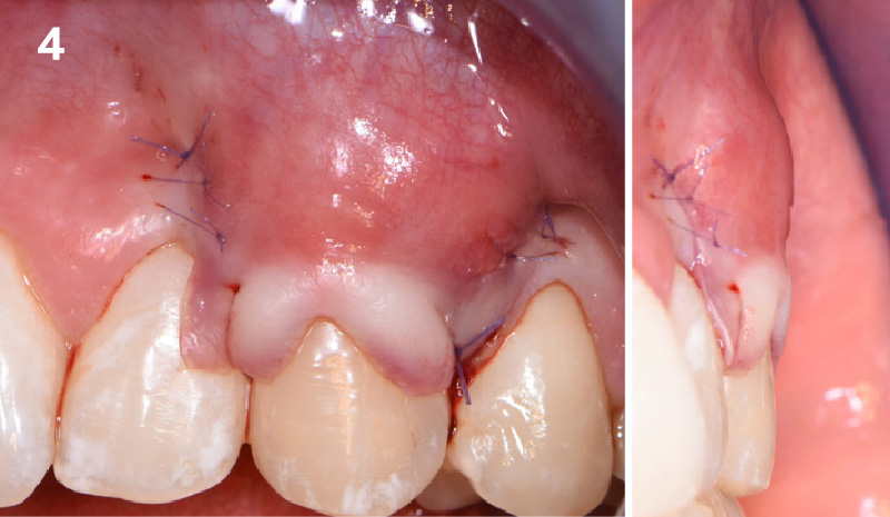
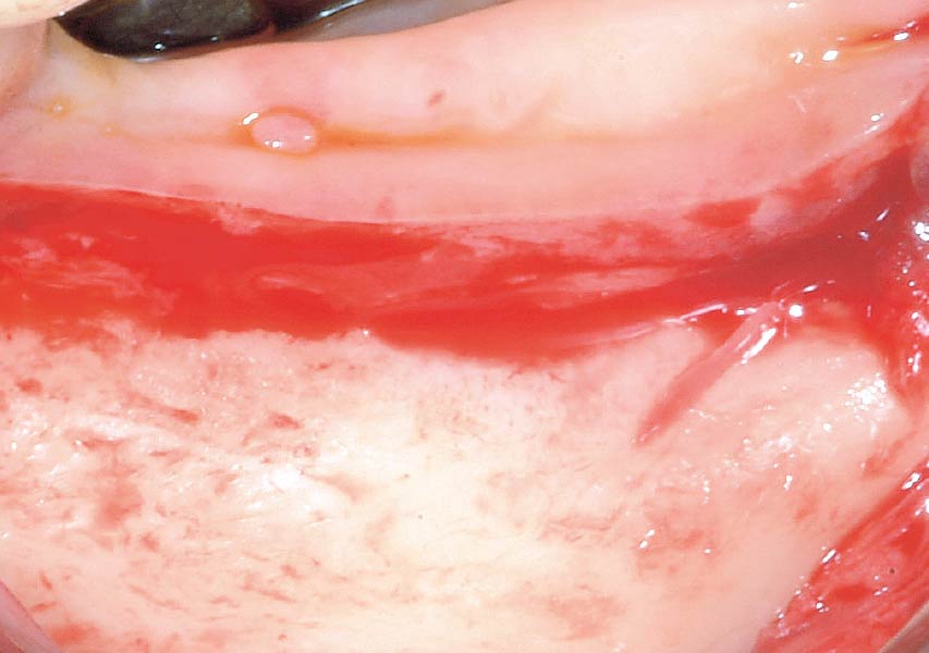
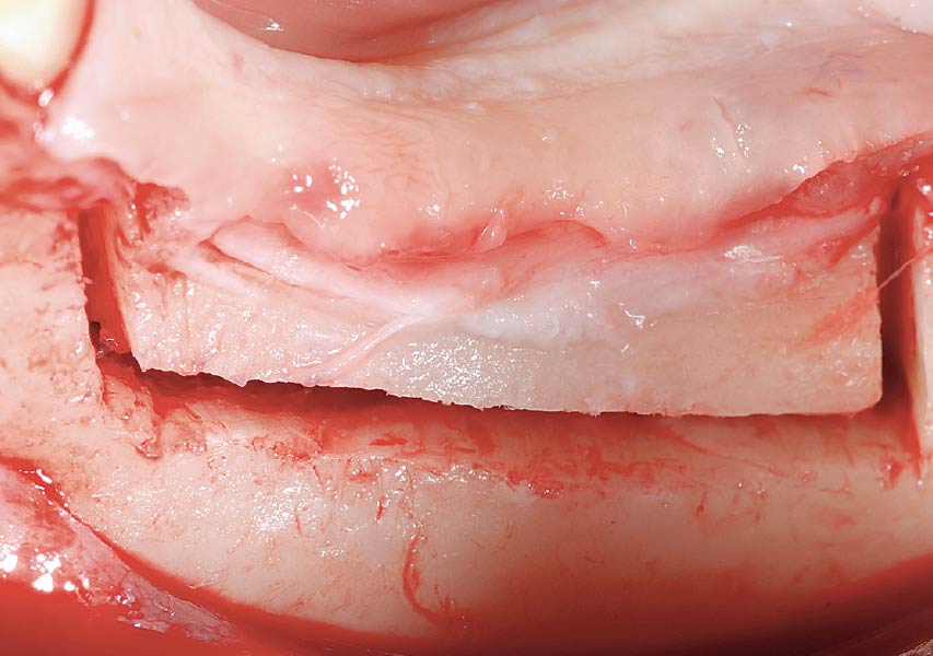
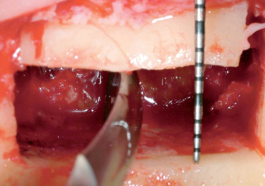
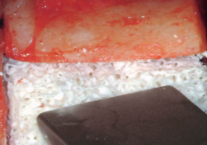
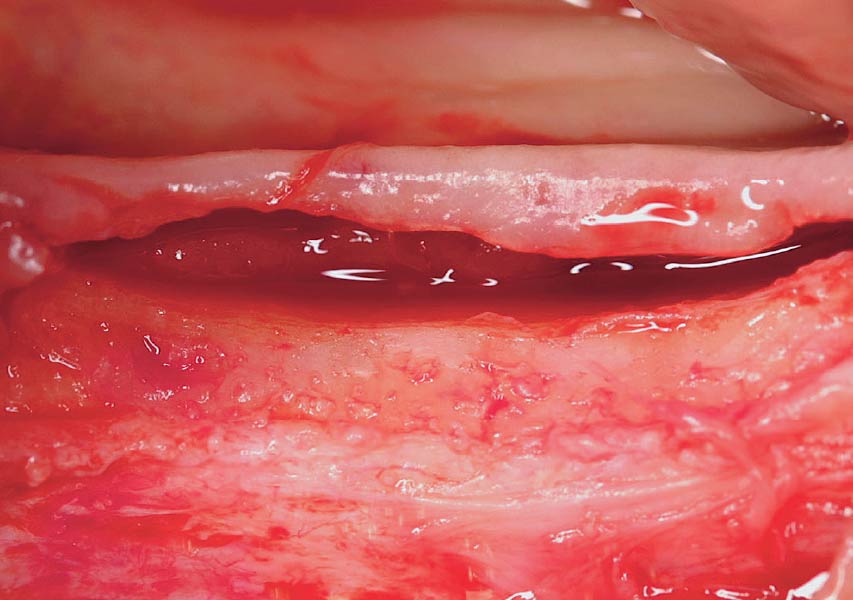
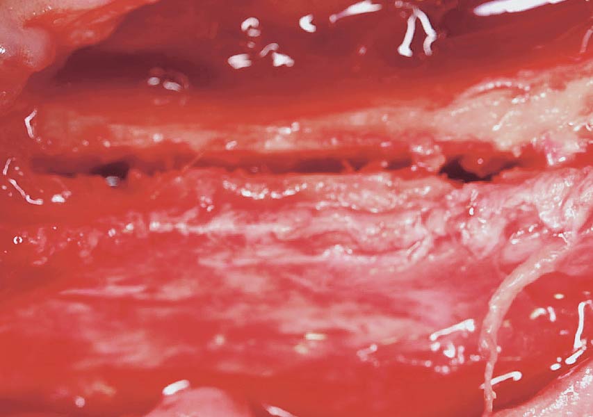
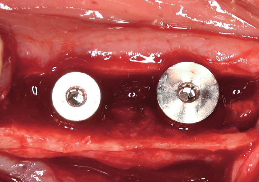
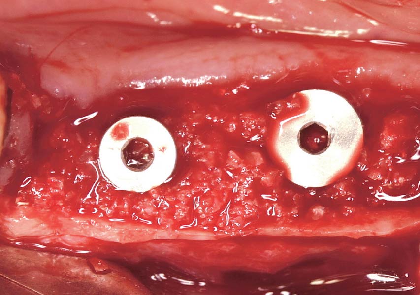
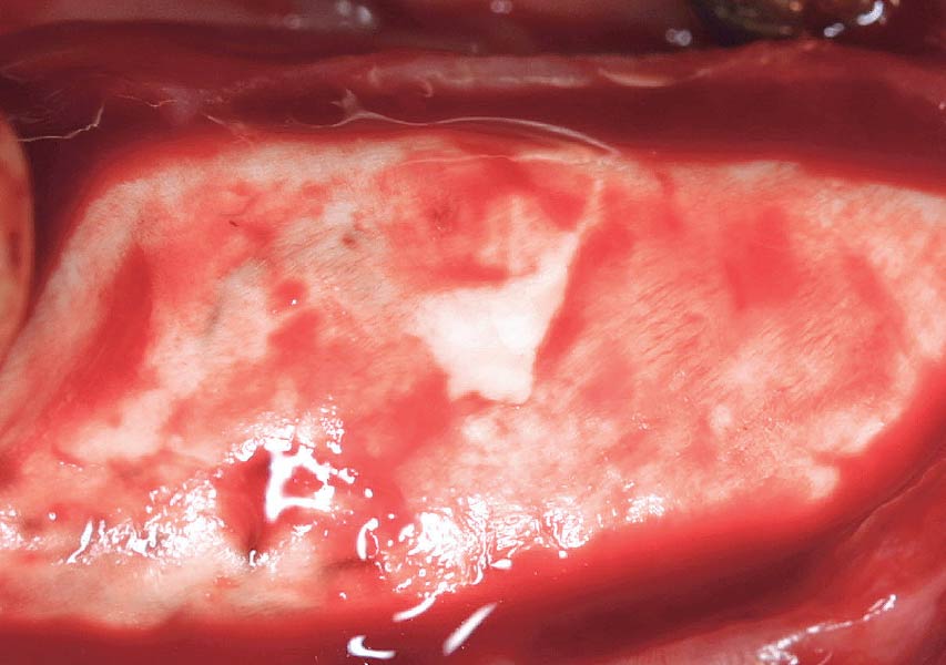
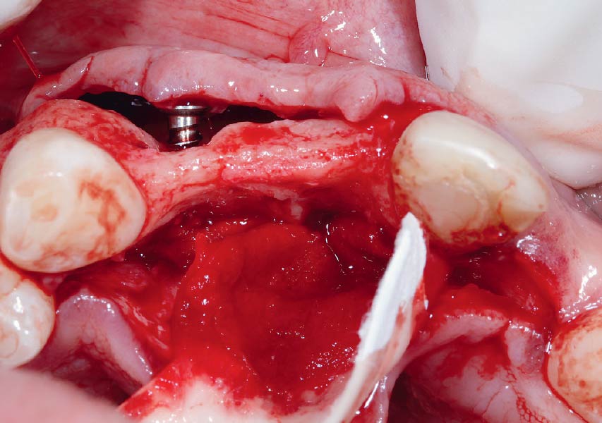
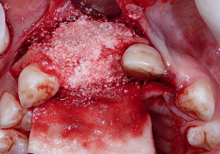
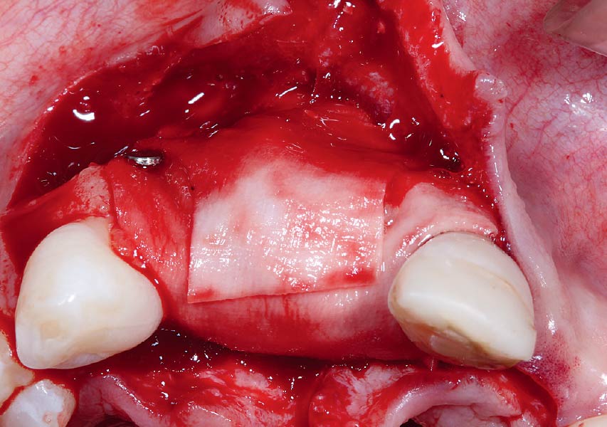
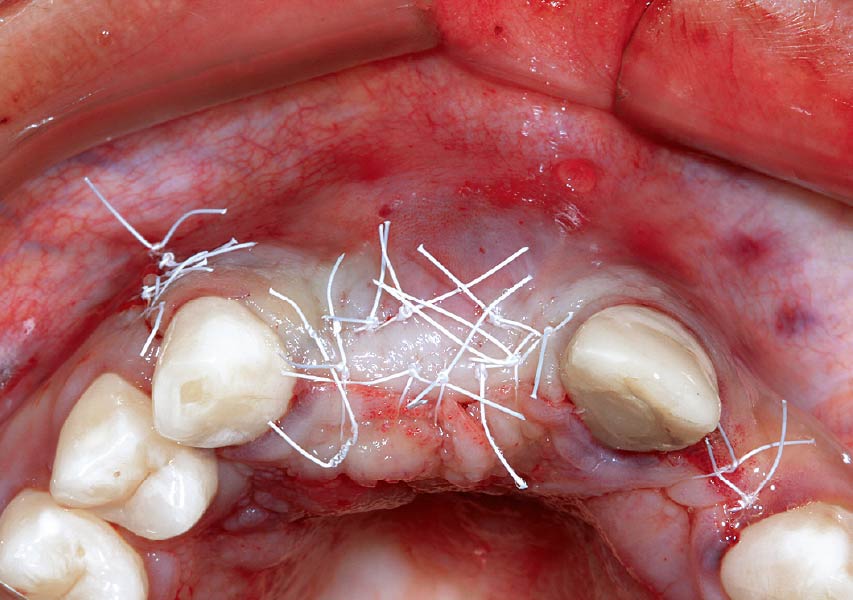
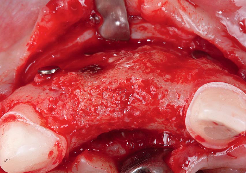
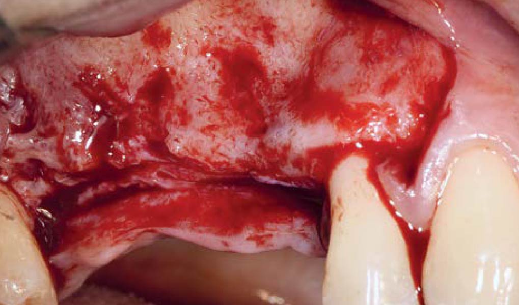
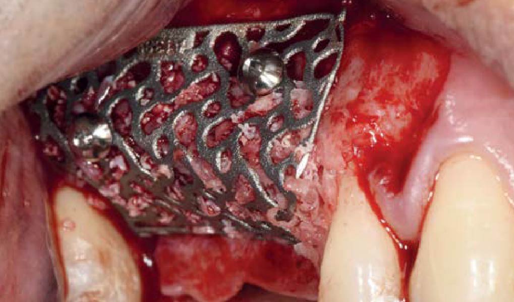
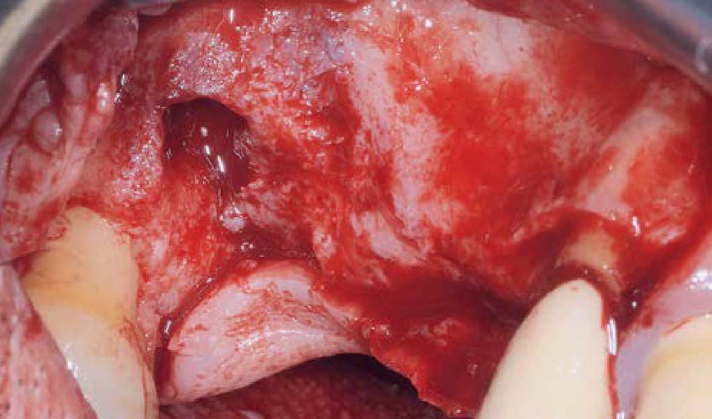
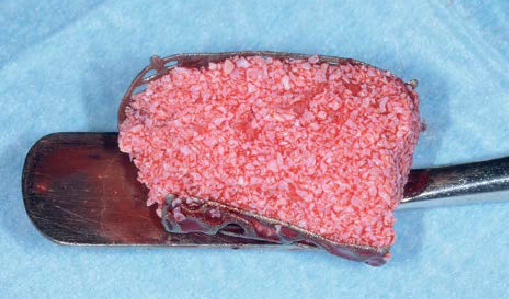
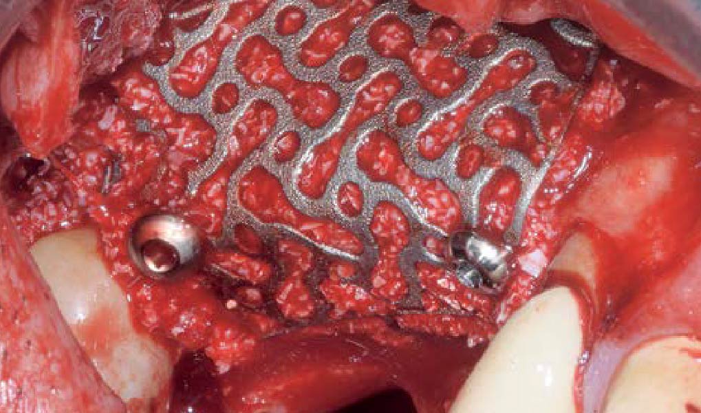
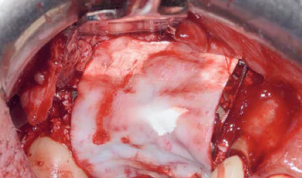
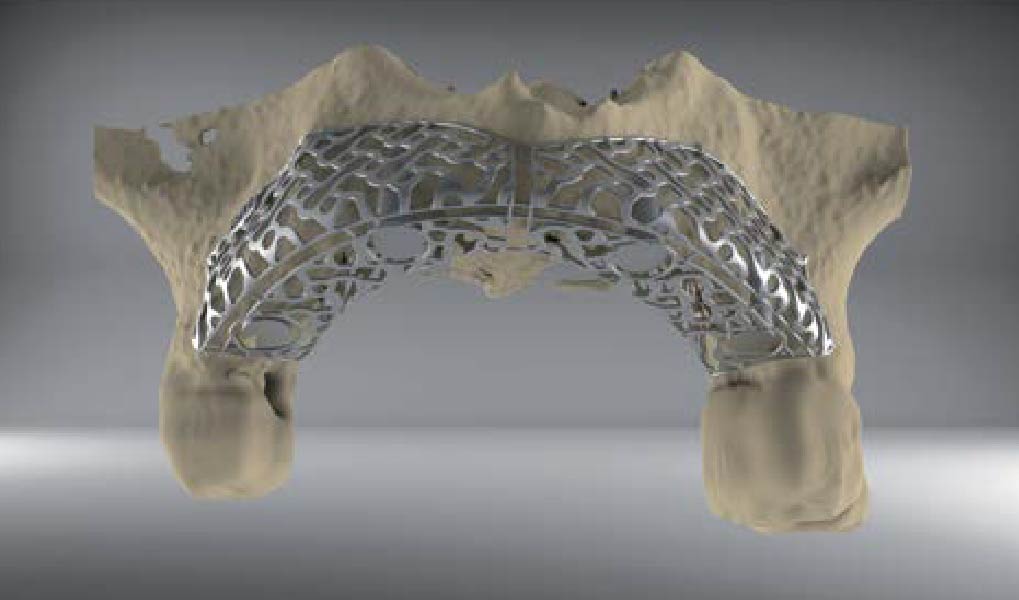
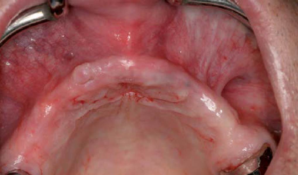
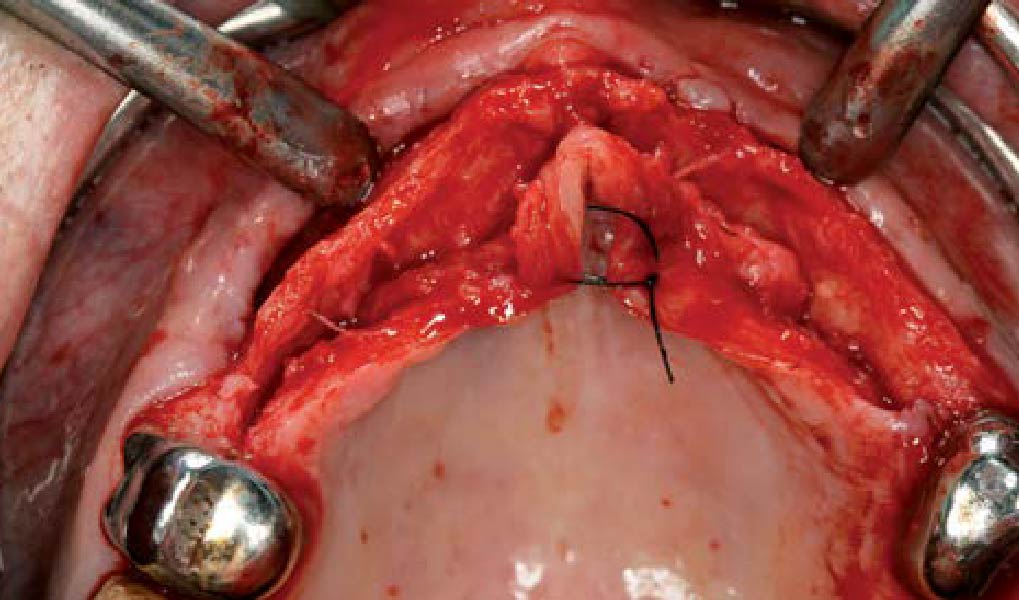
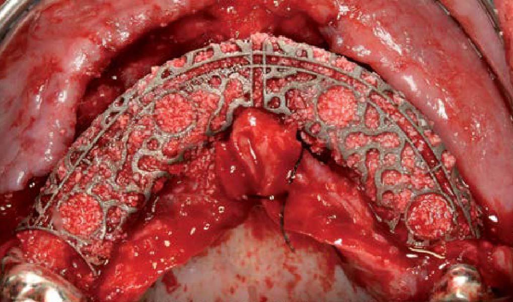
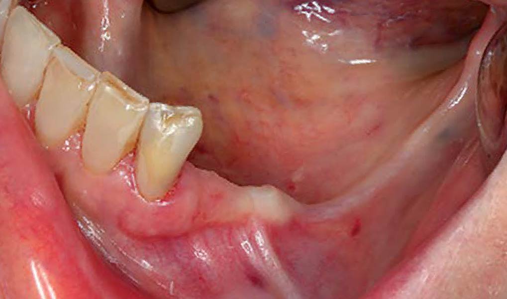
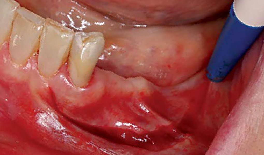
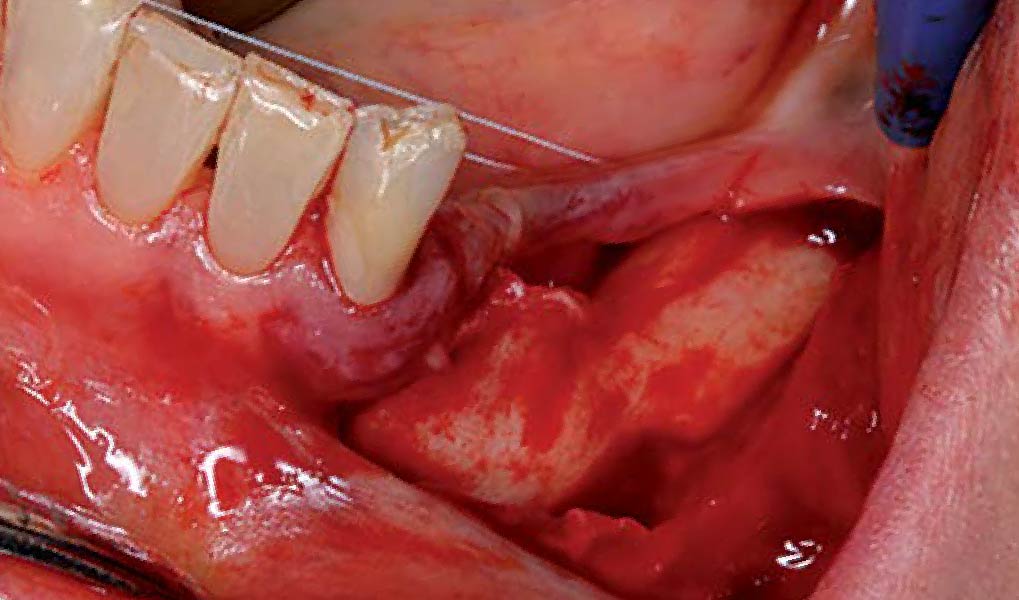
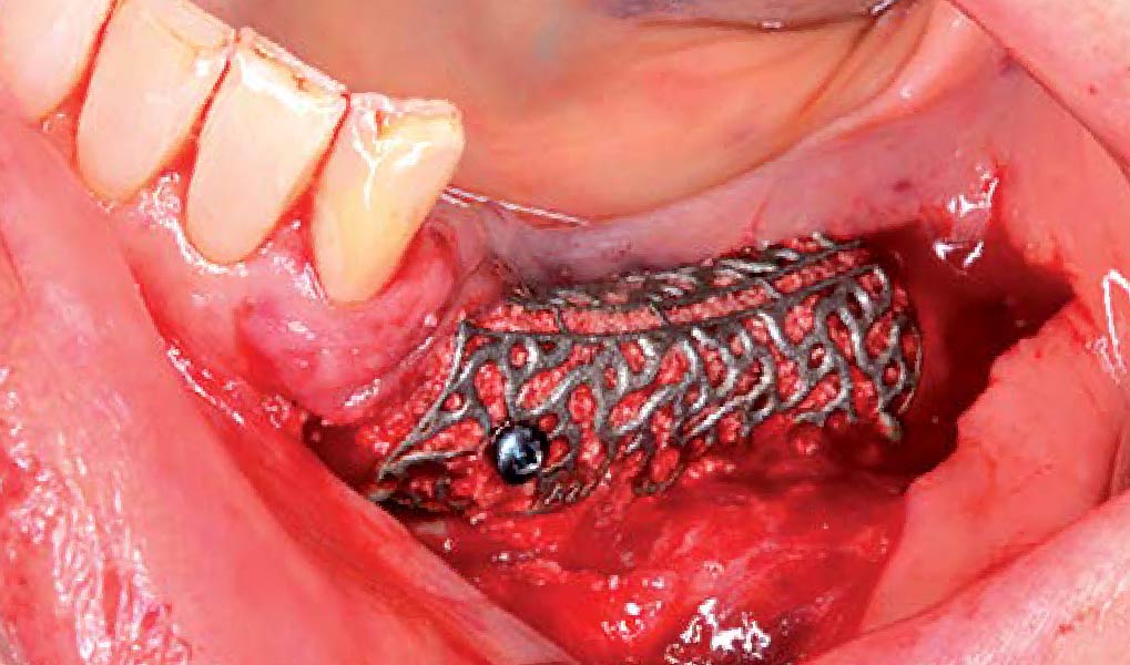
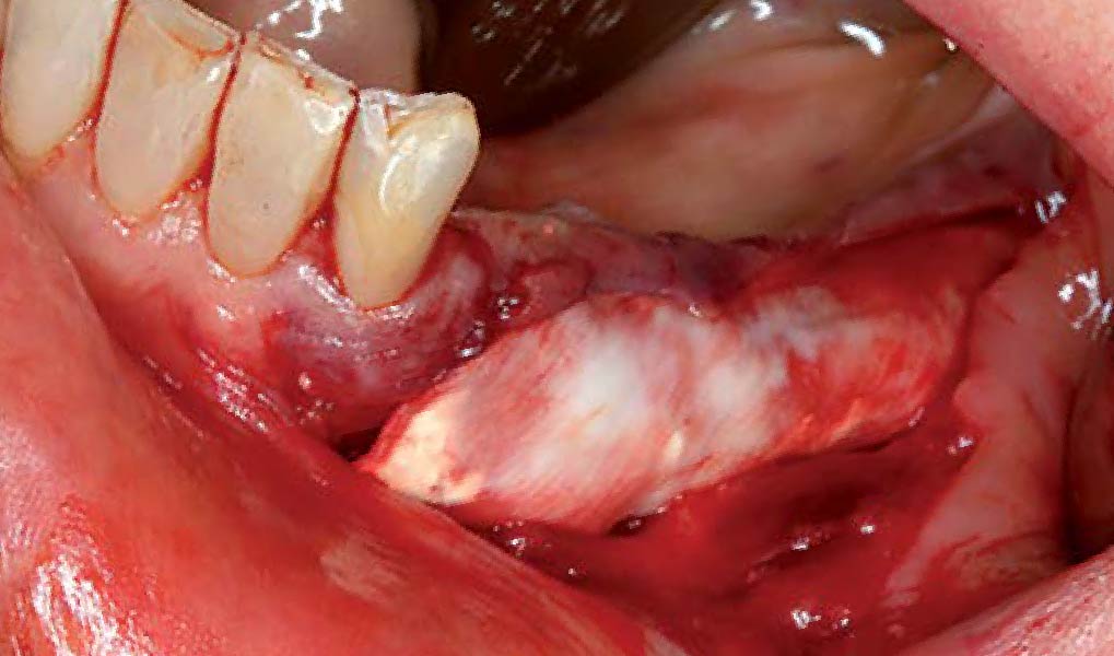
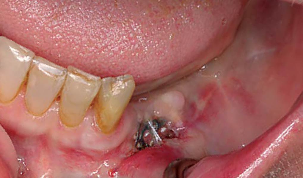
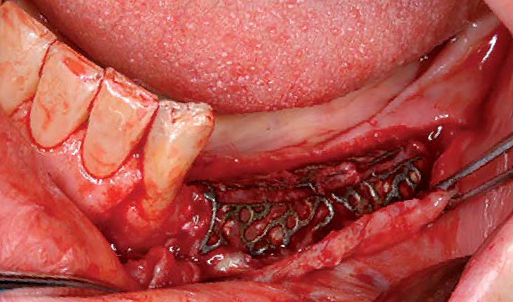
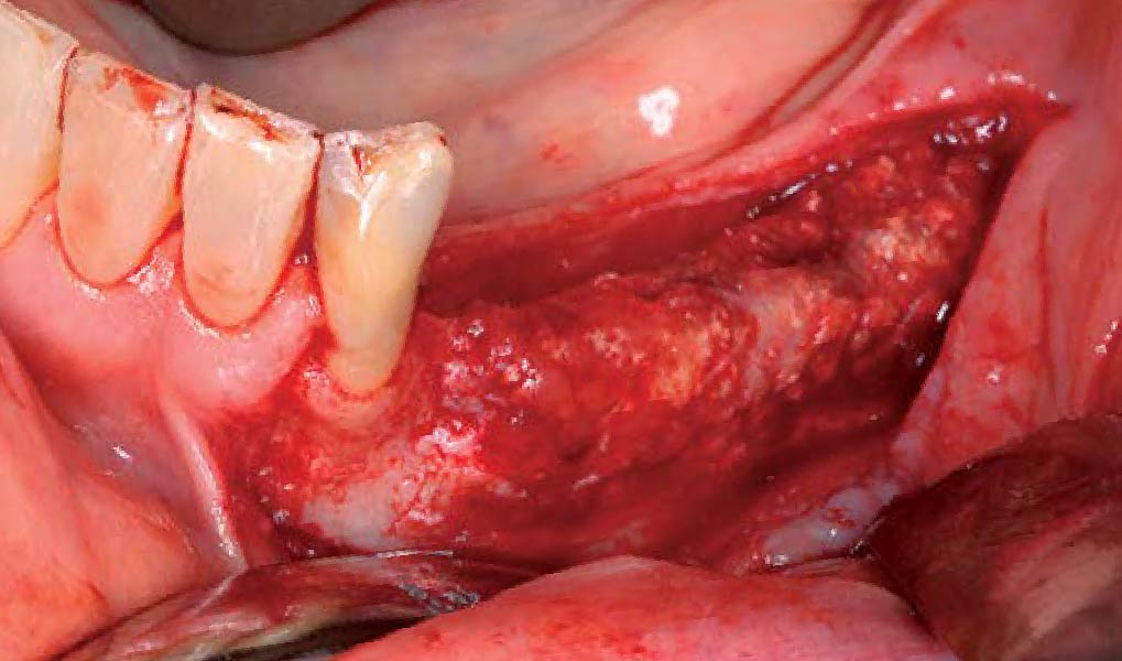
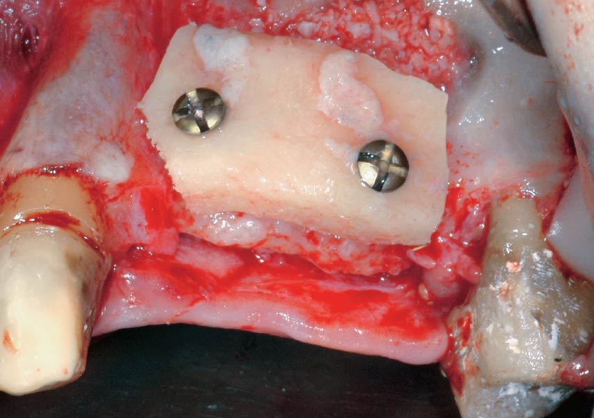
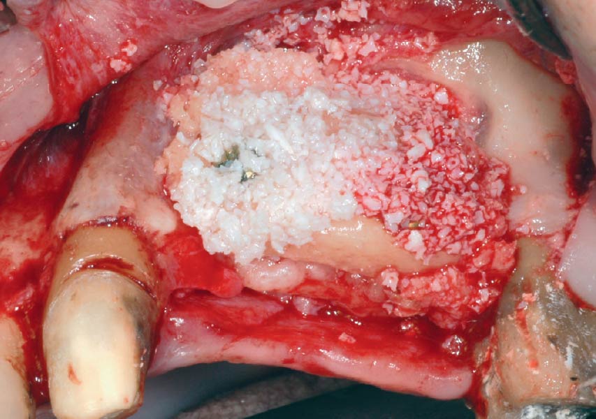
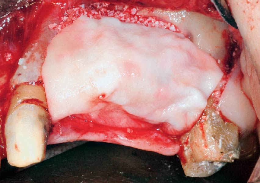
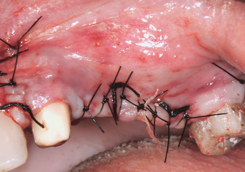
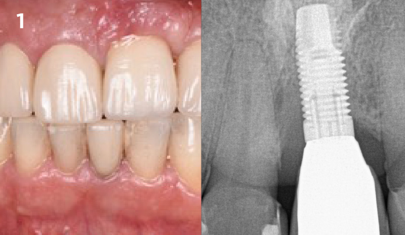
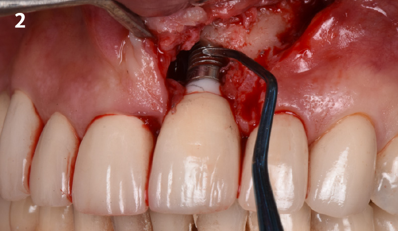
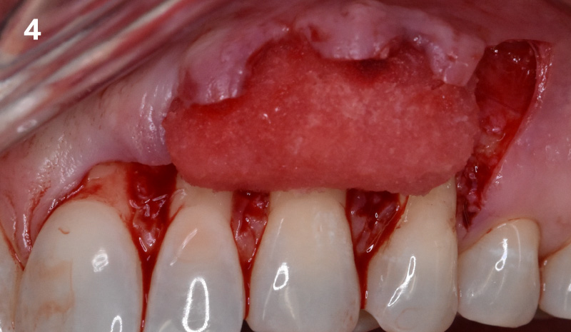
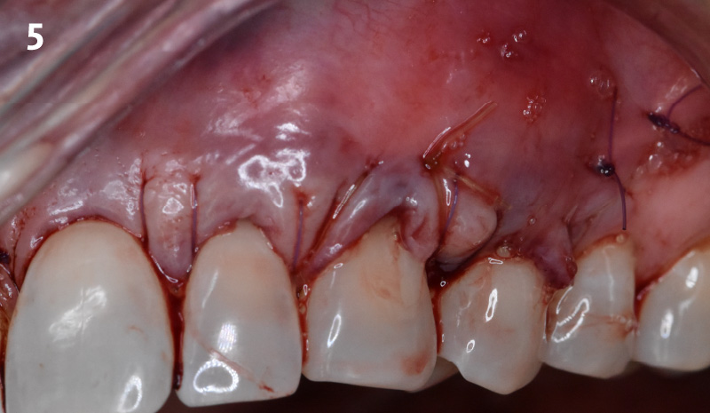
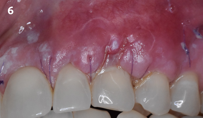
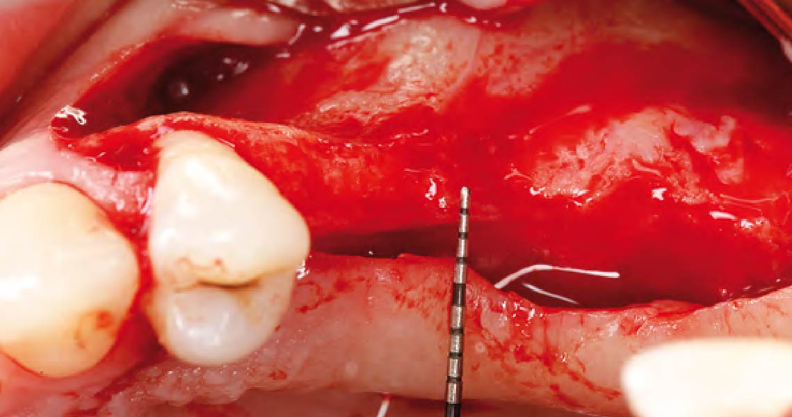
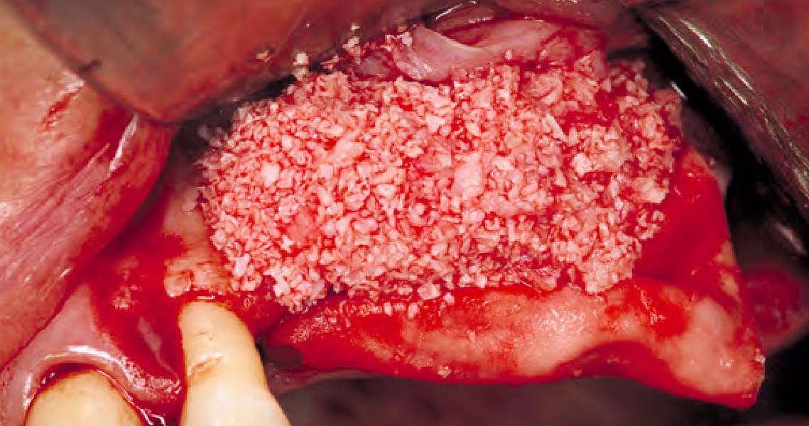
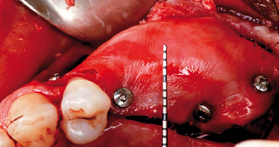
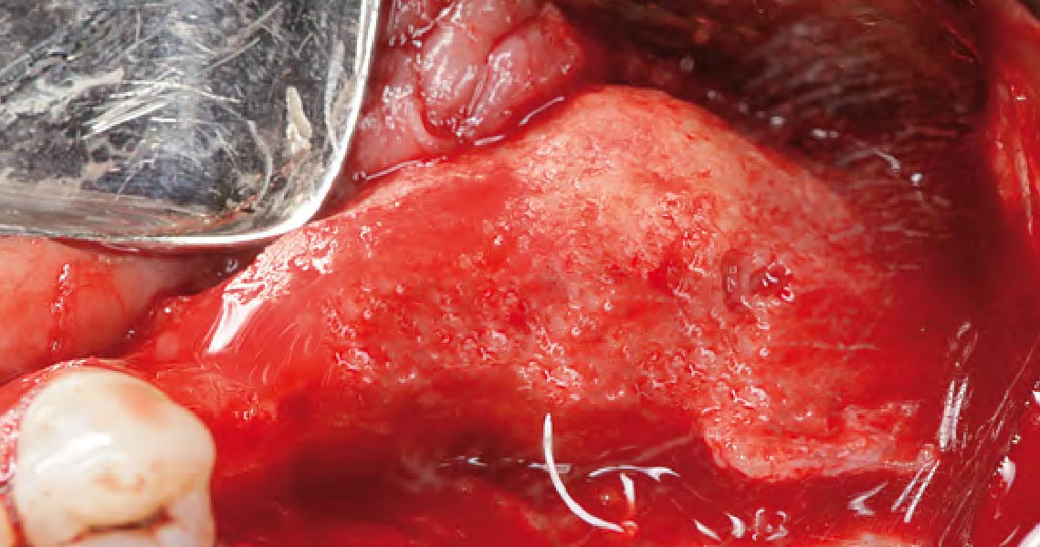
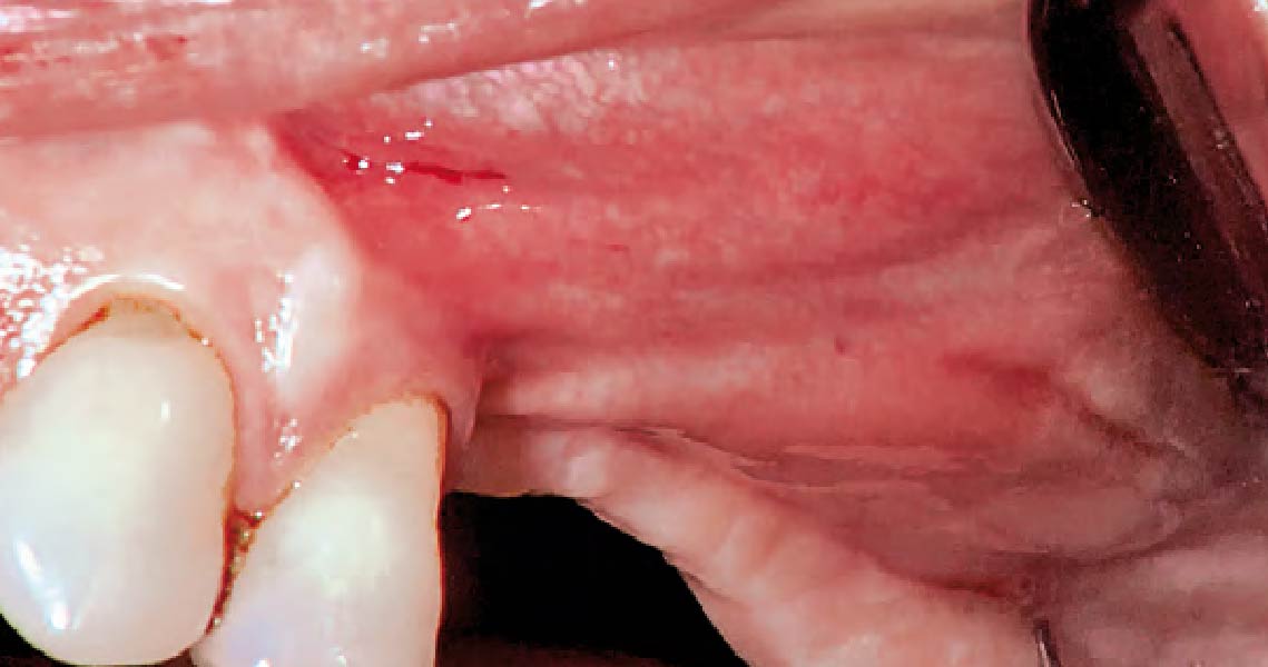
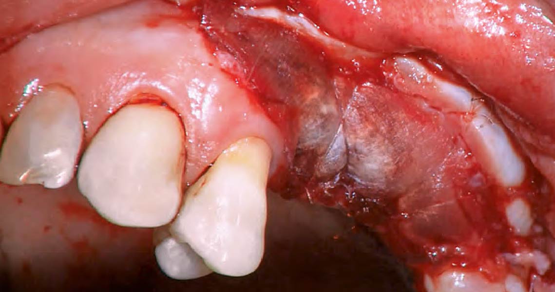
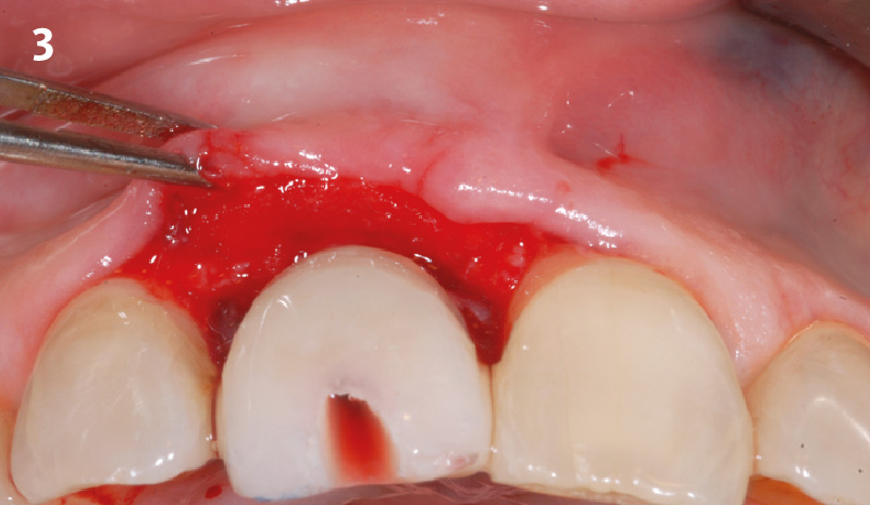
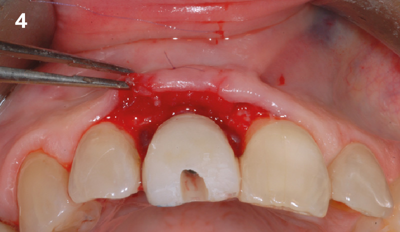
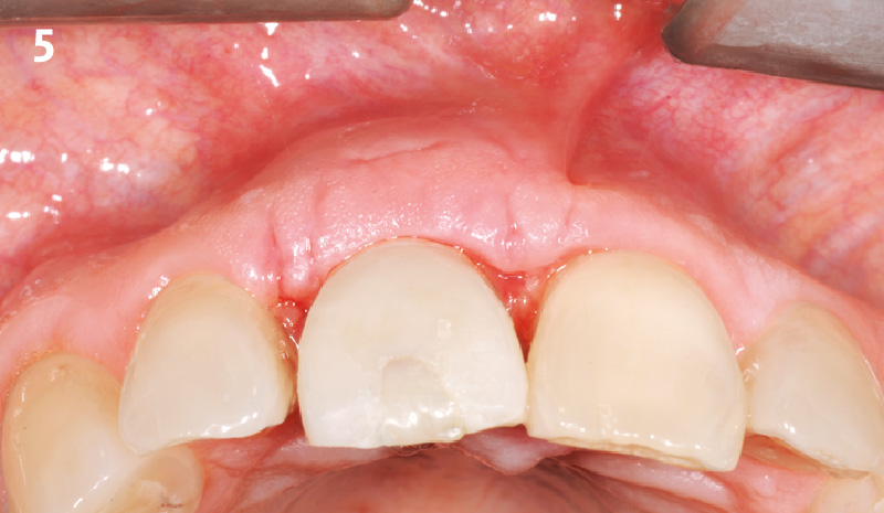
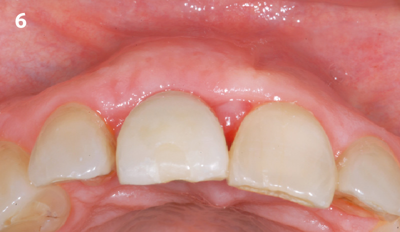
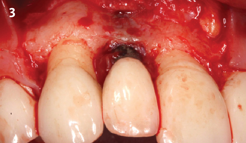
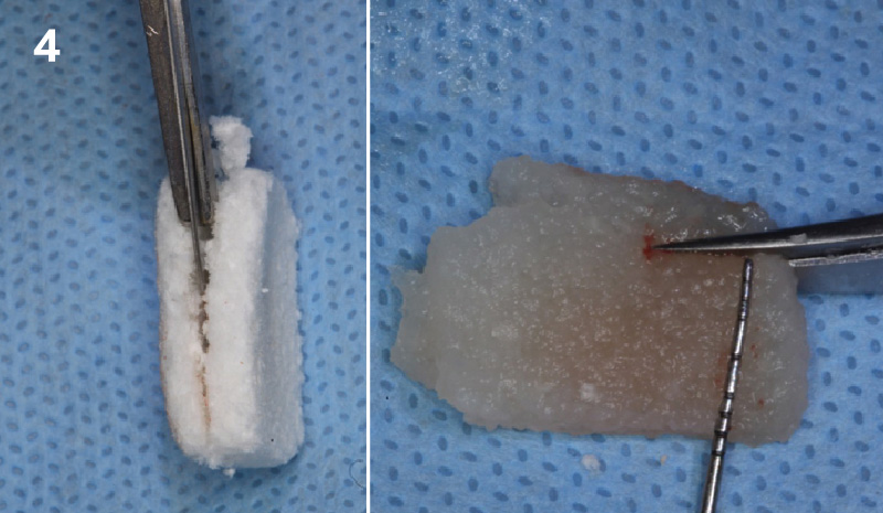
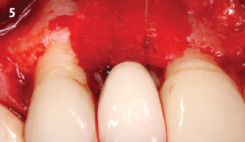
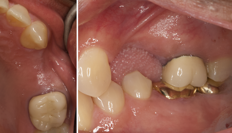
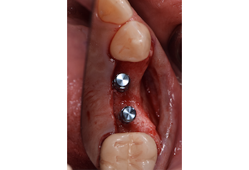
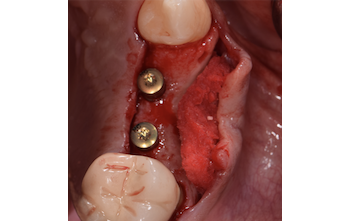
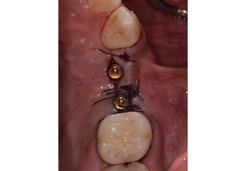
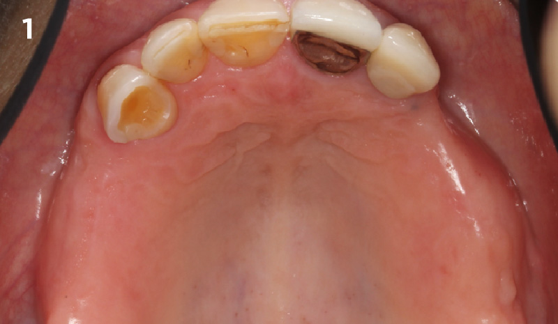
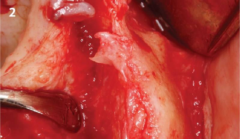
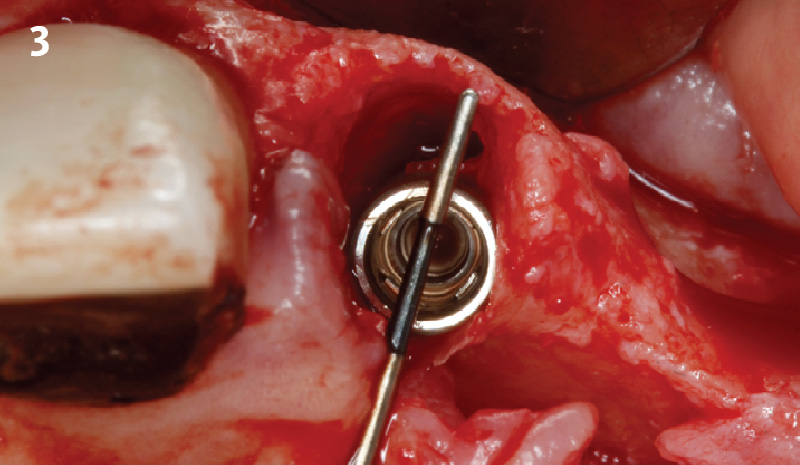
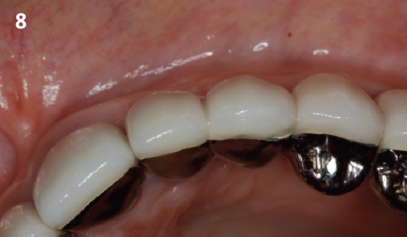
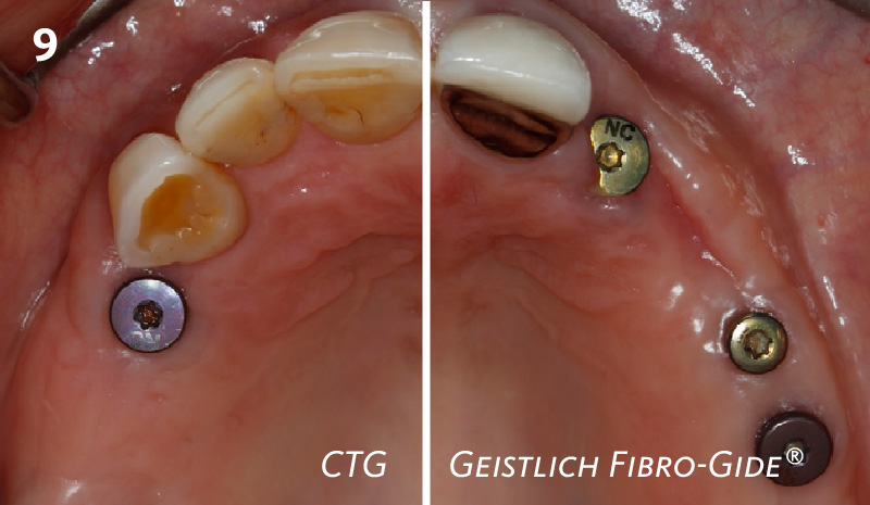
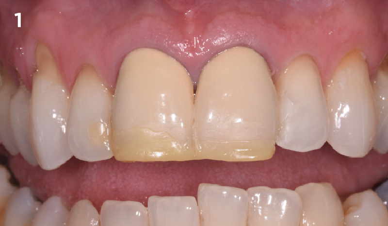
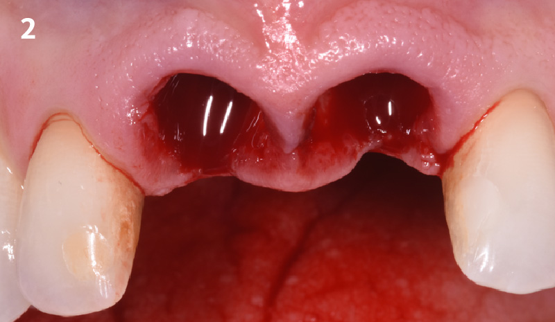
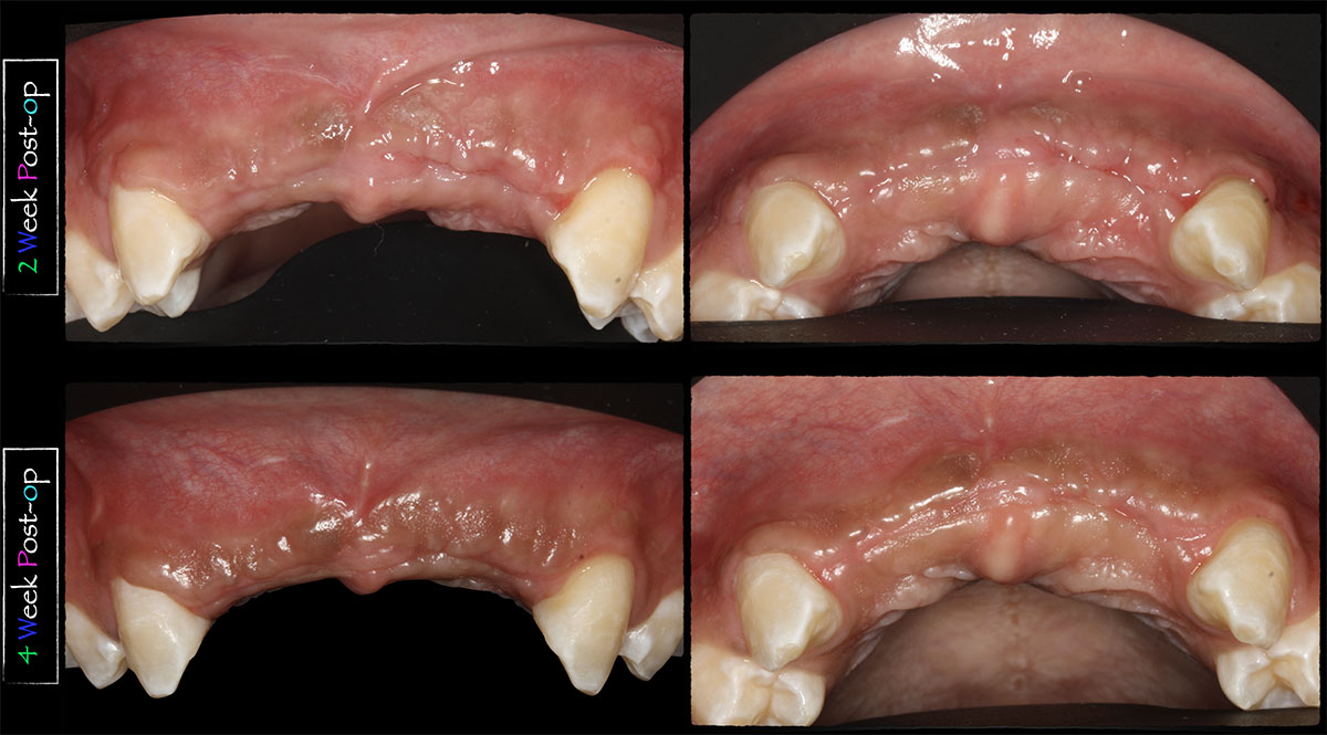
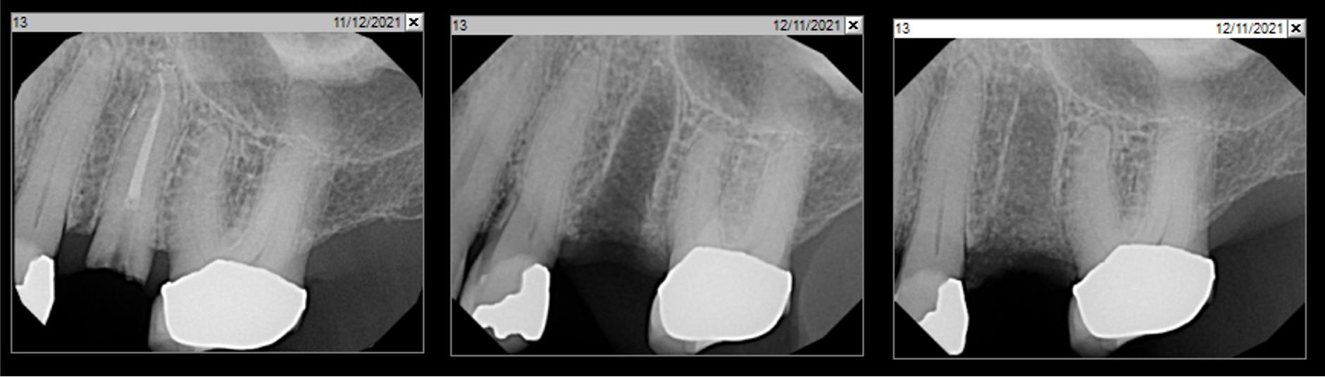
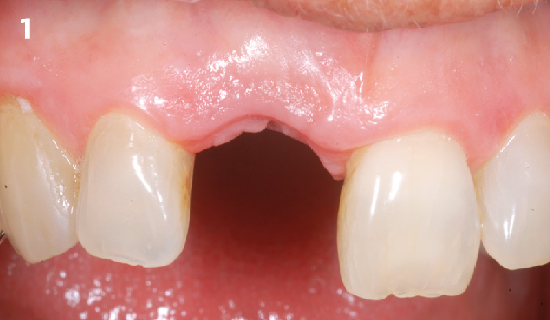
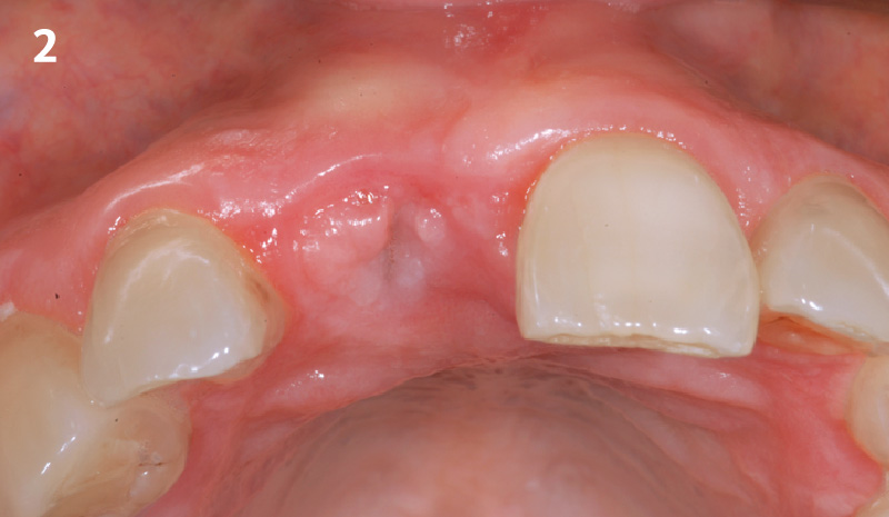
CLINICAL CASE

THE OUTCOME

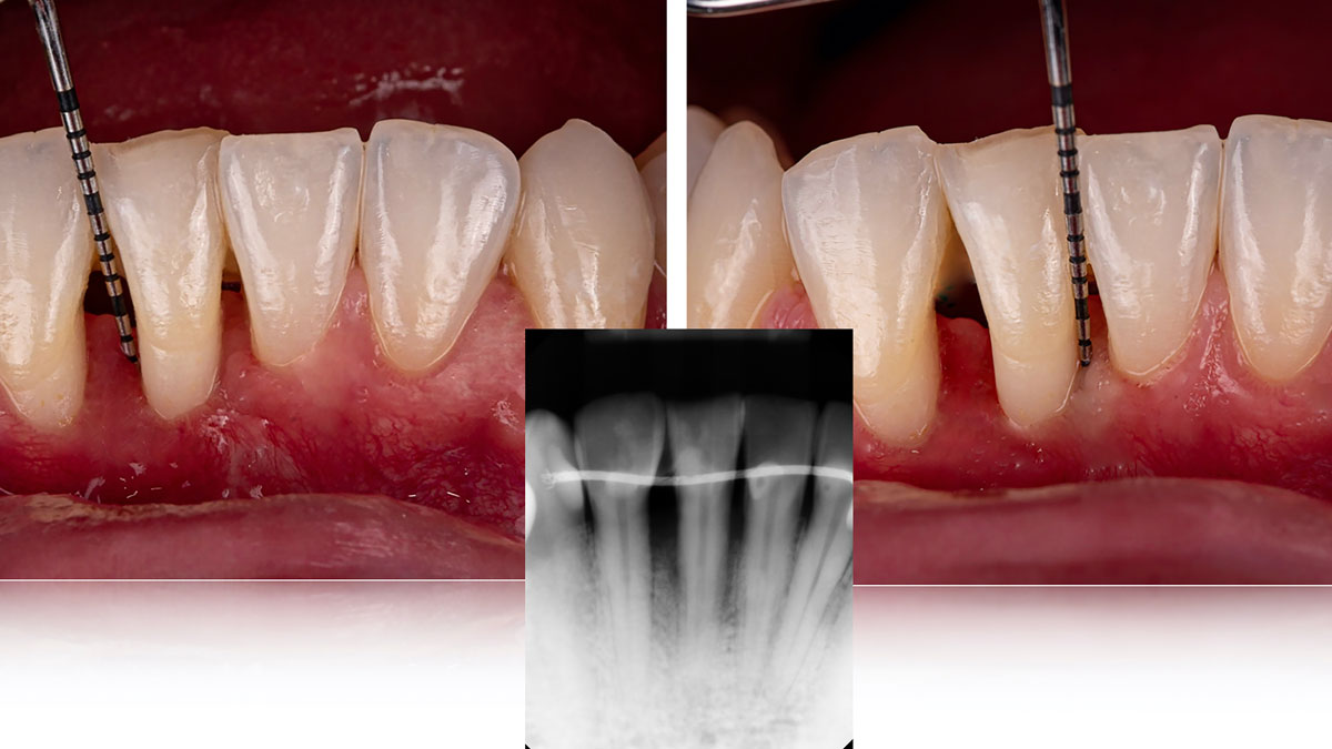
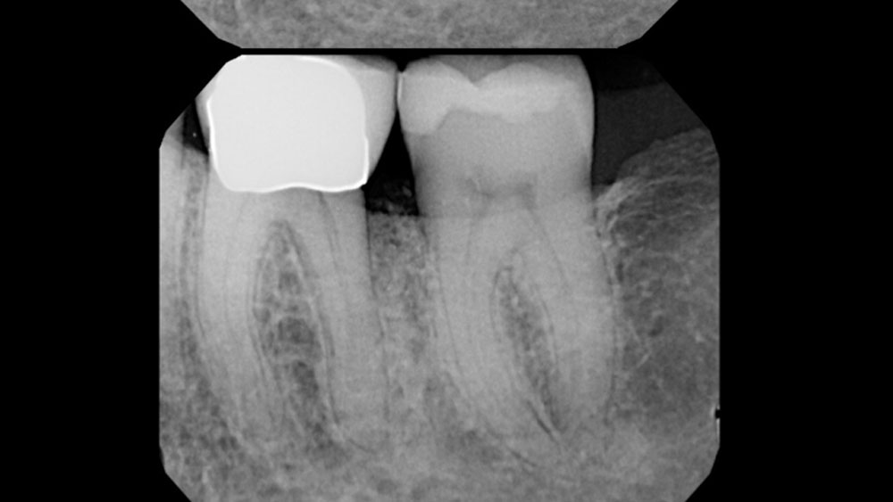
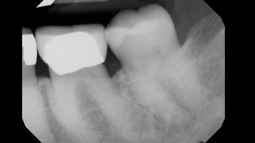
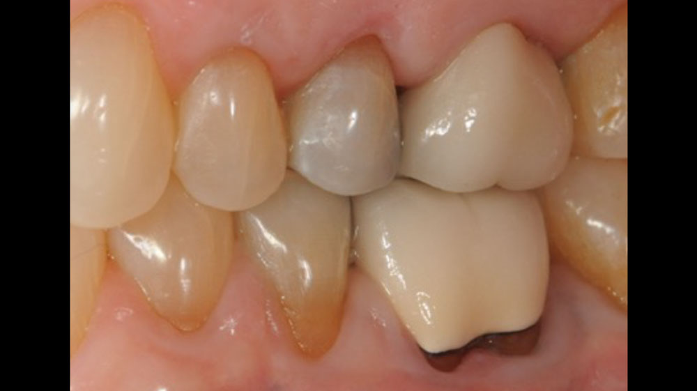
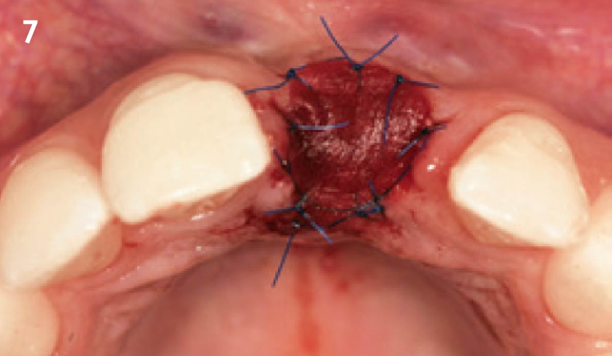
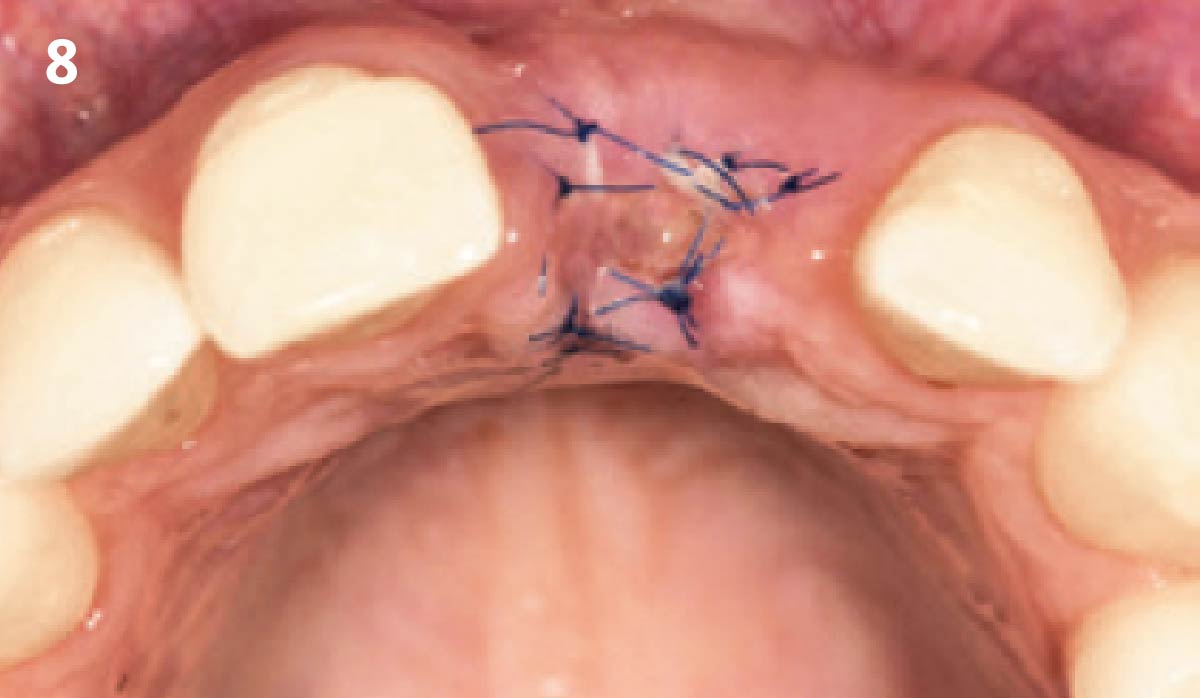
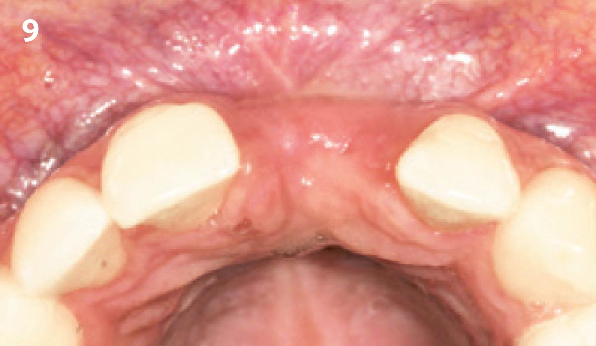
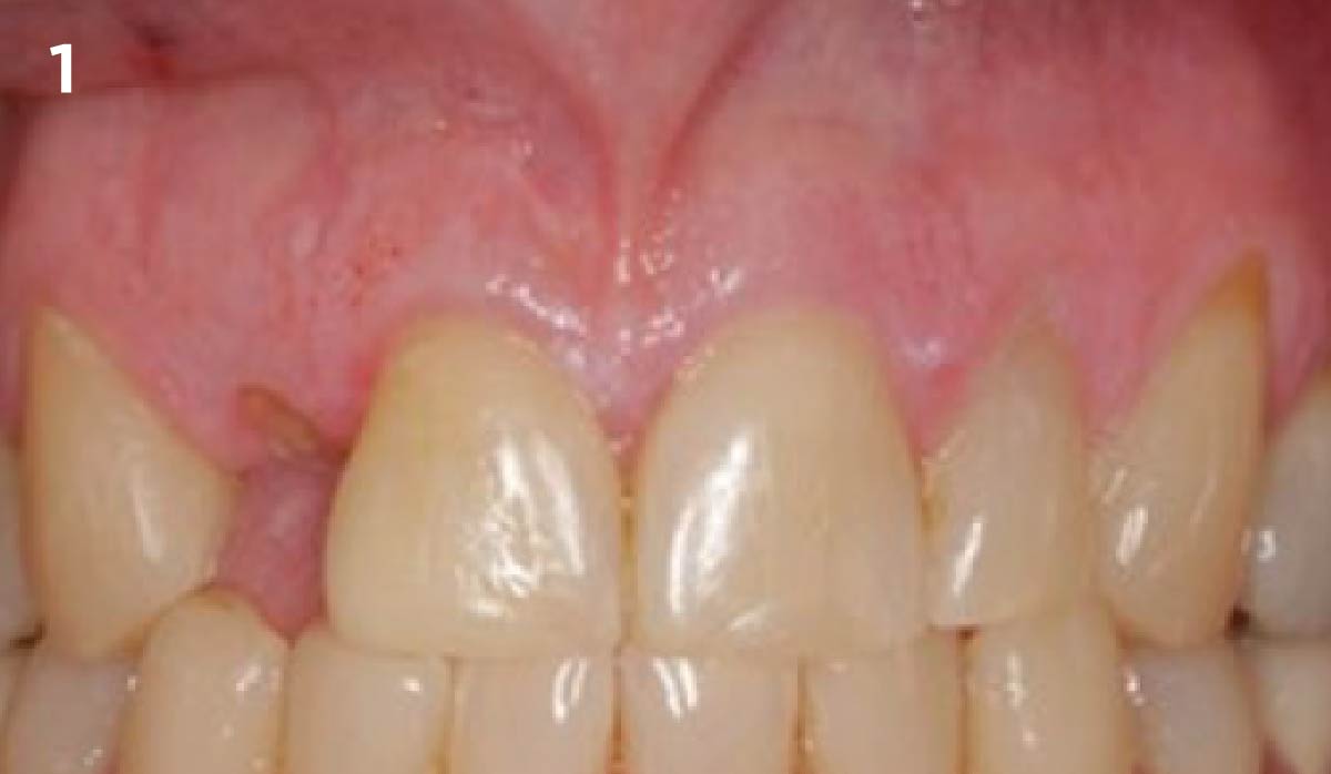
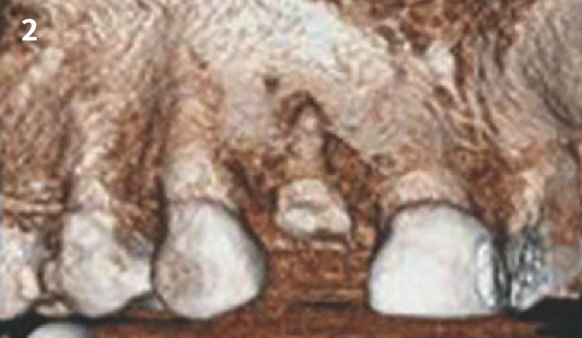
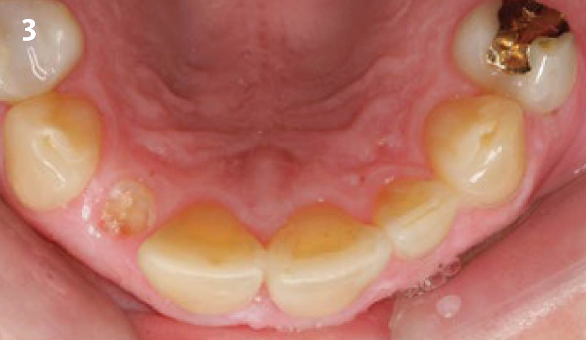
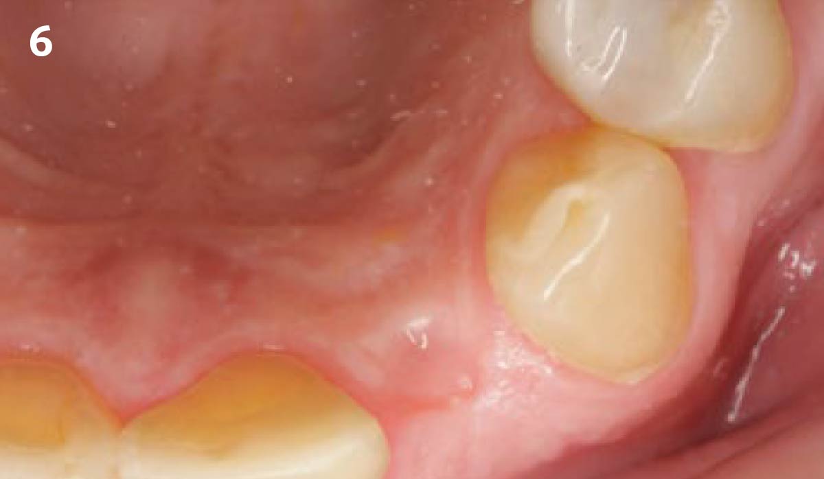
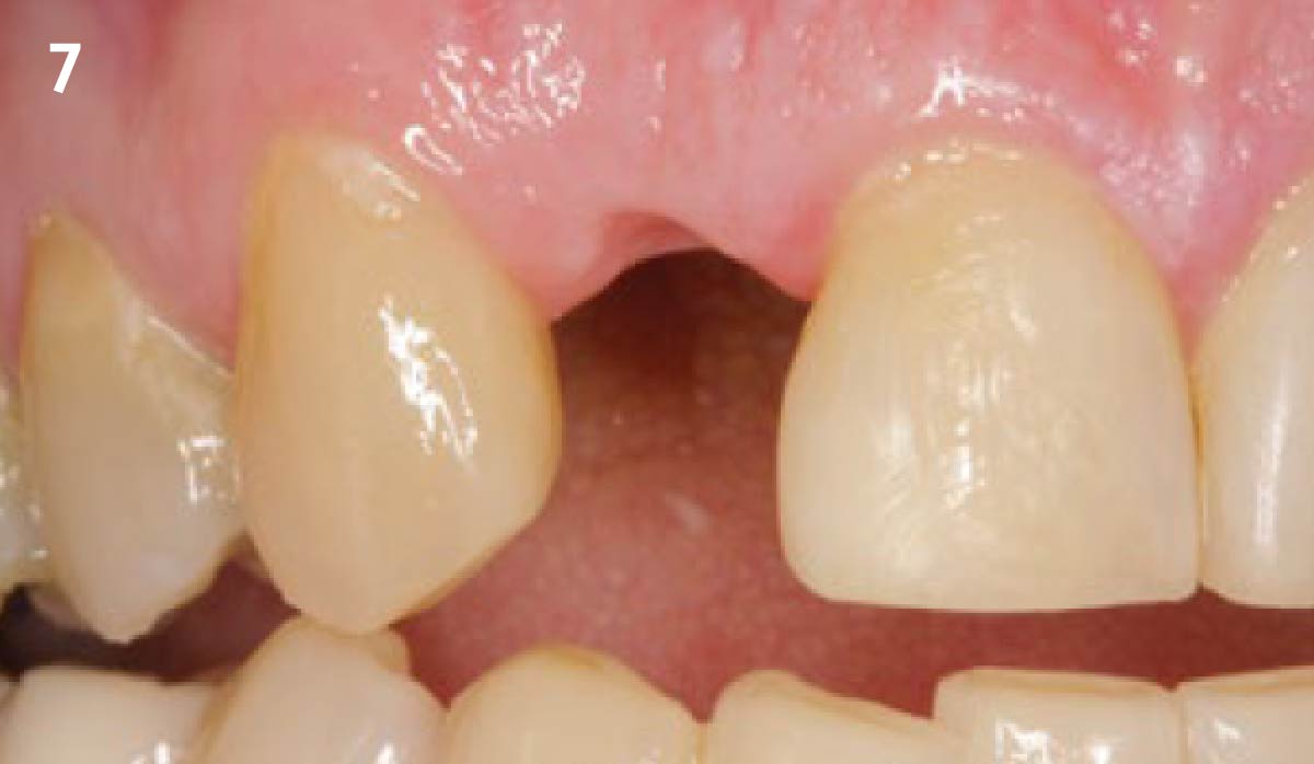
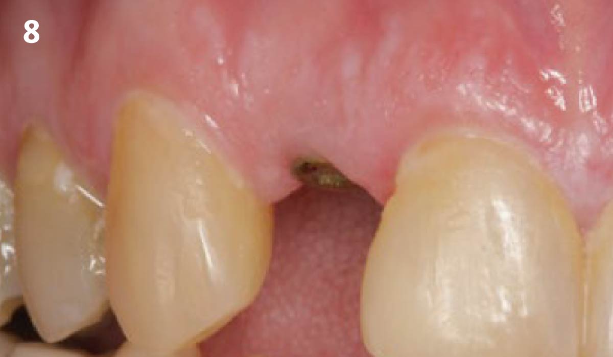
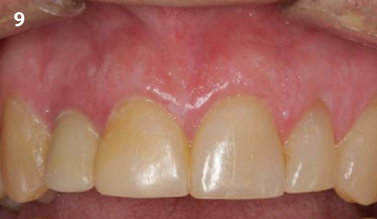
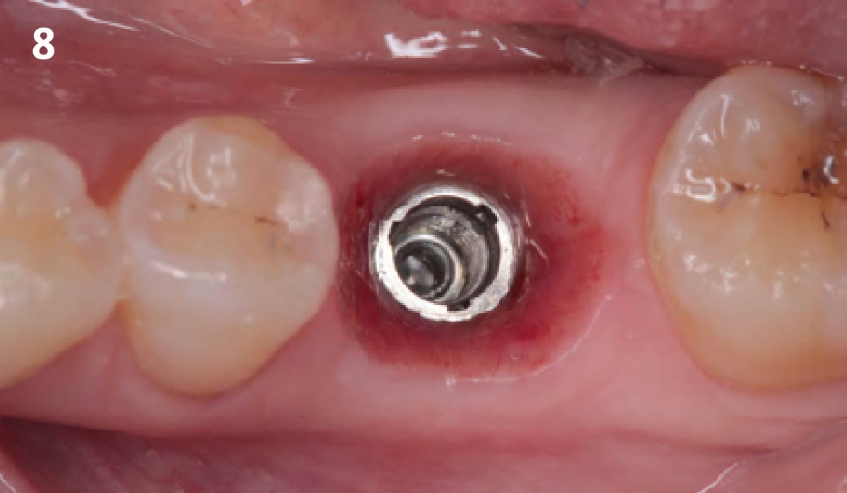
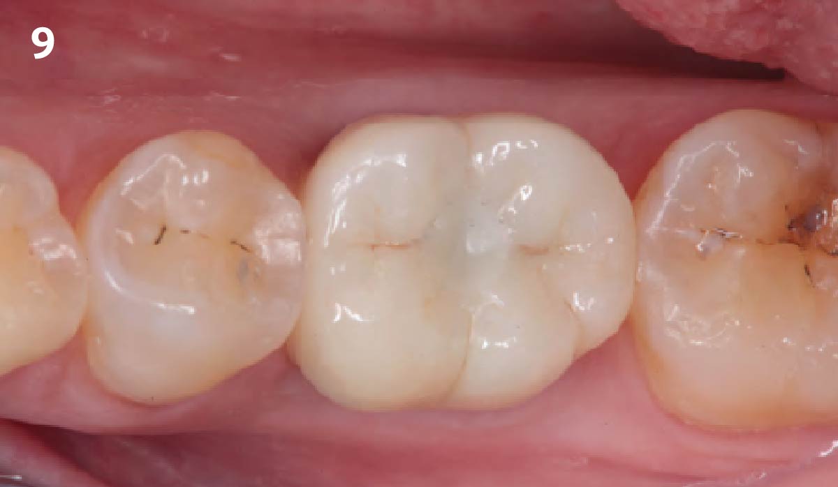
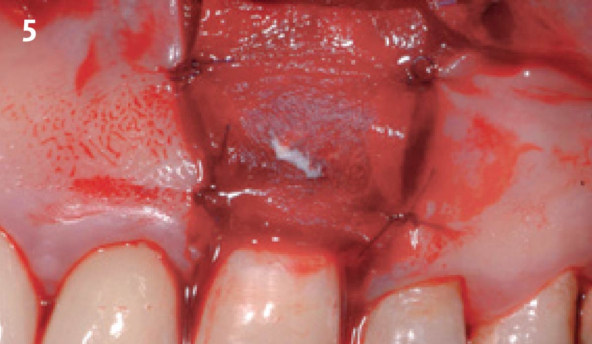
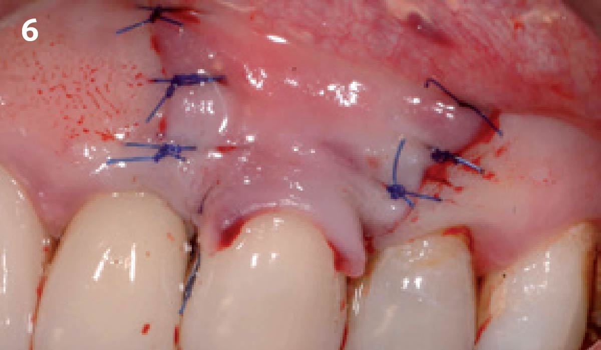
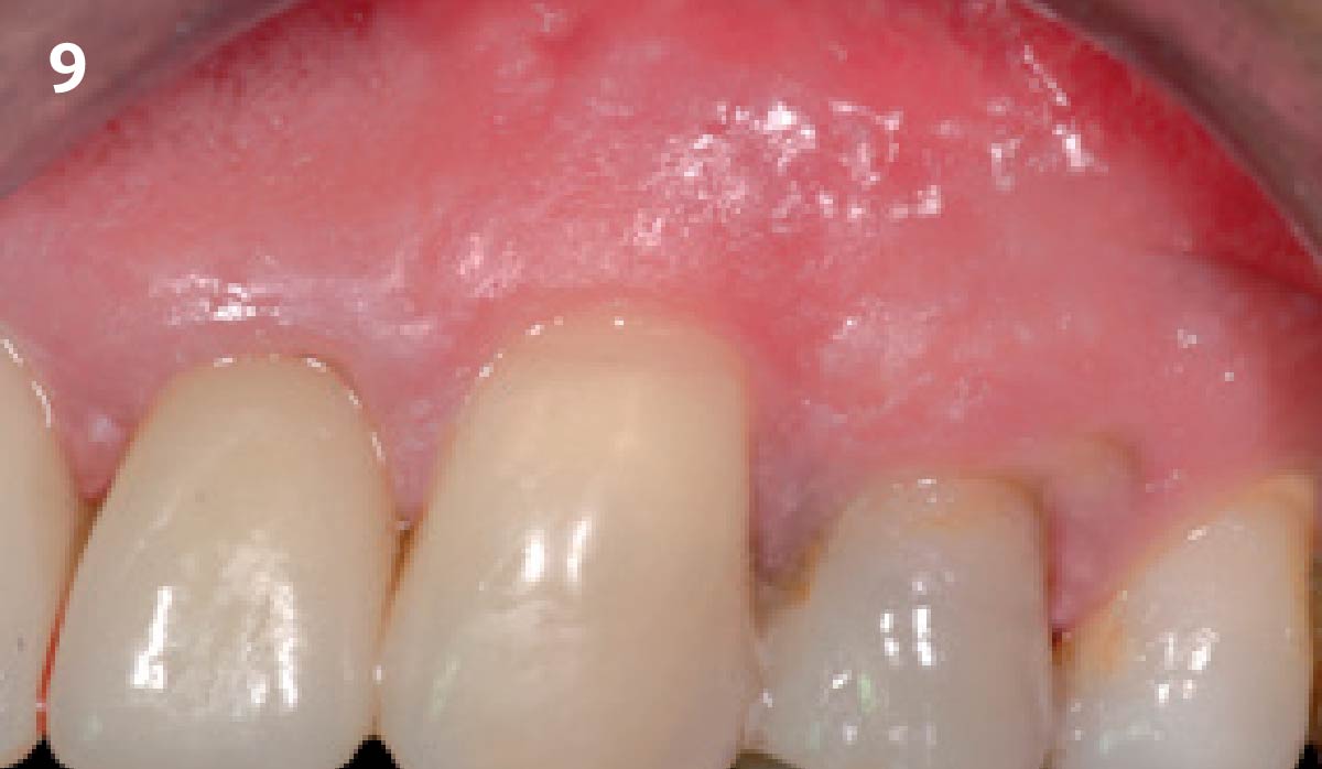
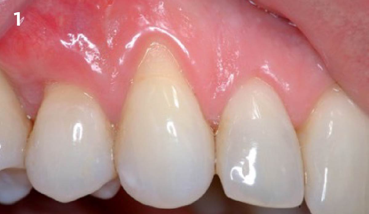
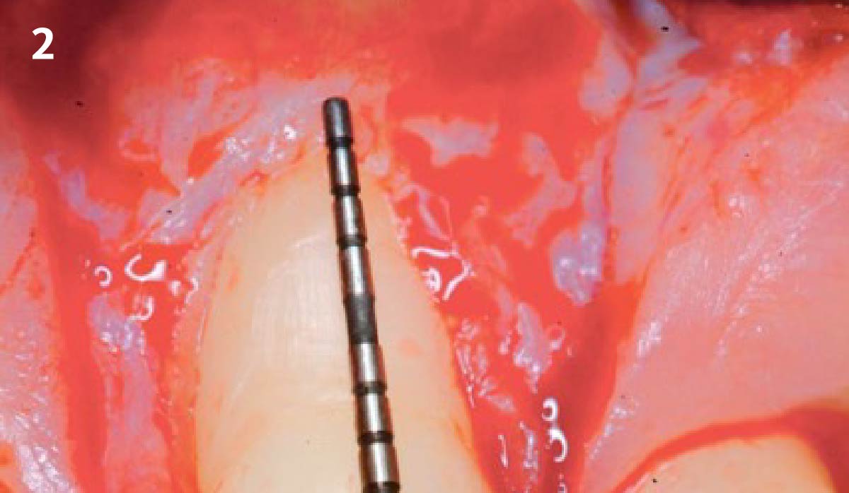
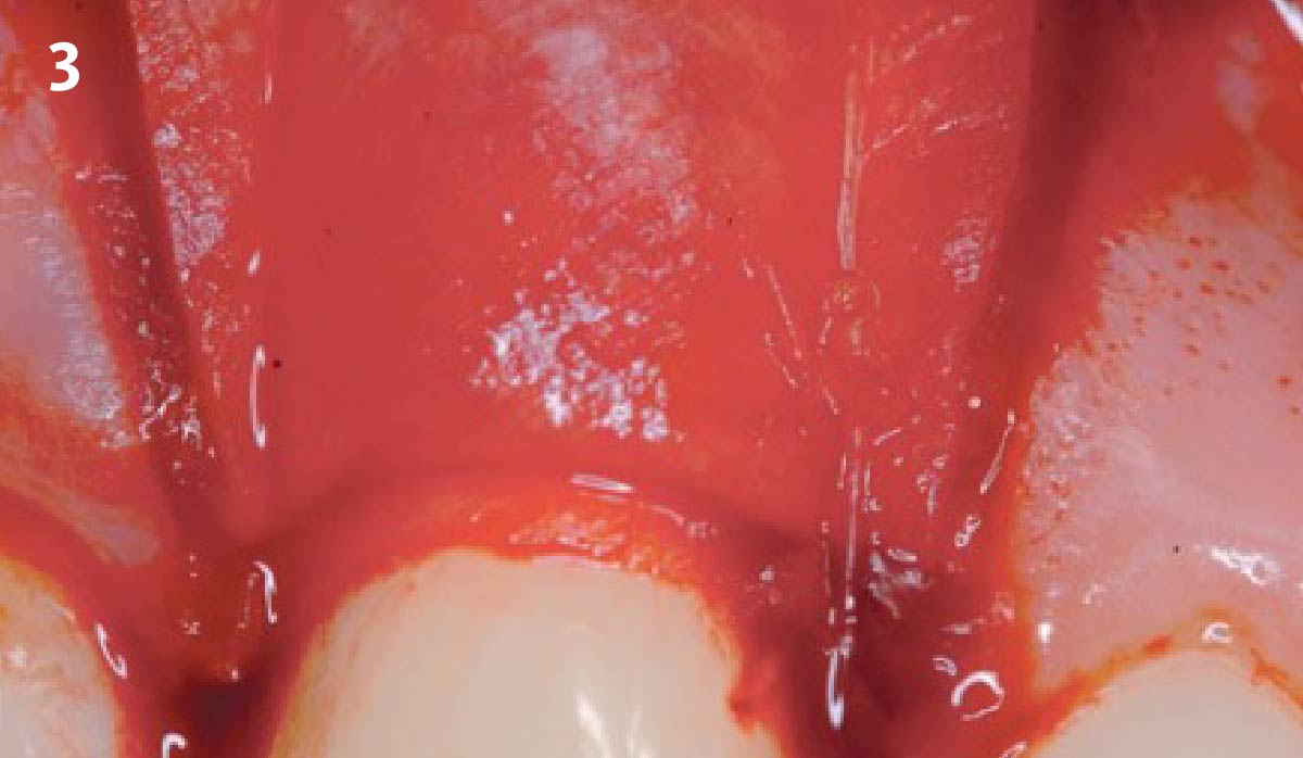
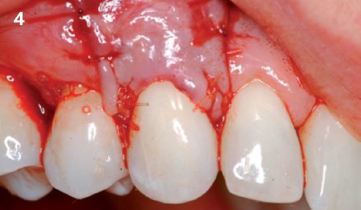
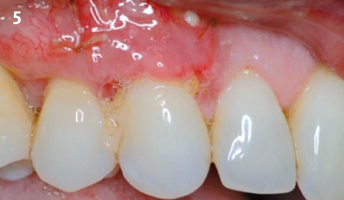
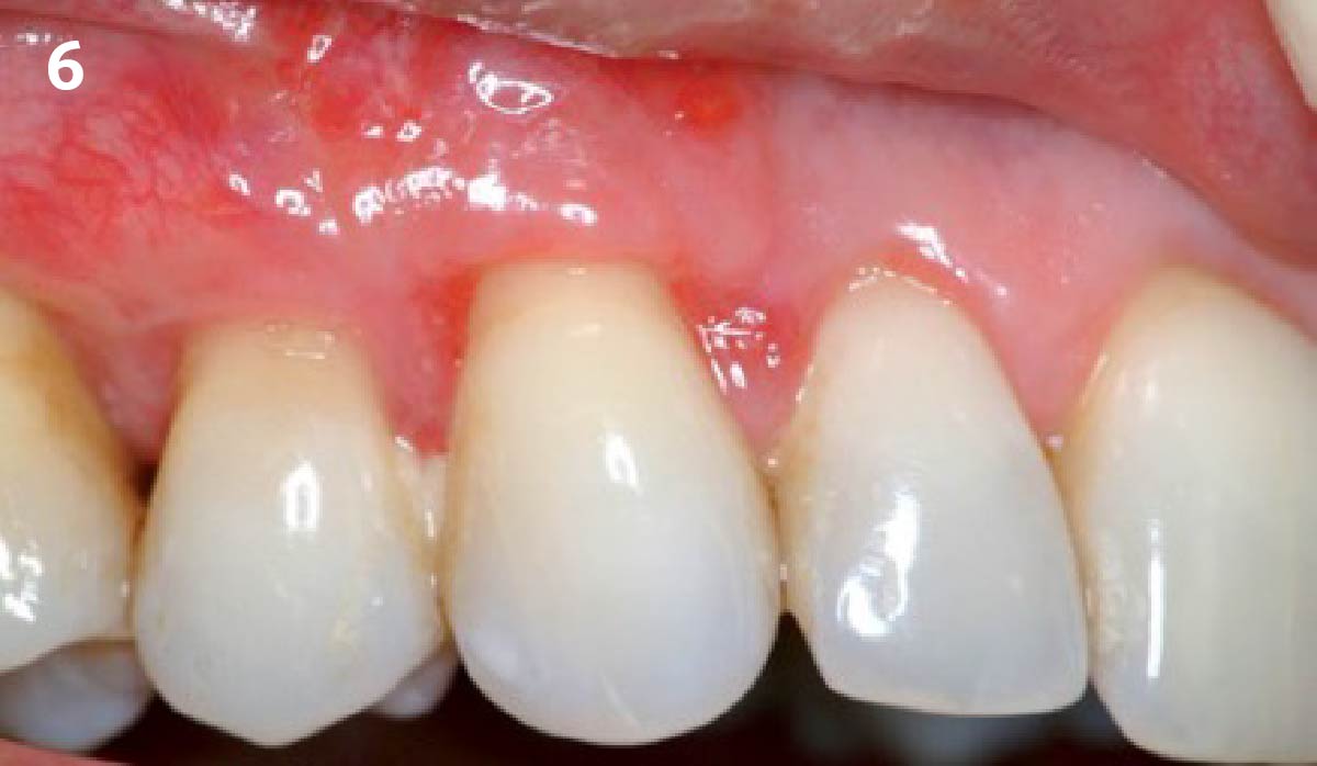
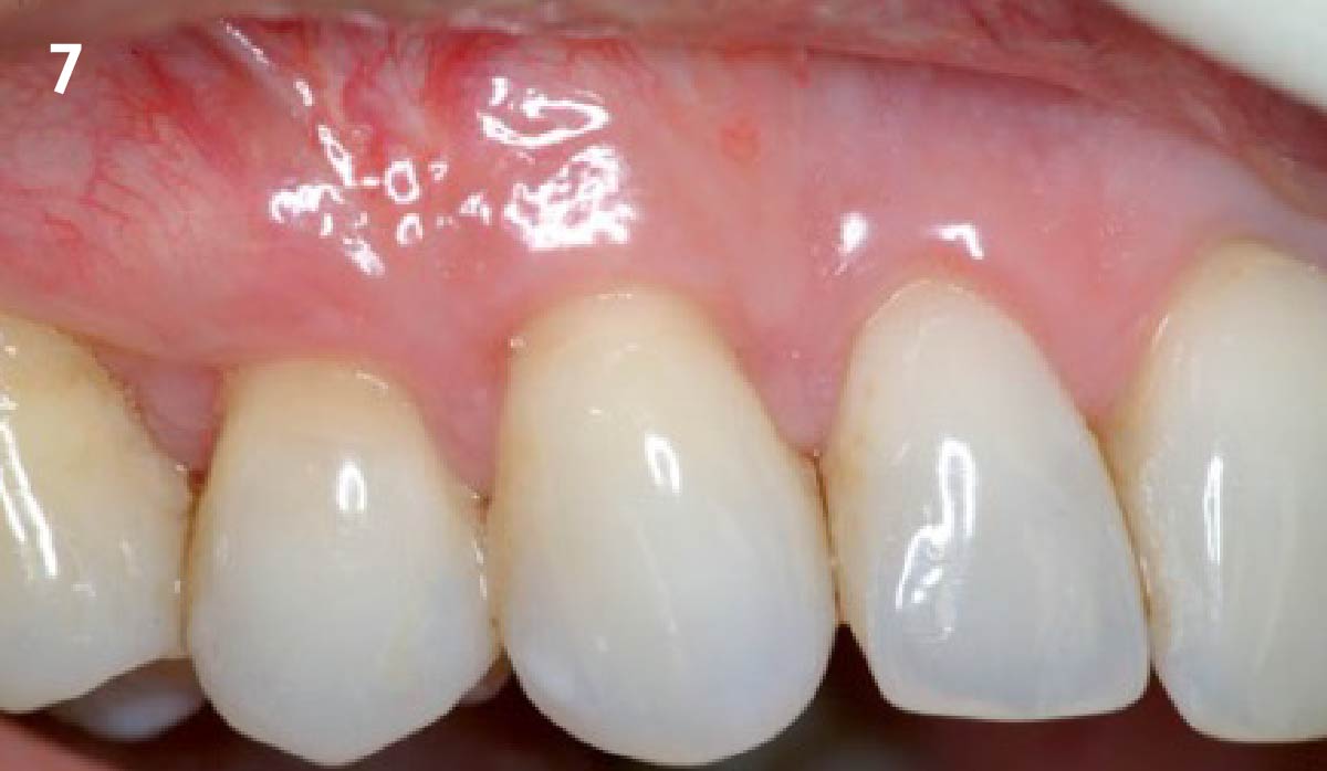
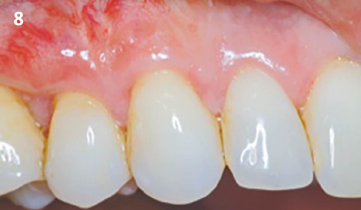
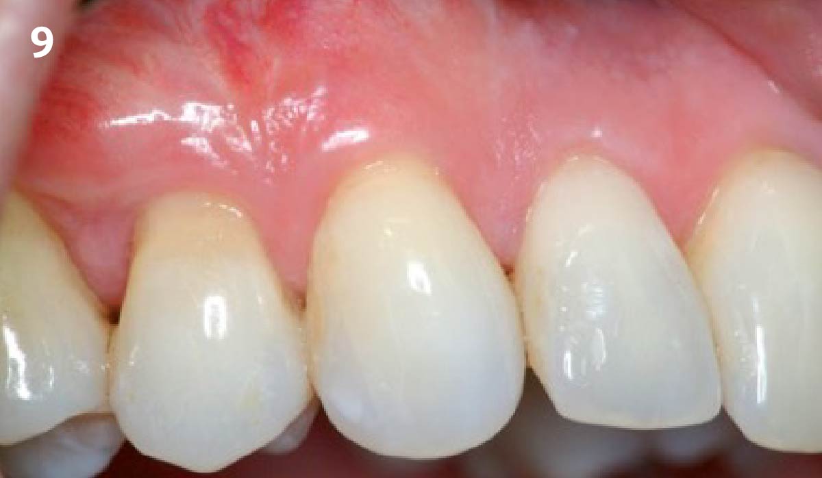
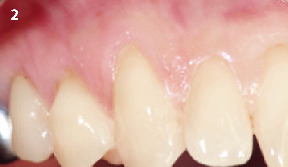
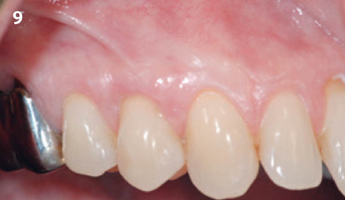
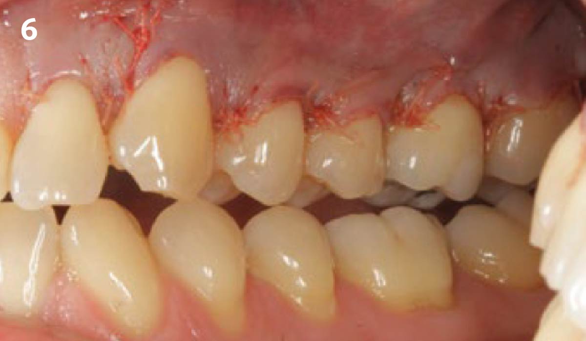
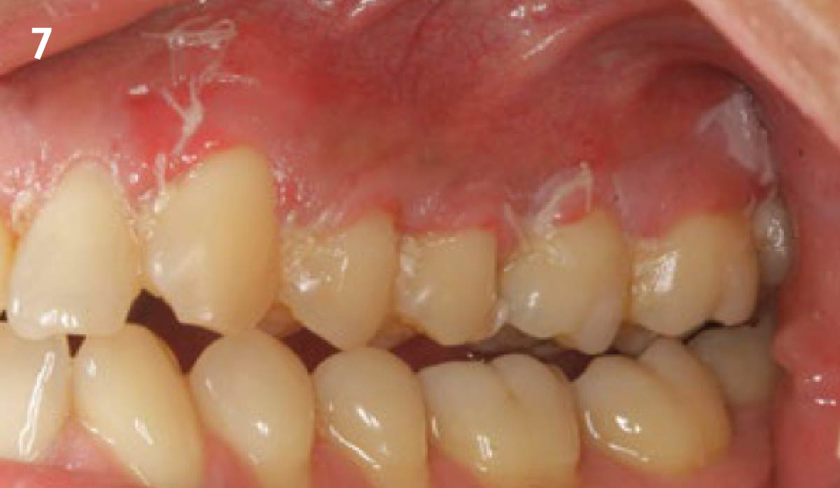
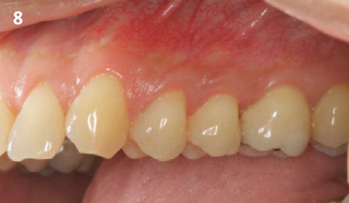
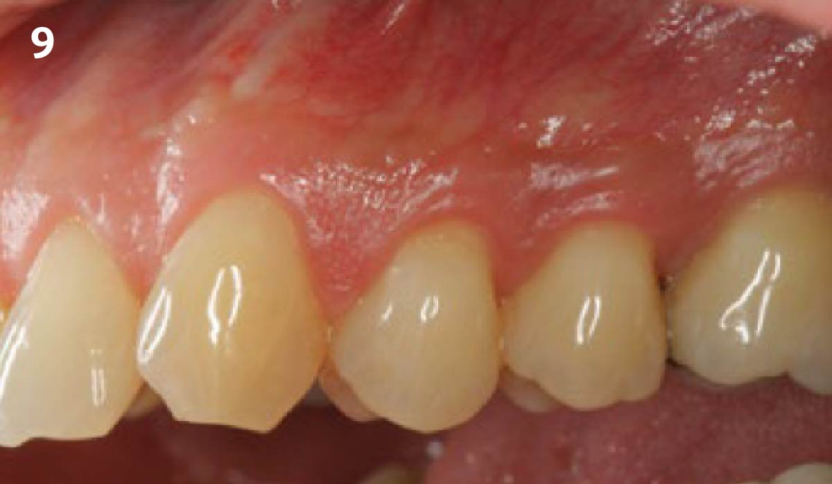
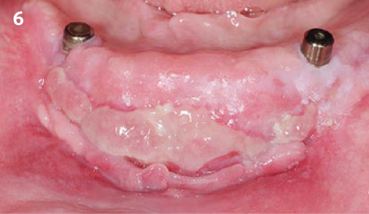
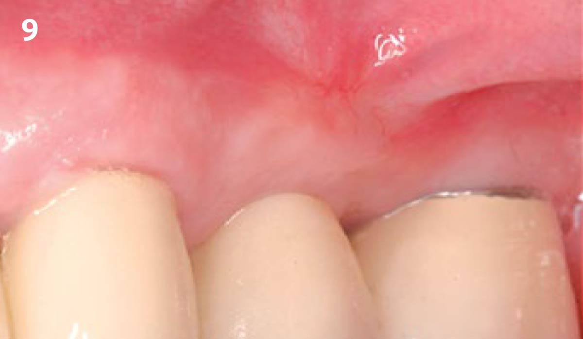
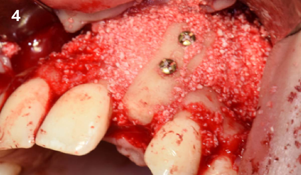
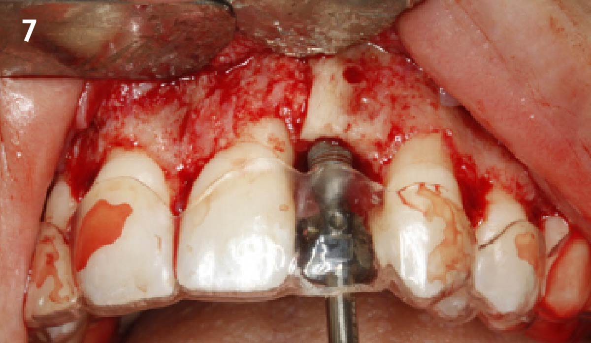
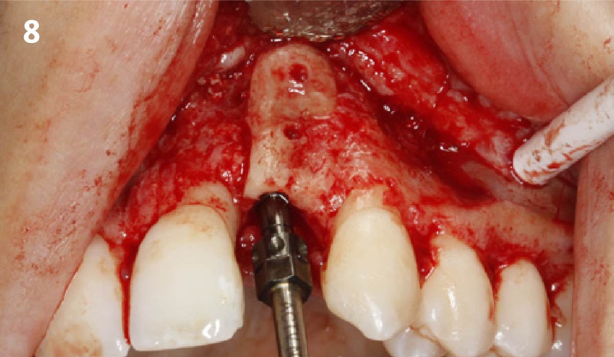
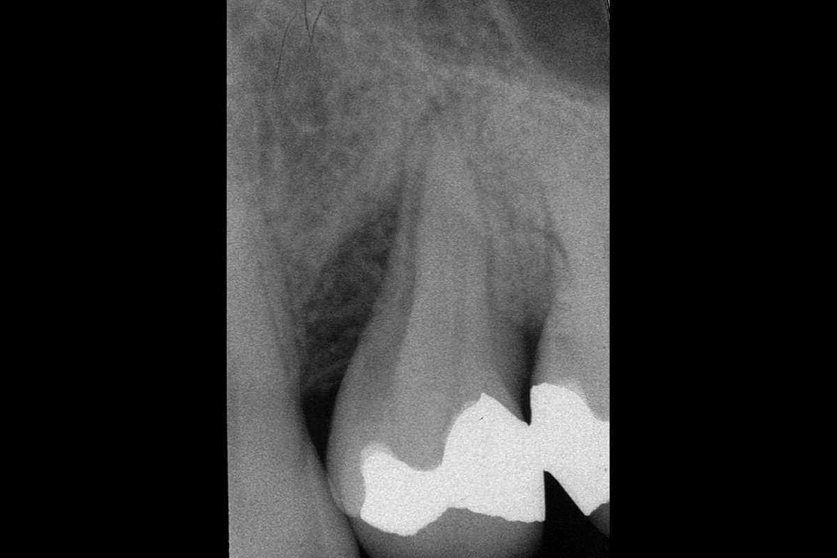
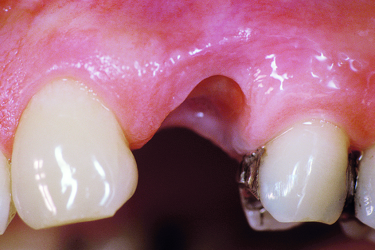
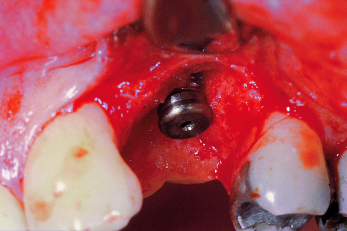
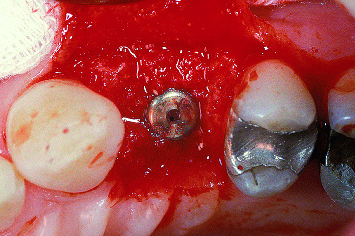
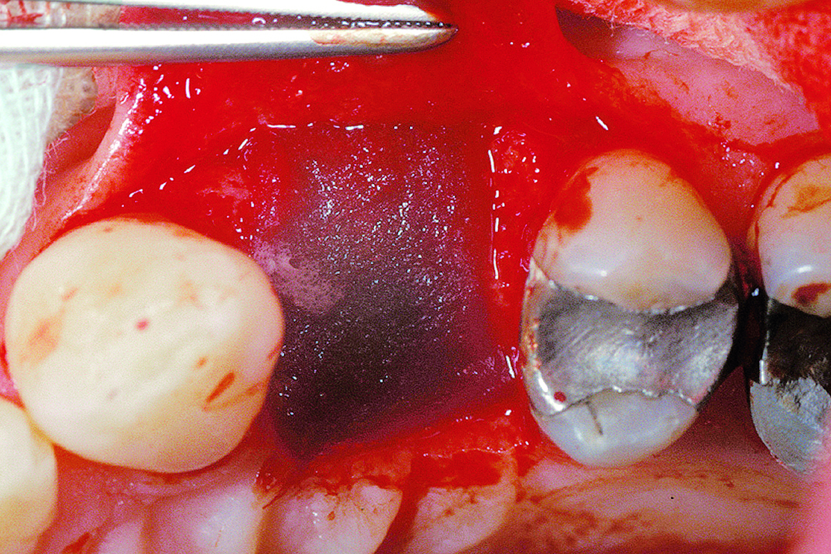
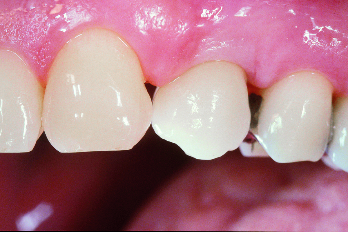
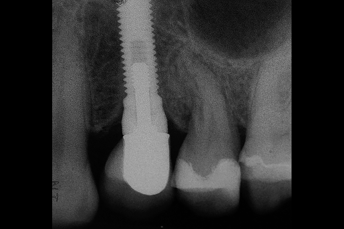
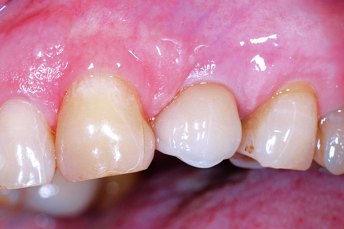
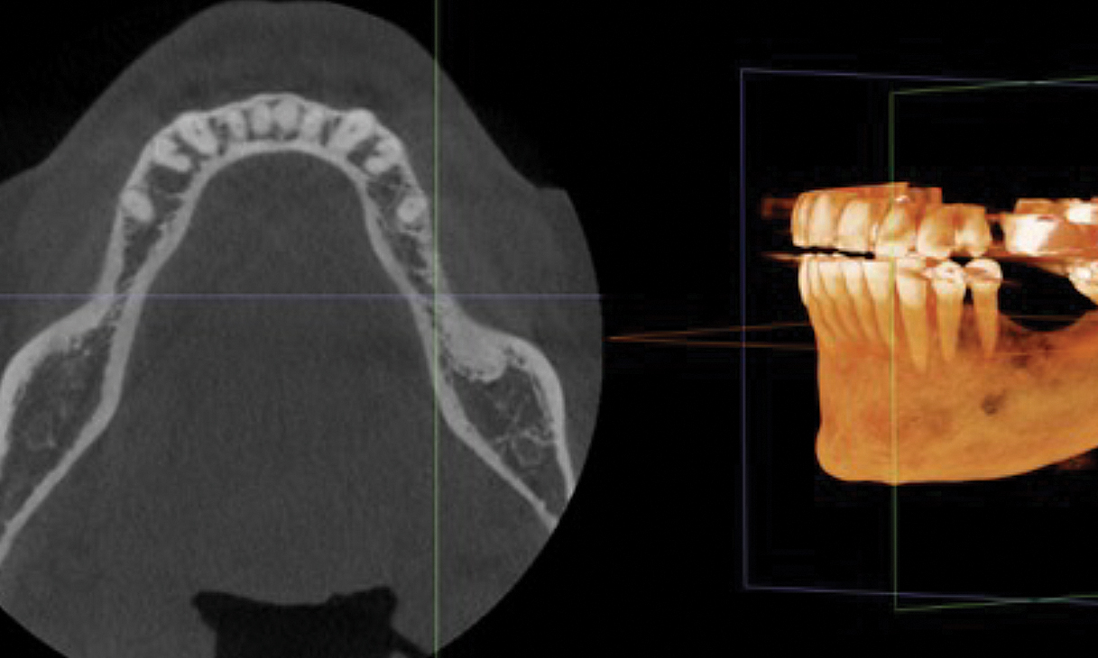
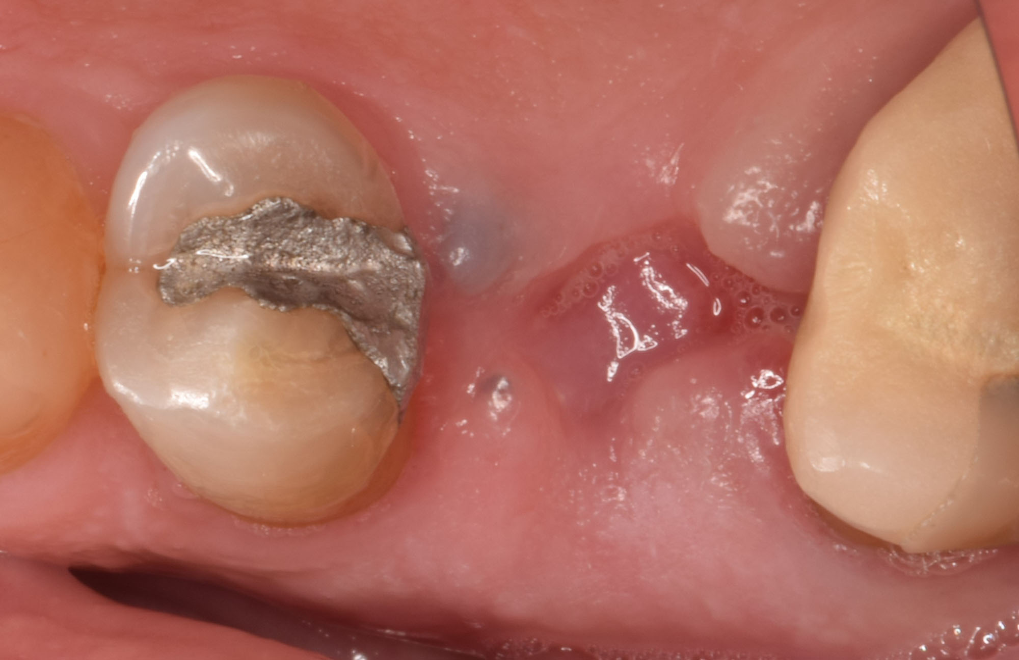
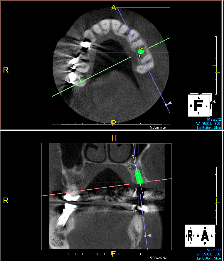
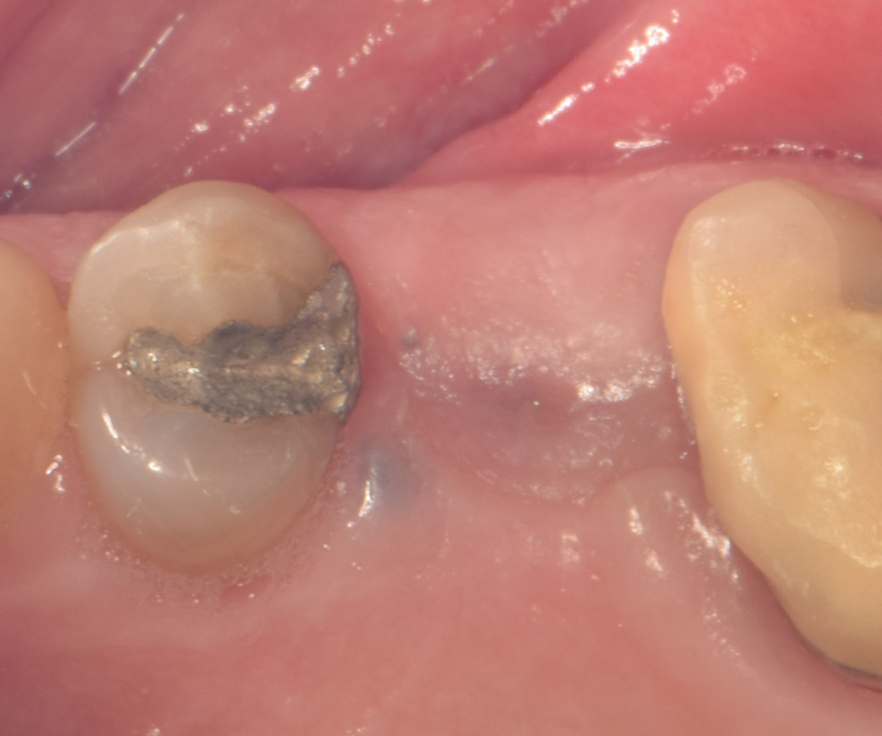
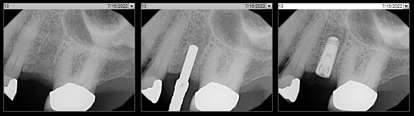
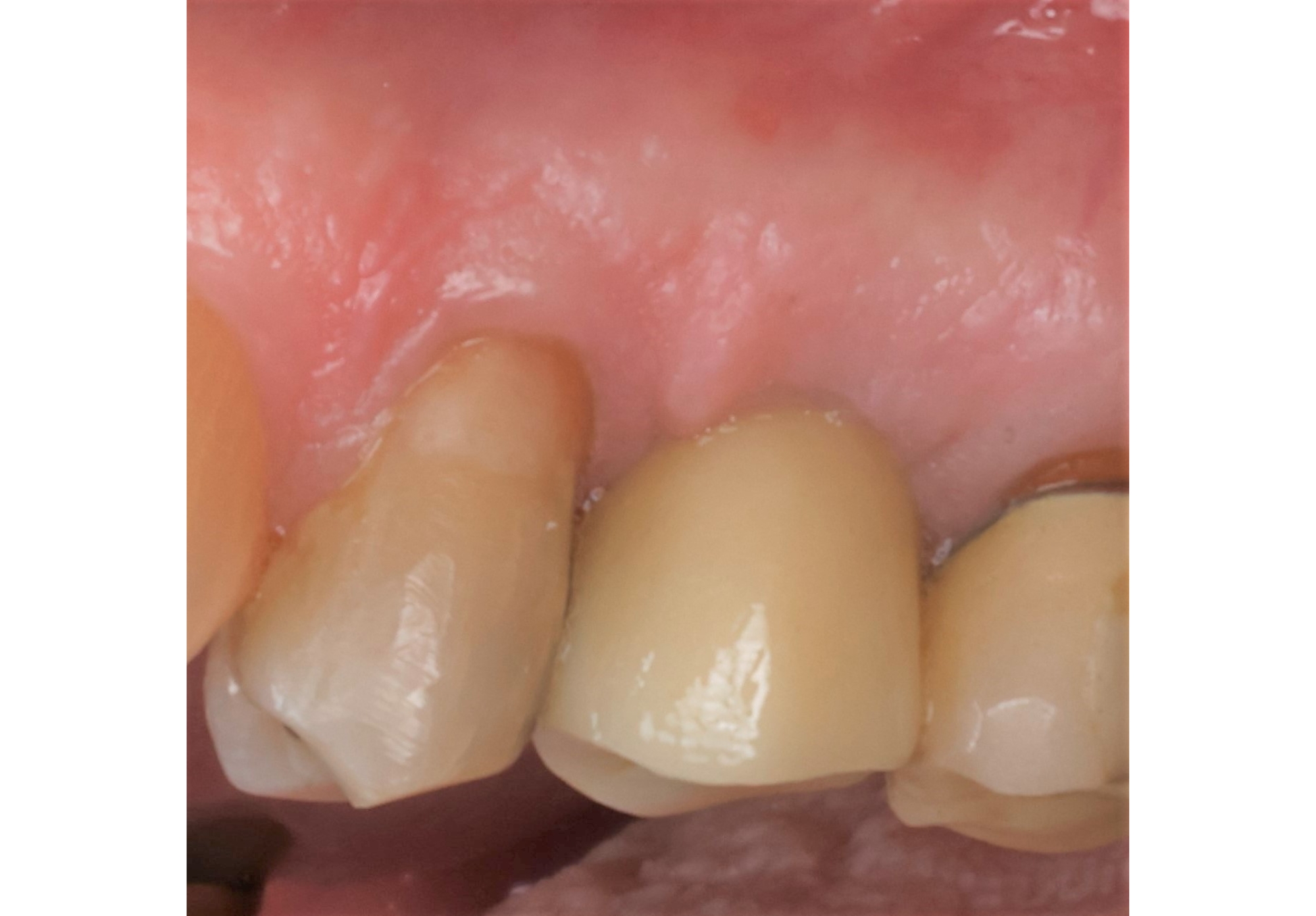
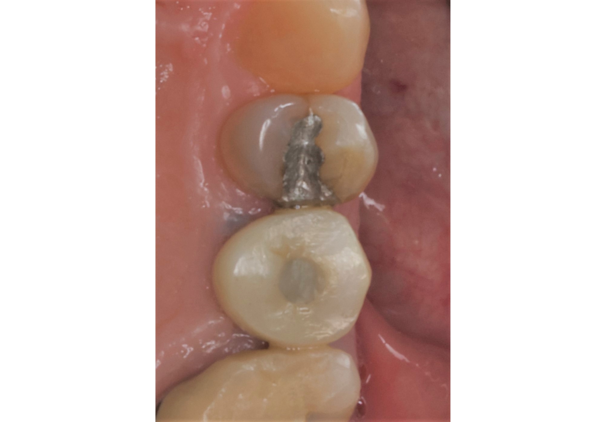
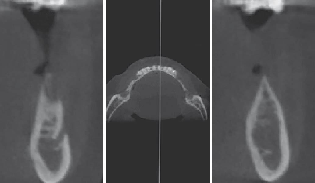
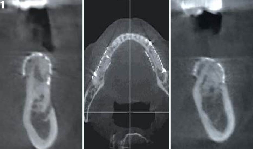
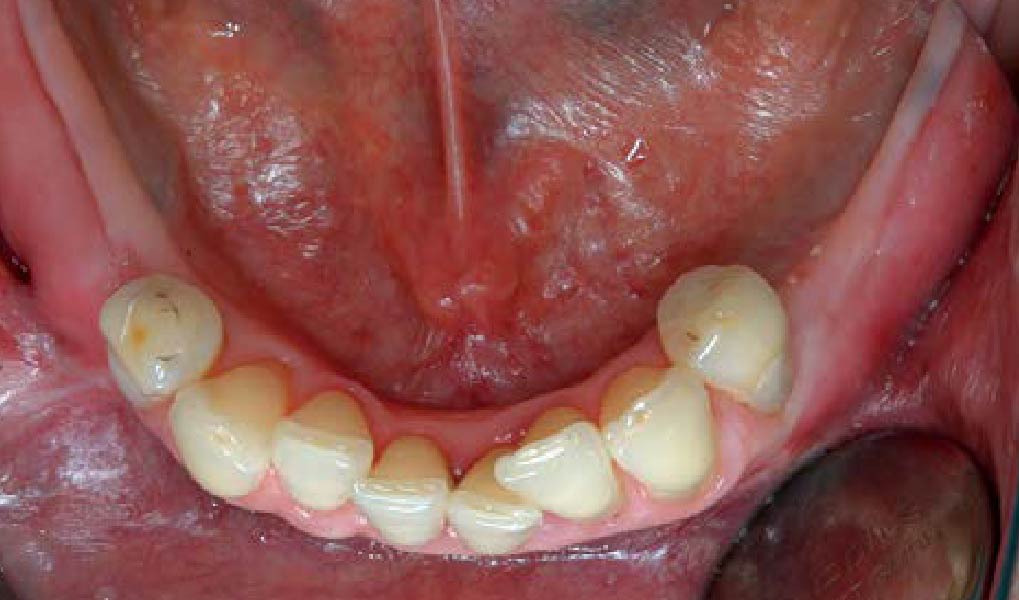
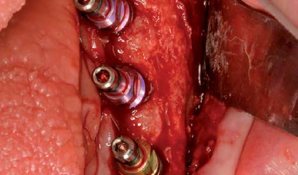
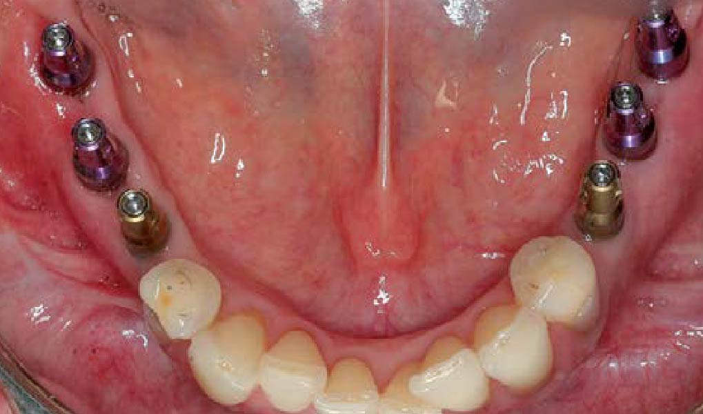
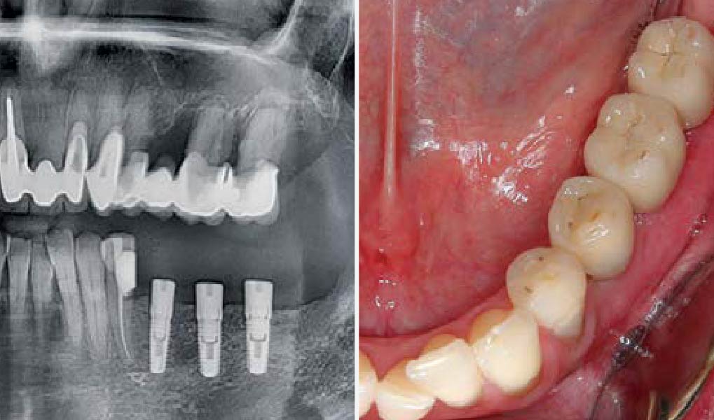
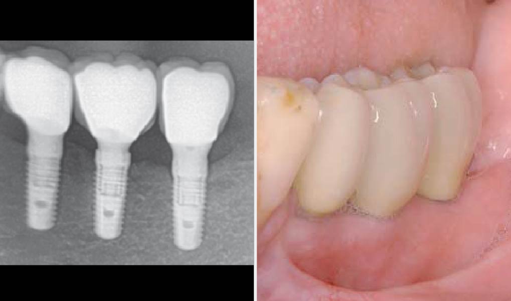
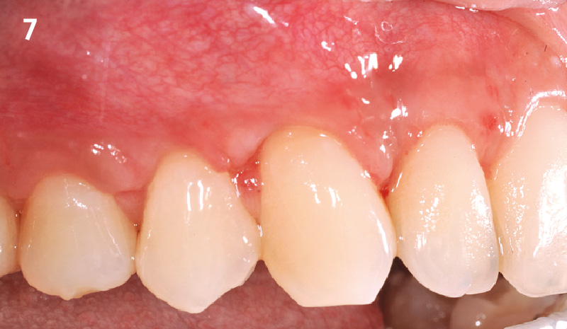
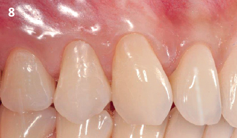
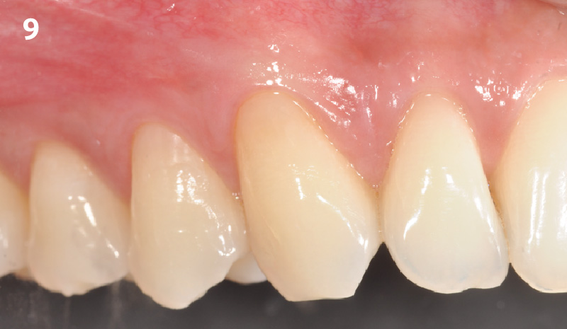
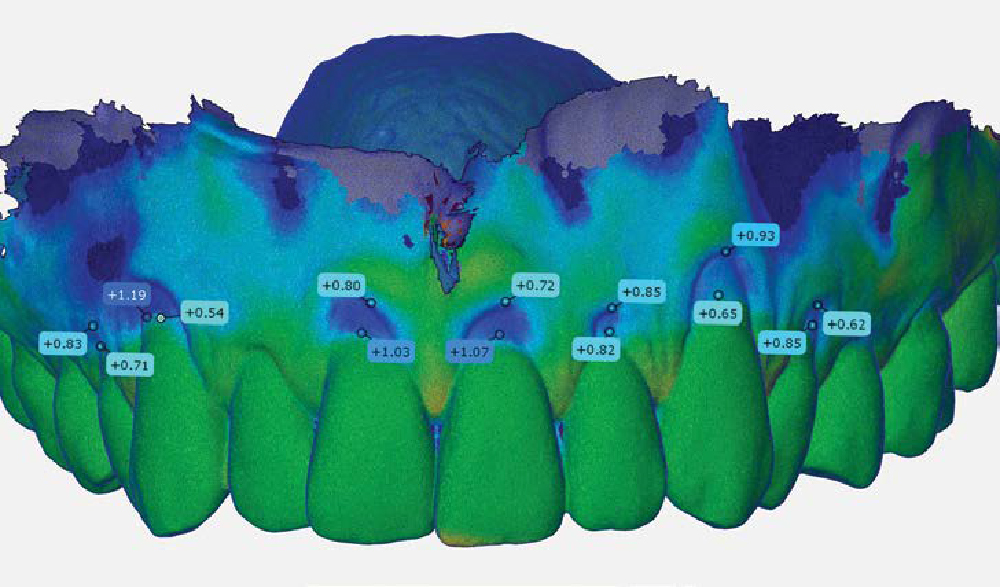
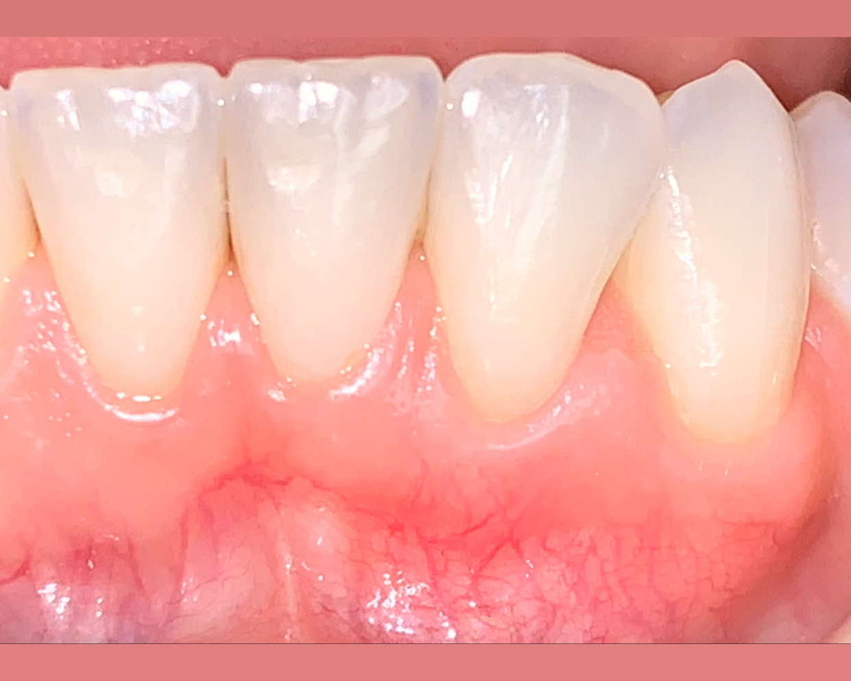
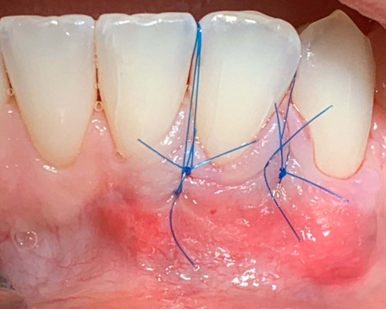
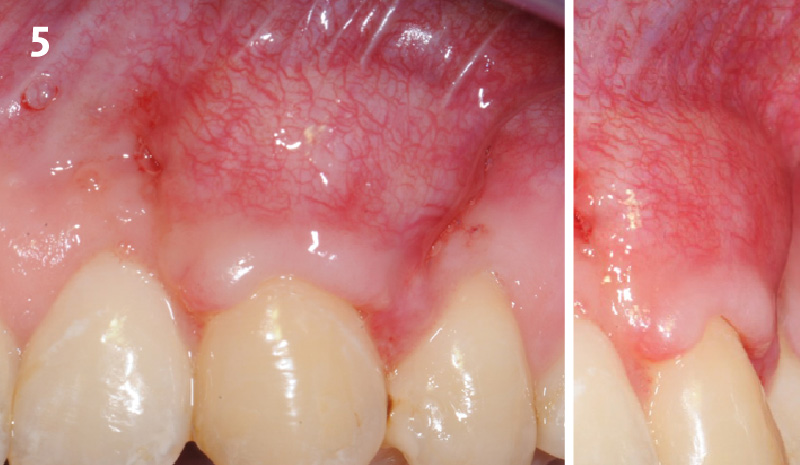
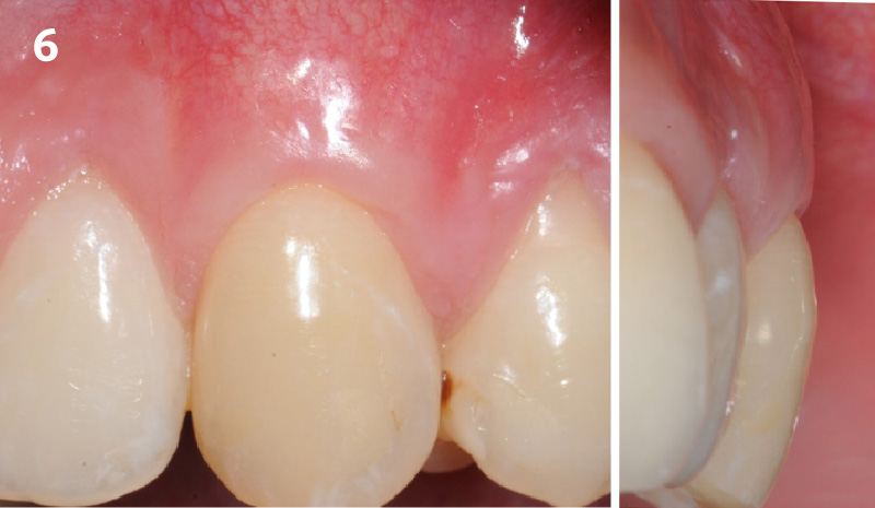
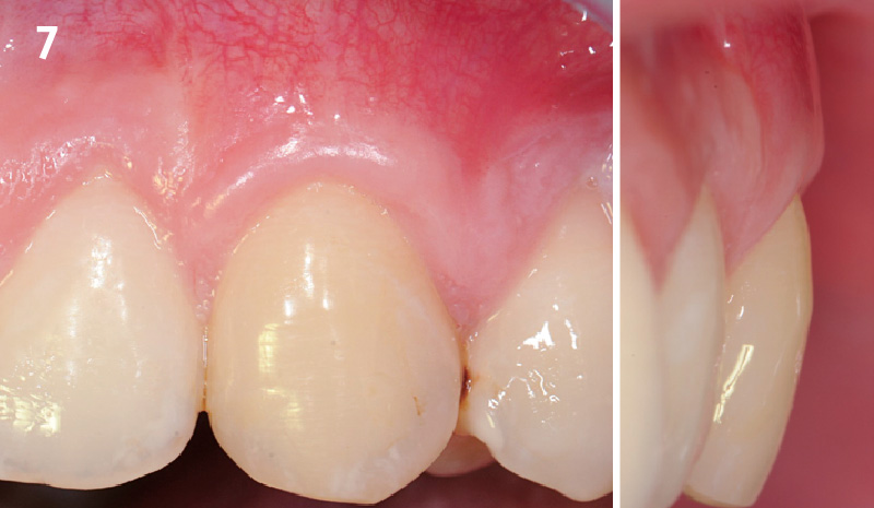
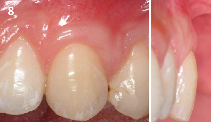
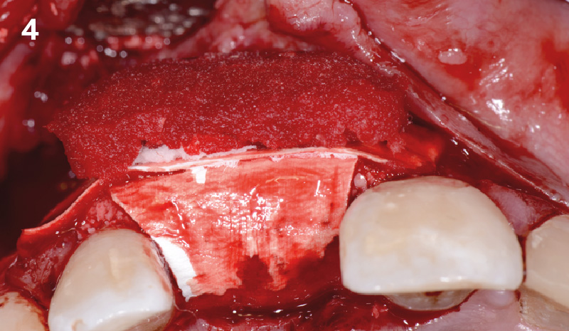
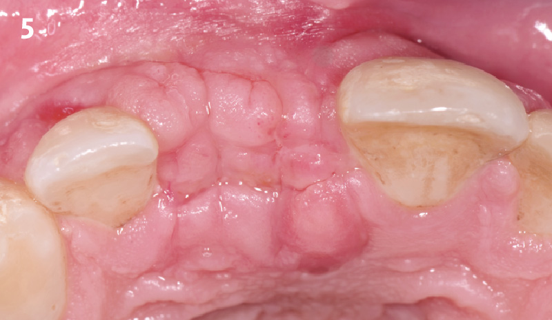
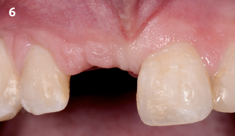
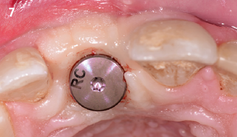
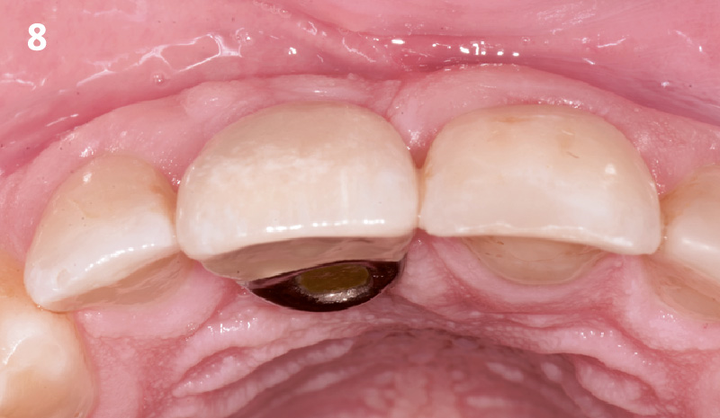
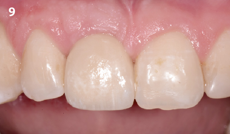
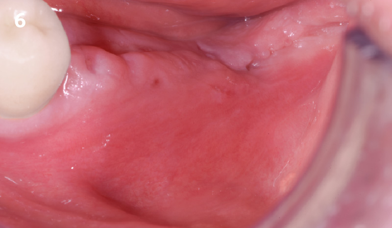
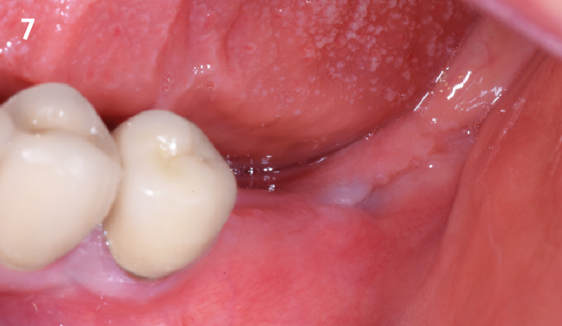
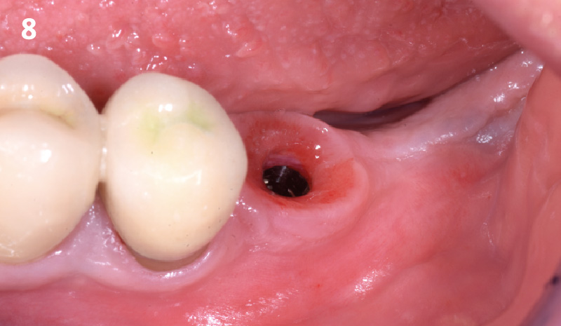
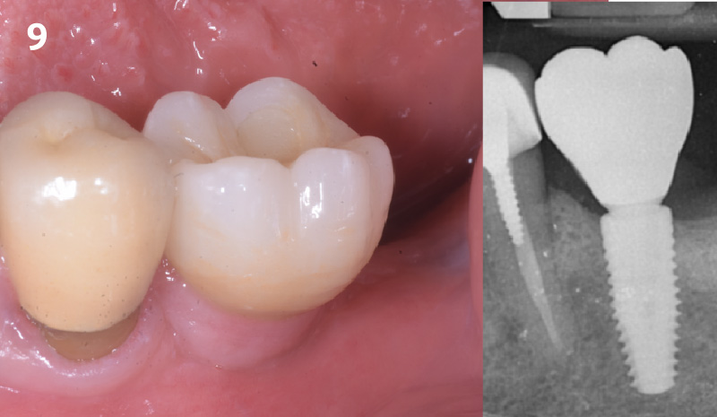
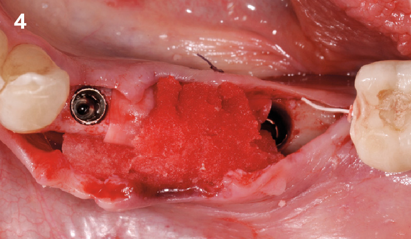
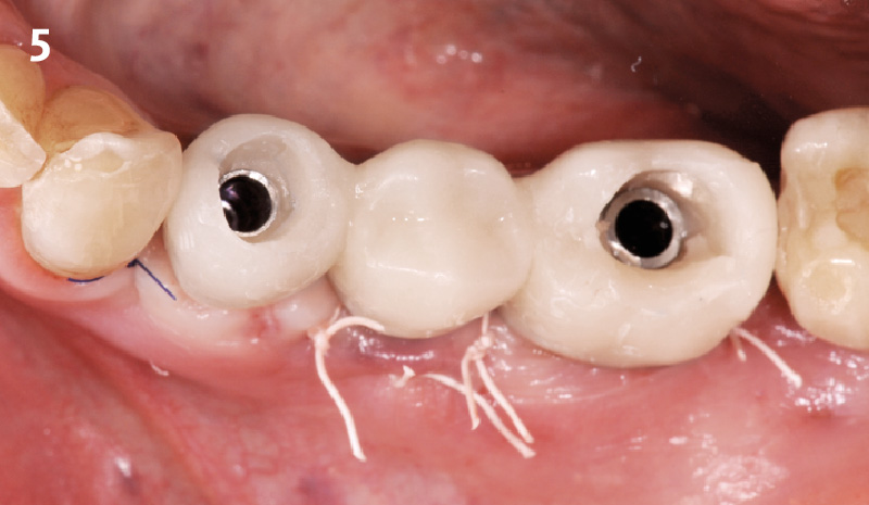
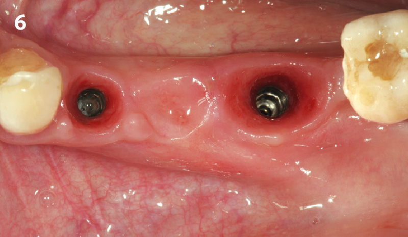
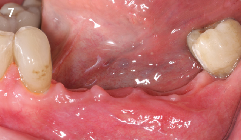
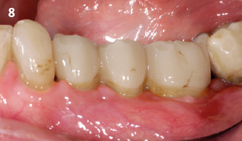
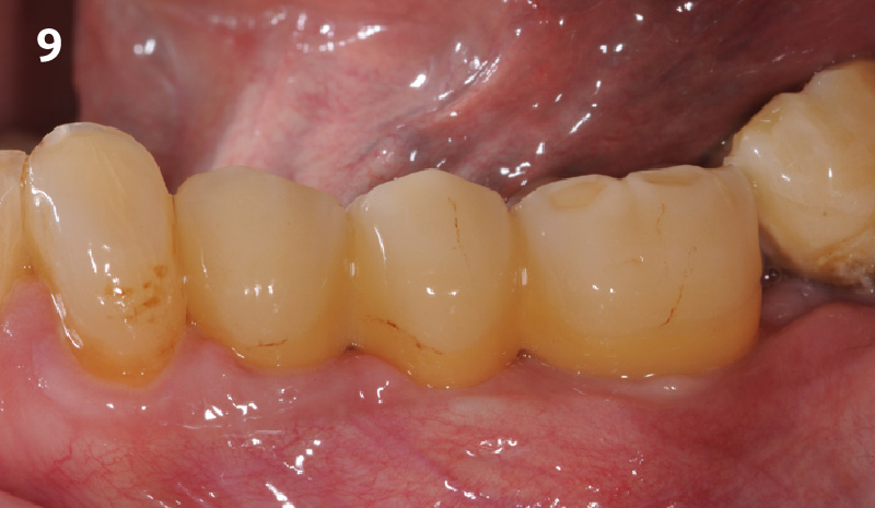
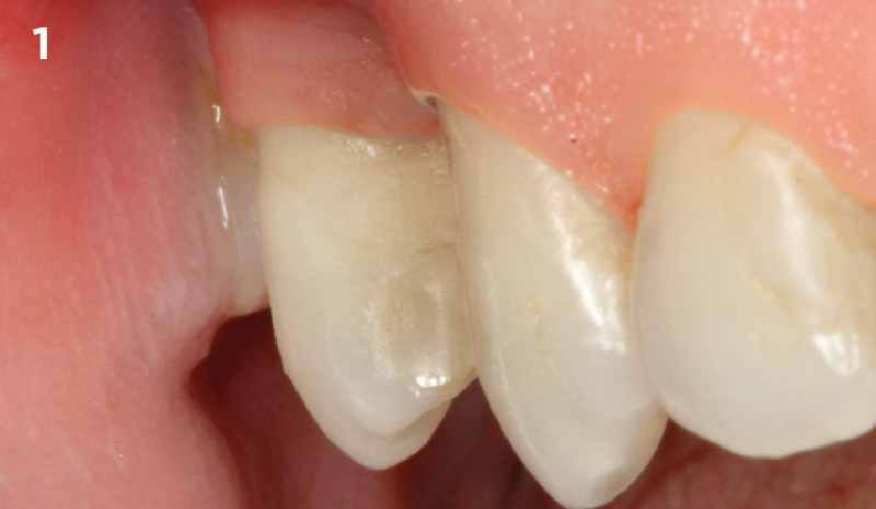
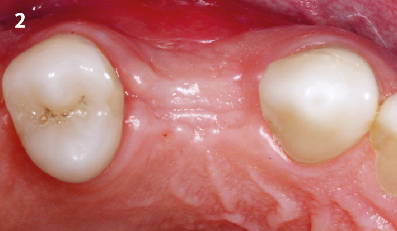
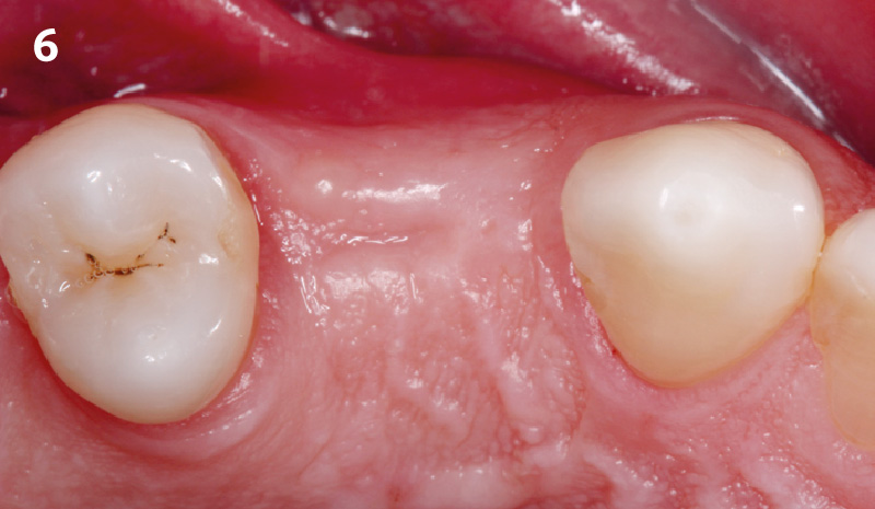
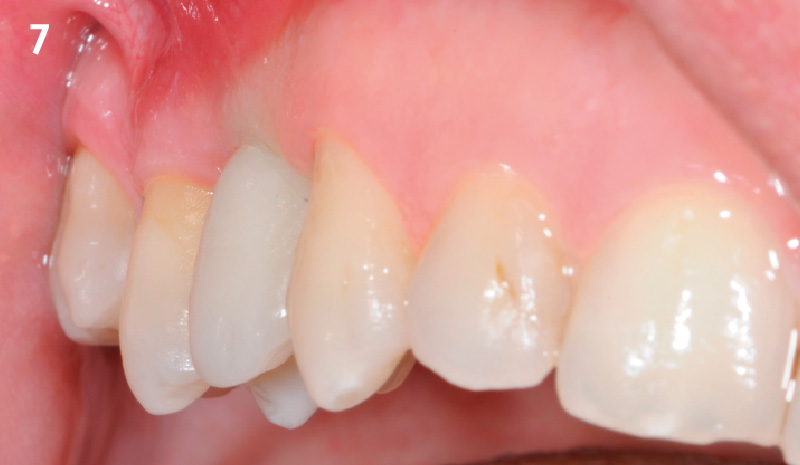
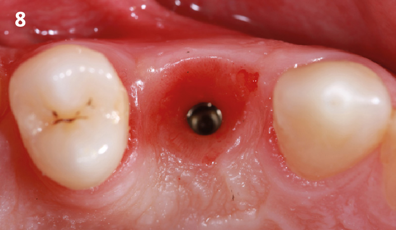
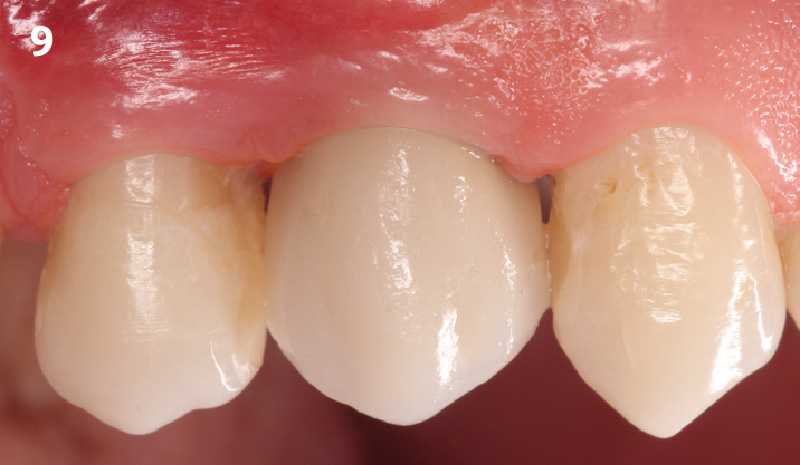
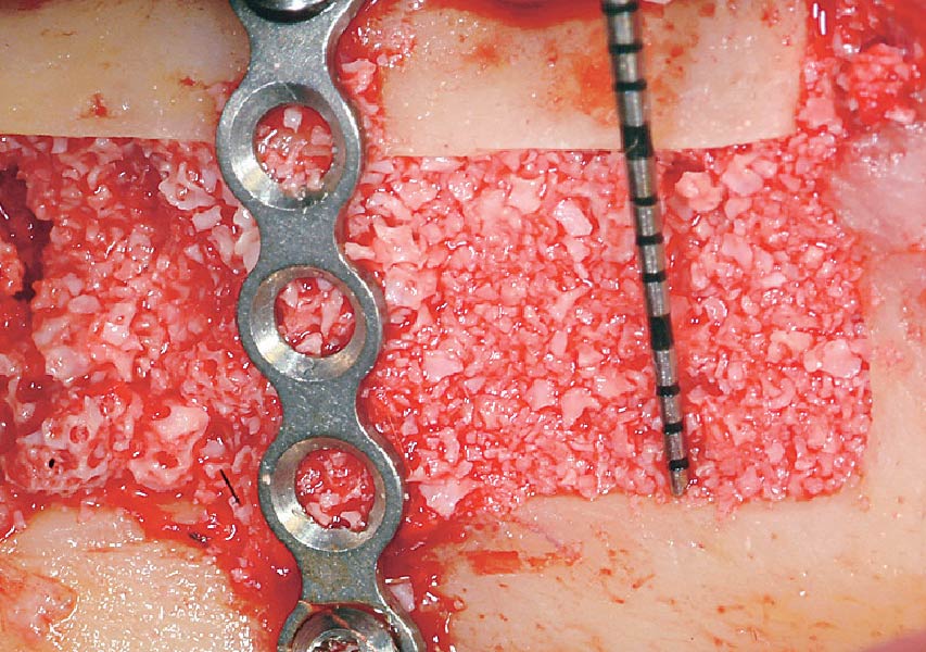
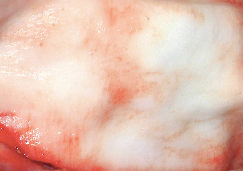
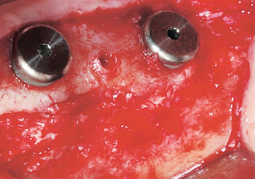
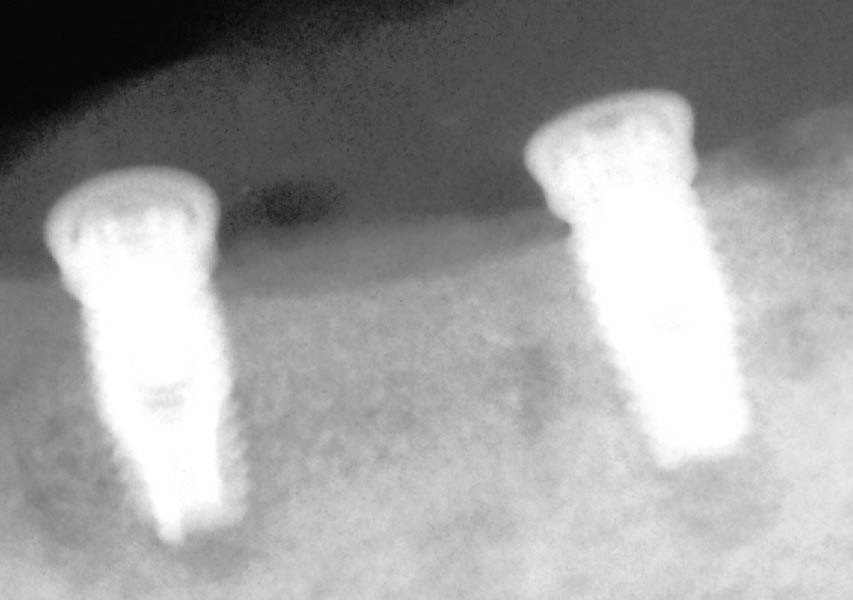
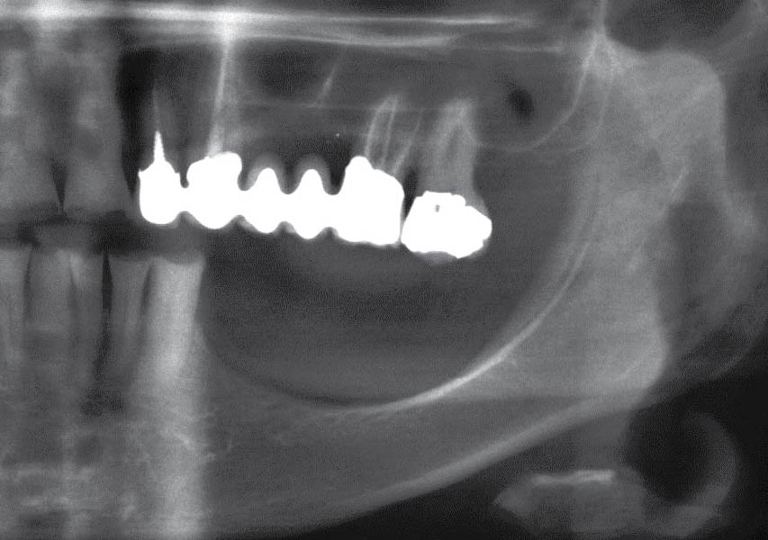
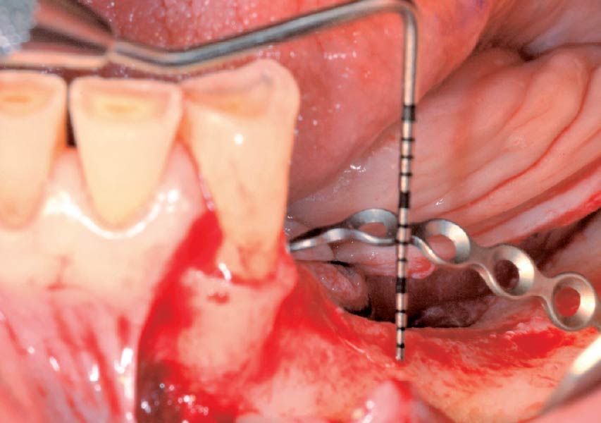
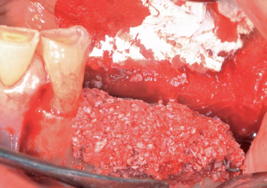
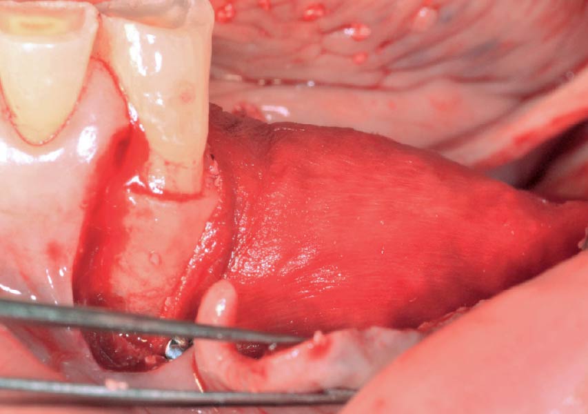
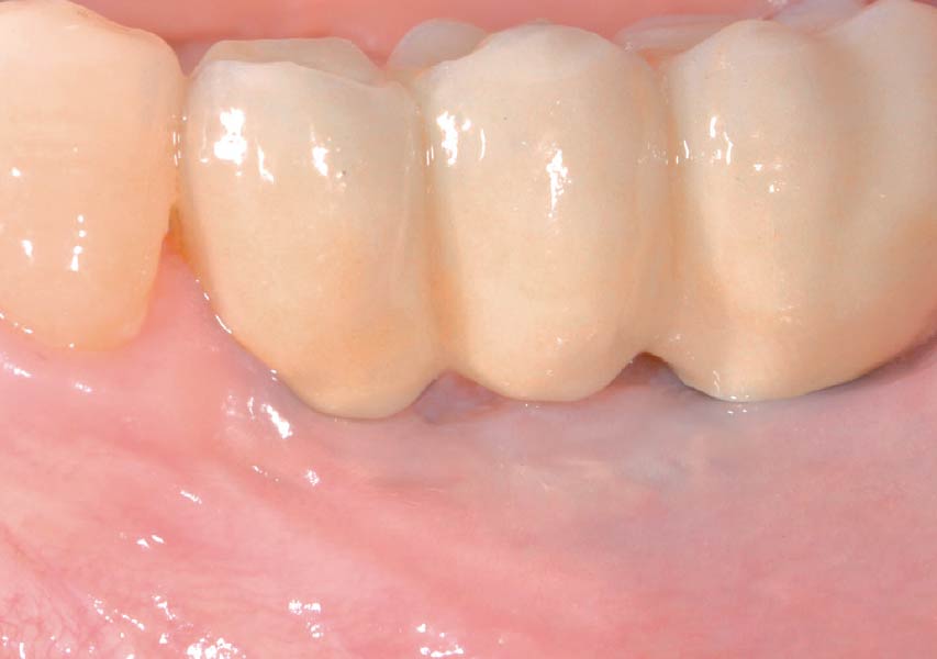
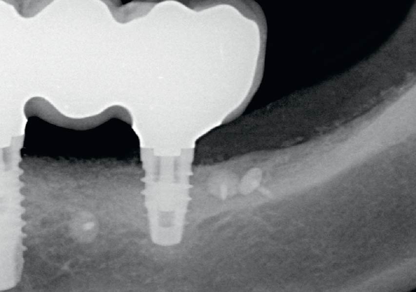
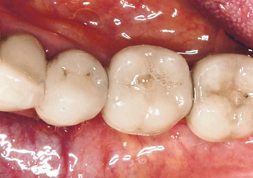
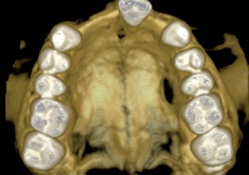
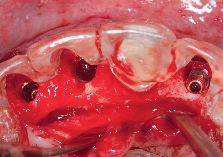
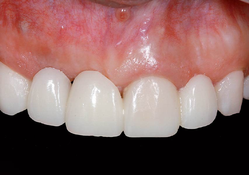
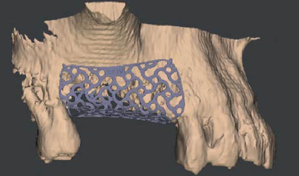
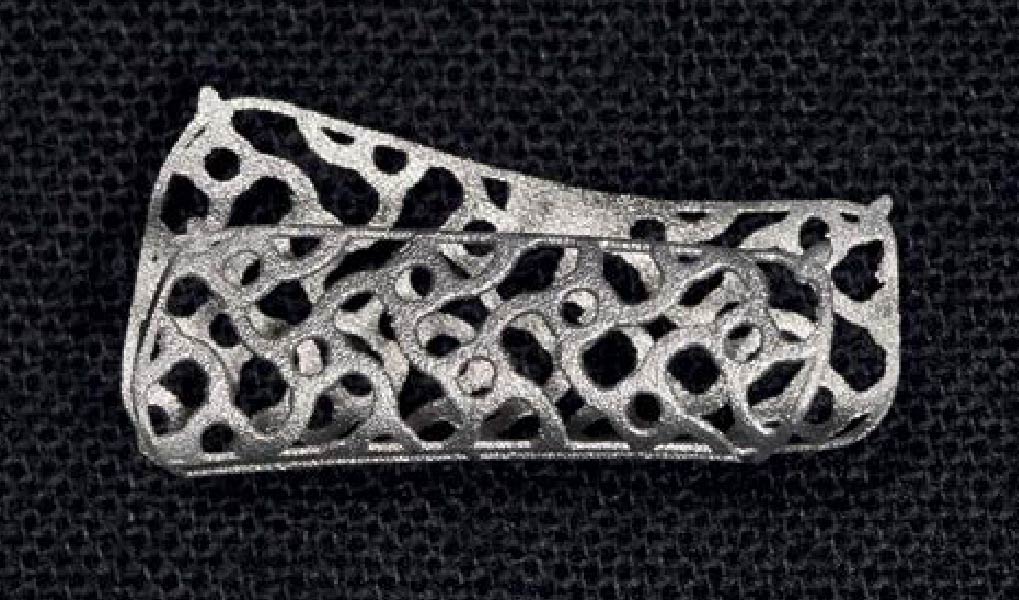
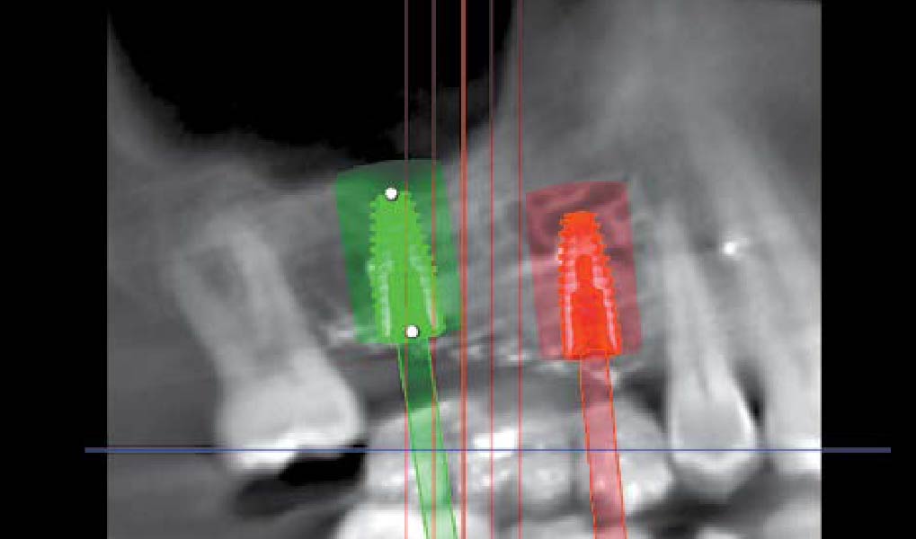
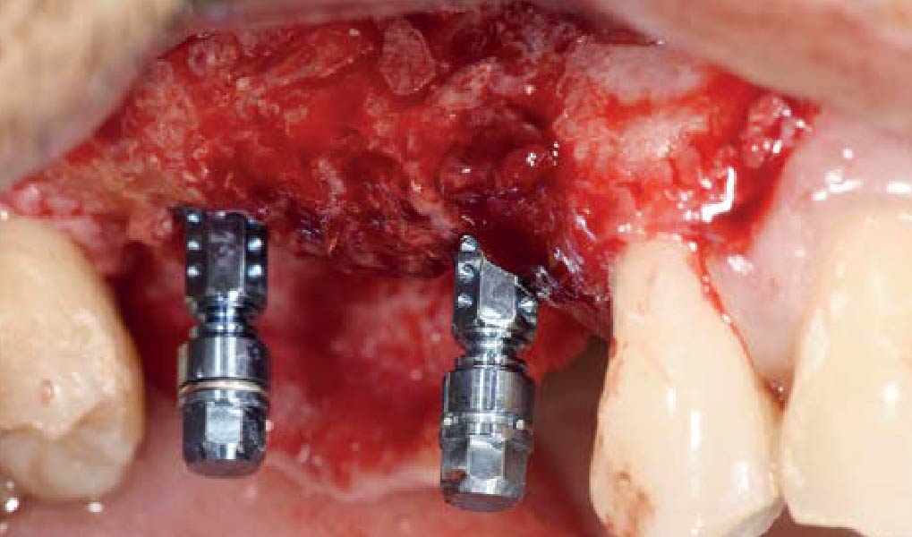
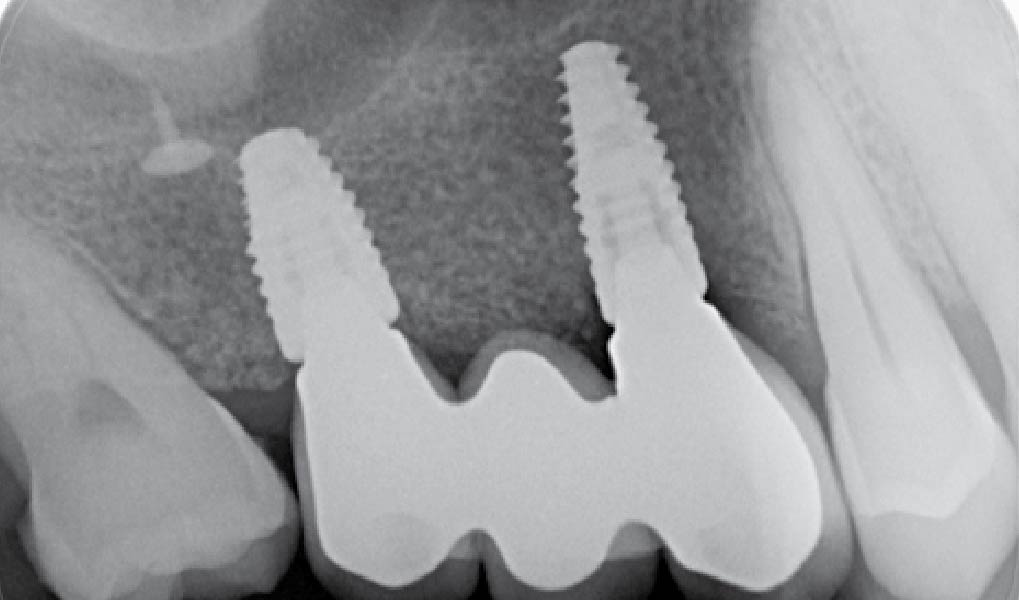
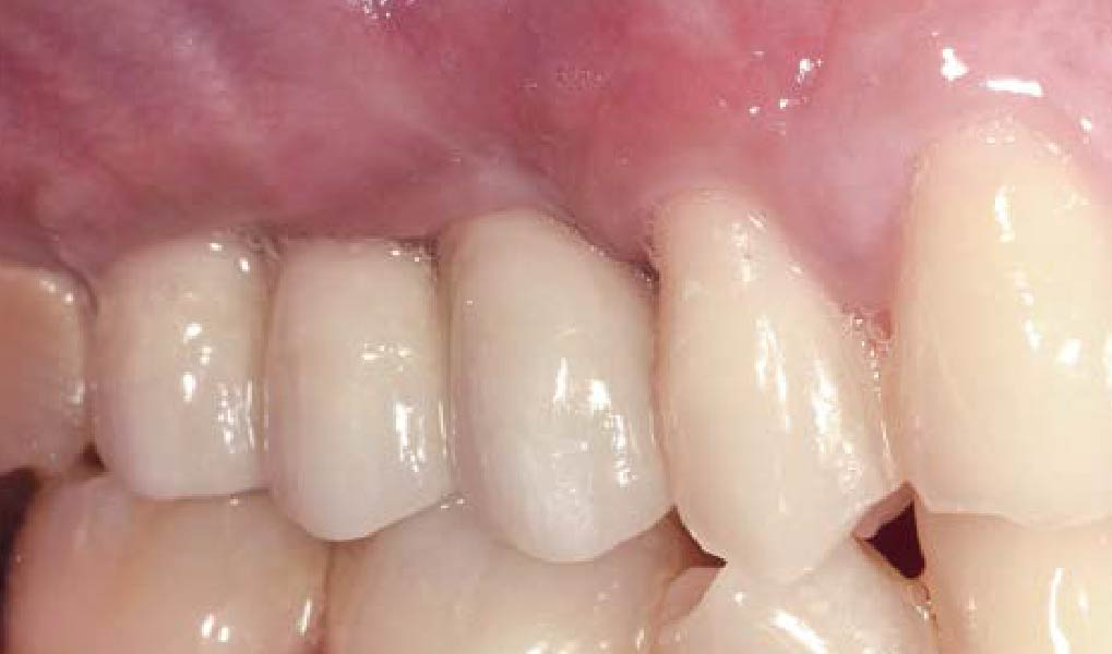
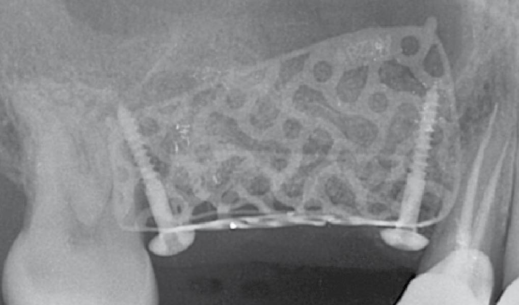
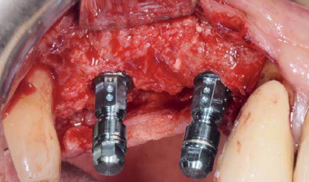
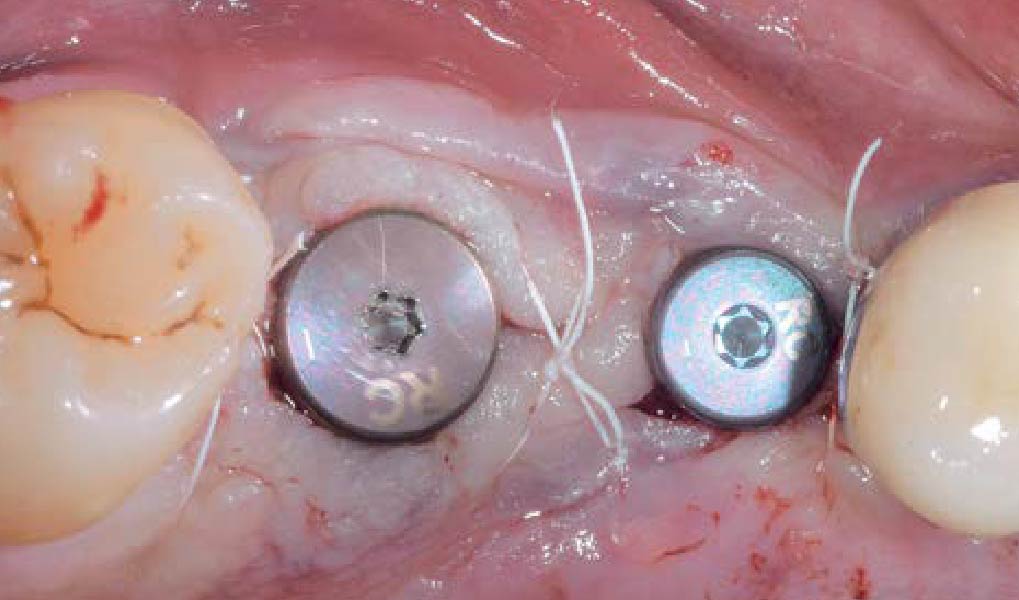
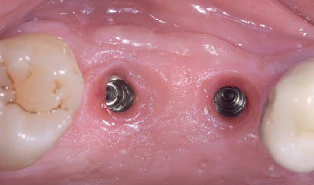
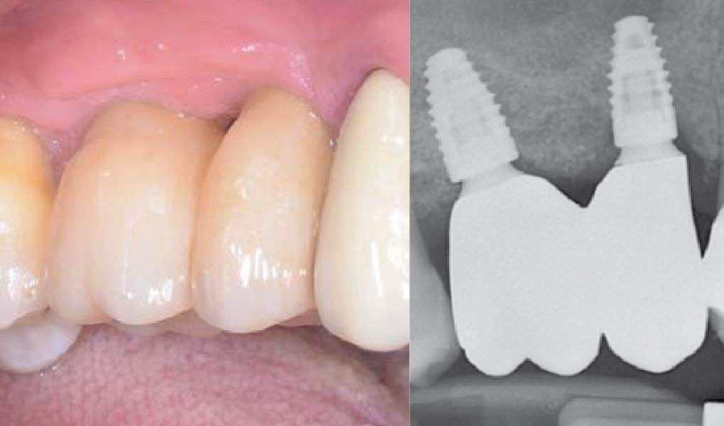
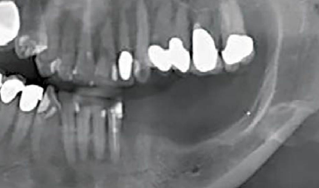
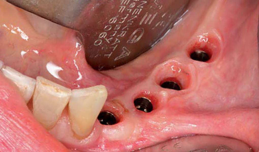
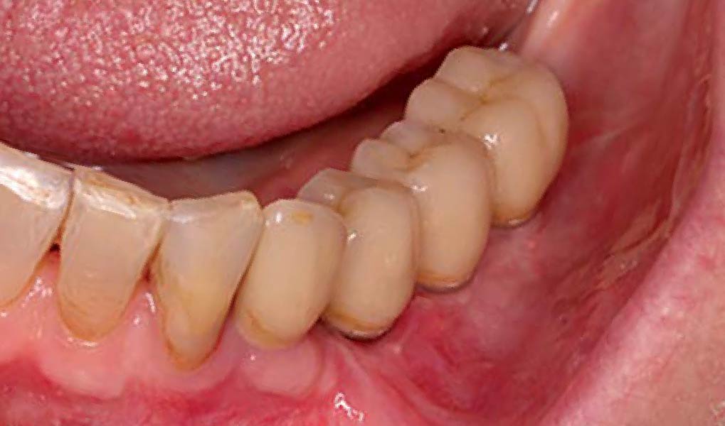
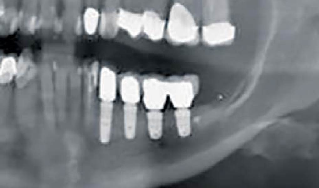
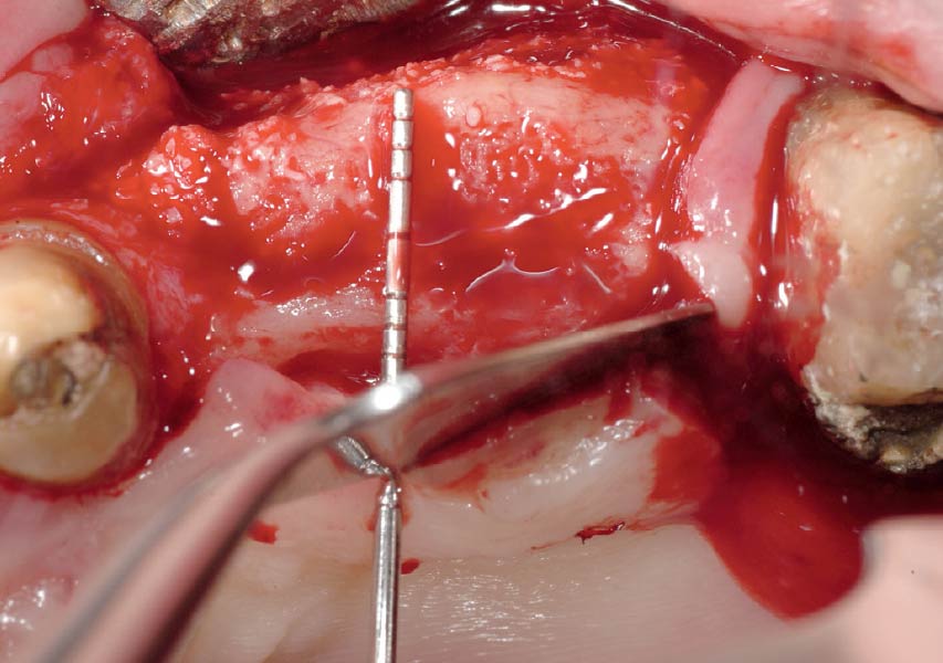
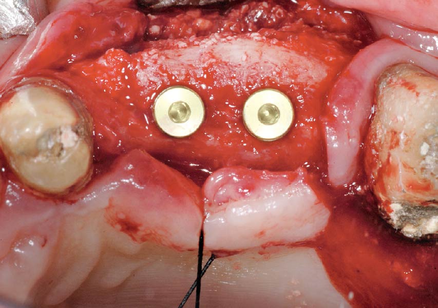
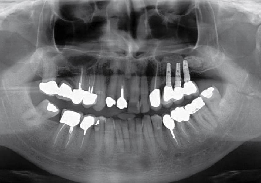
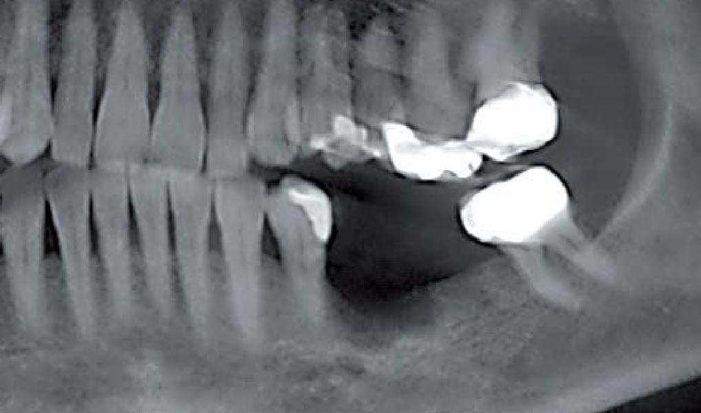
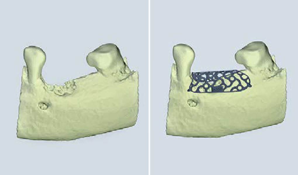
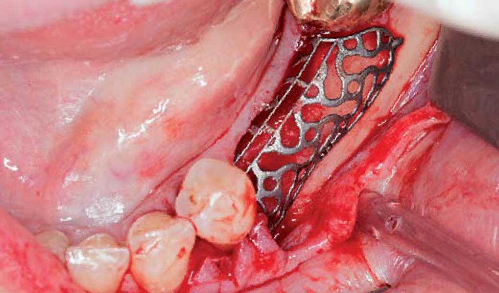
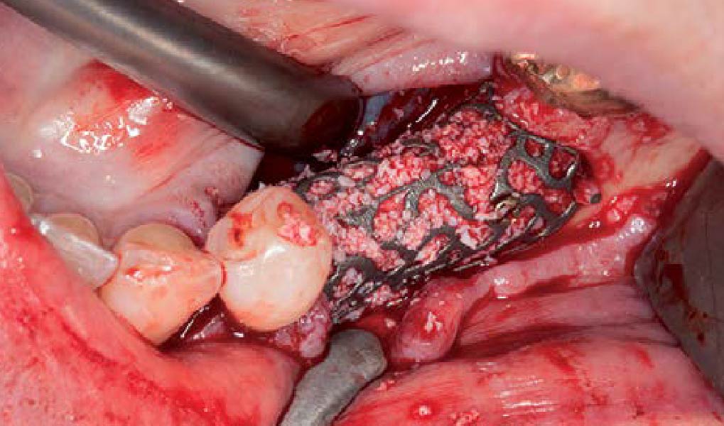
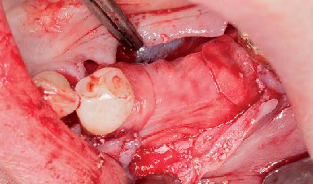
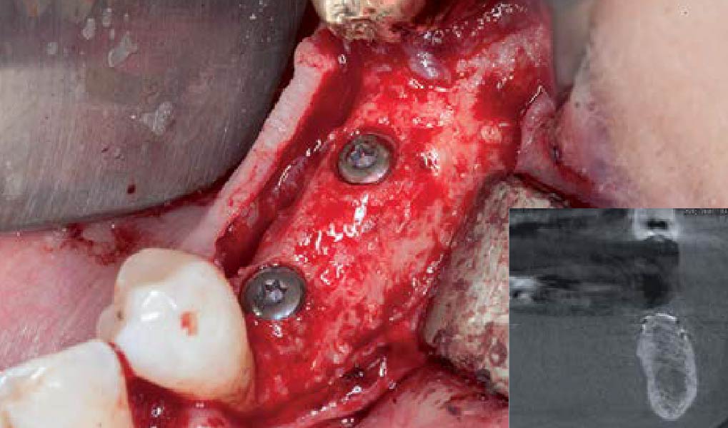
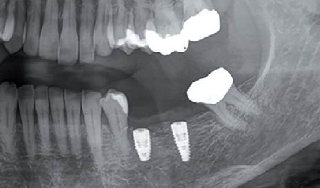
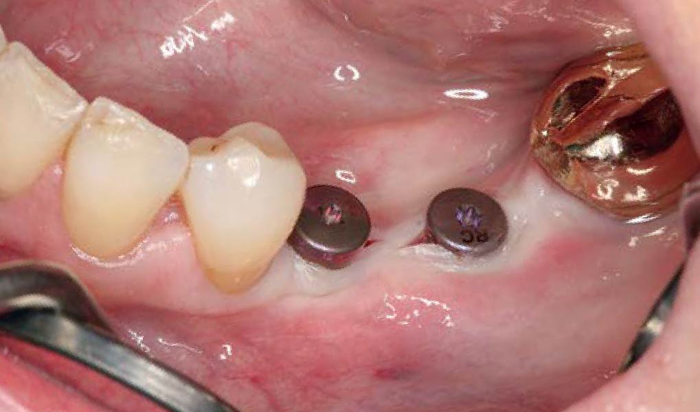
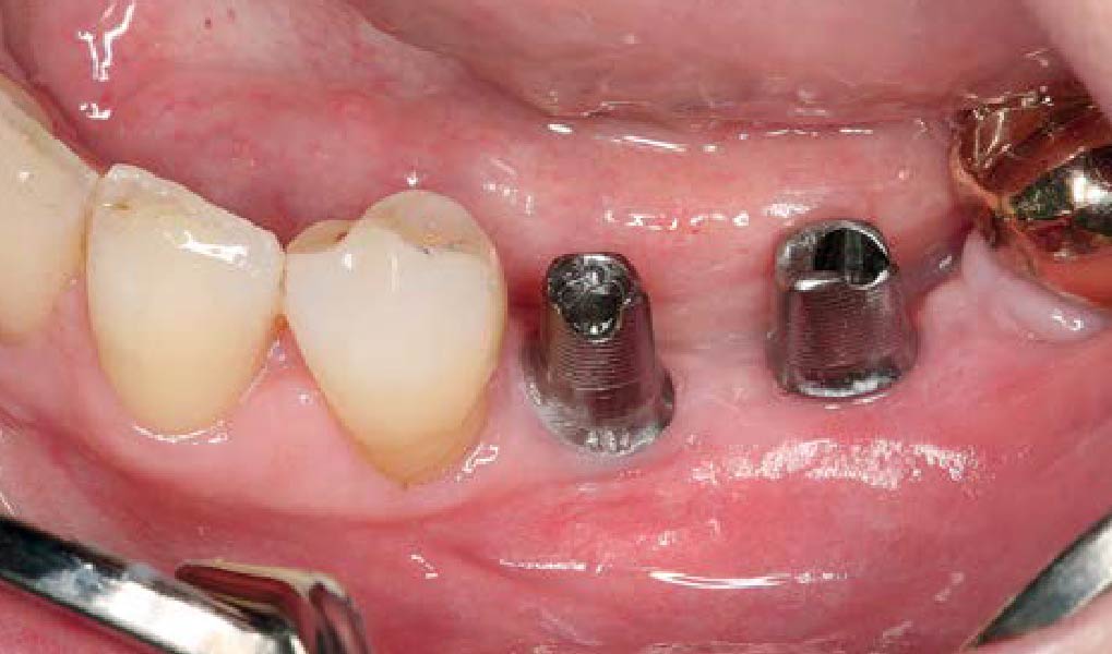
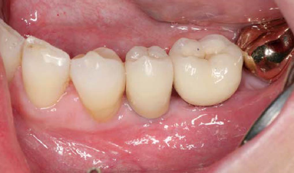
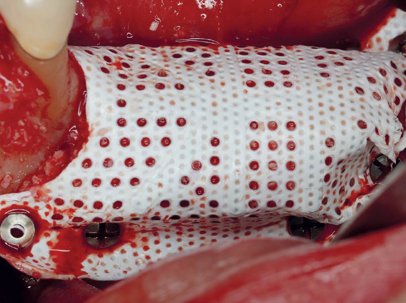
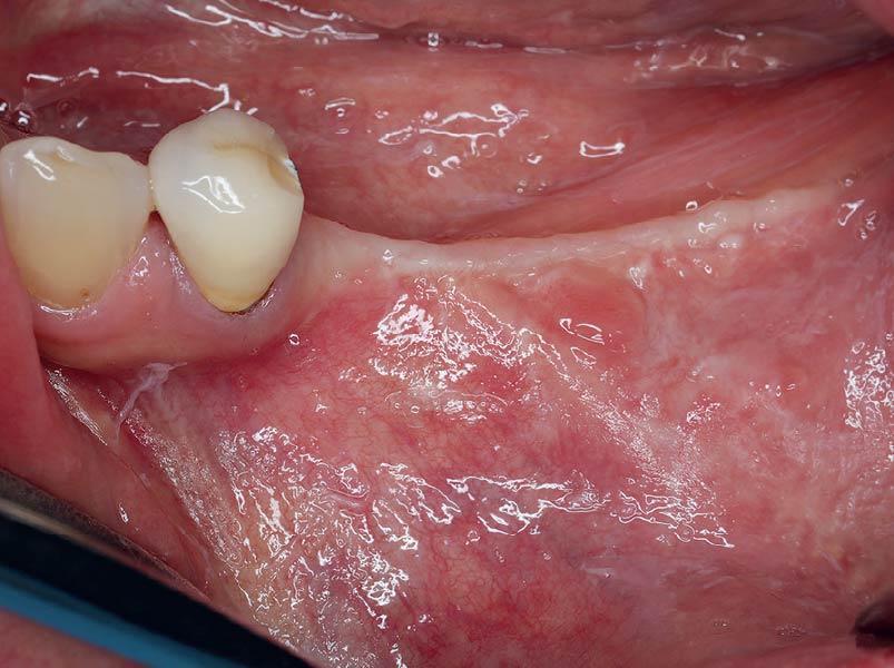
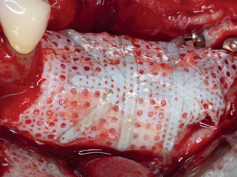
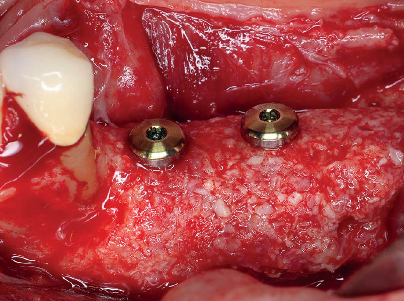
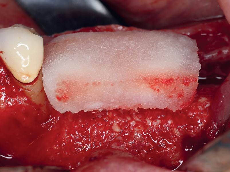
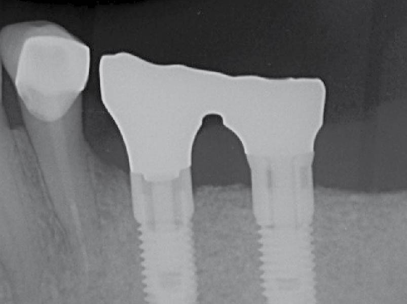
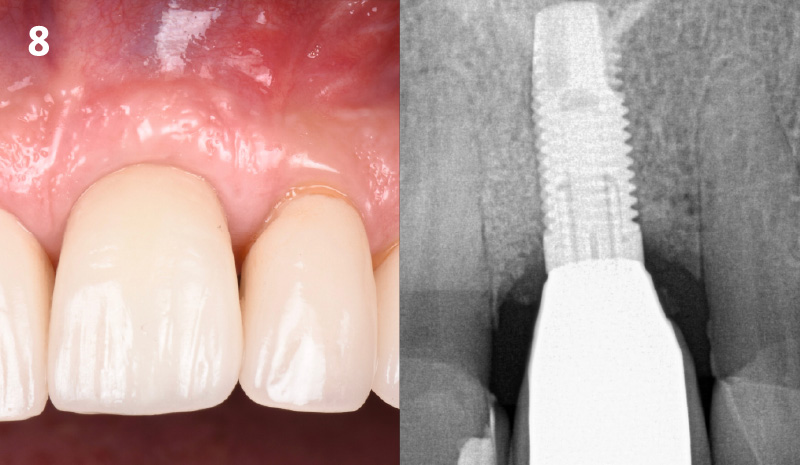
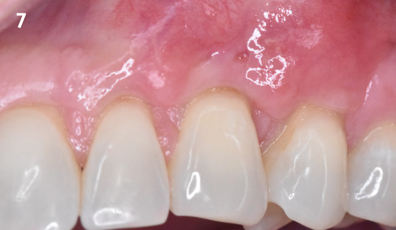
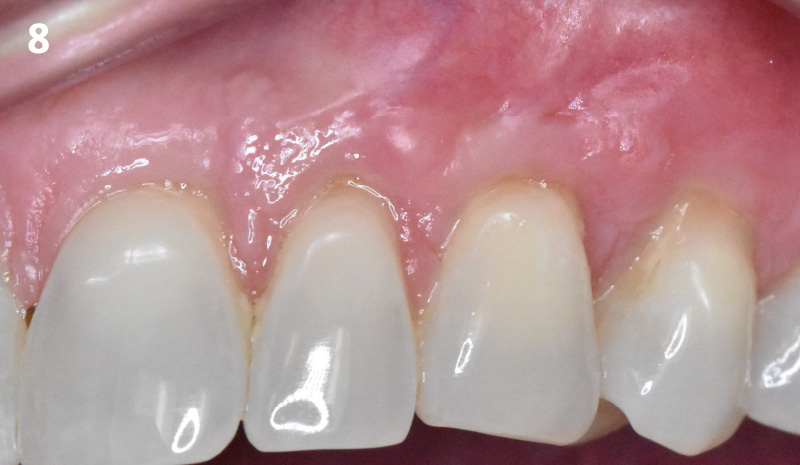
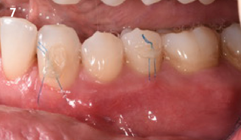
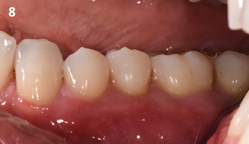
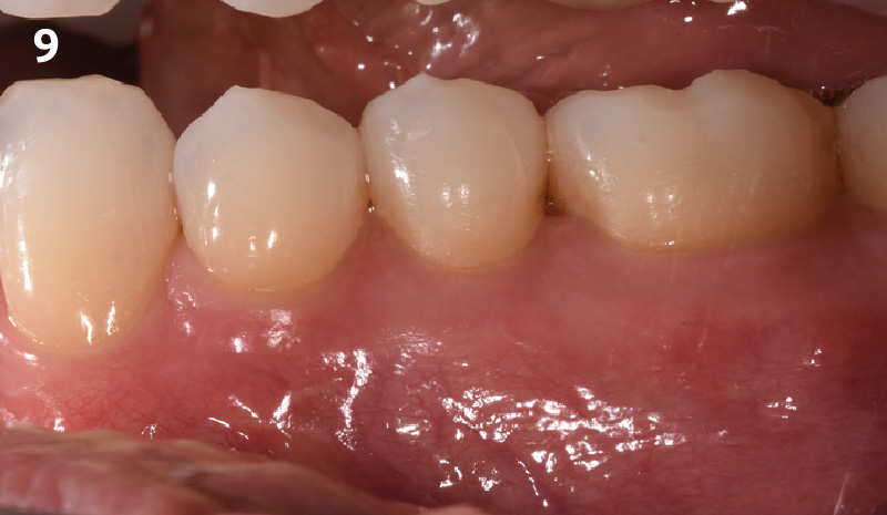
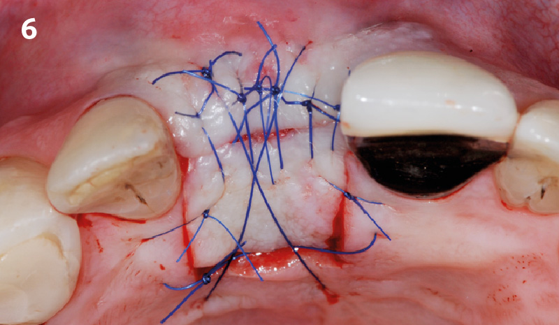
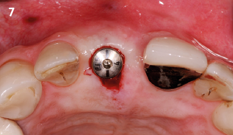
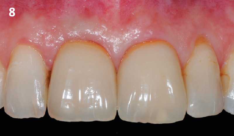
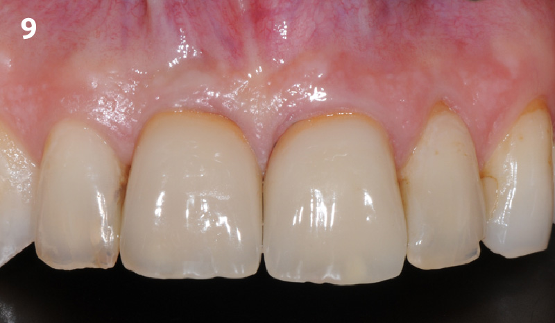
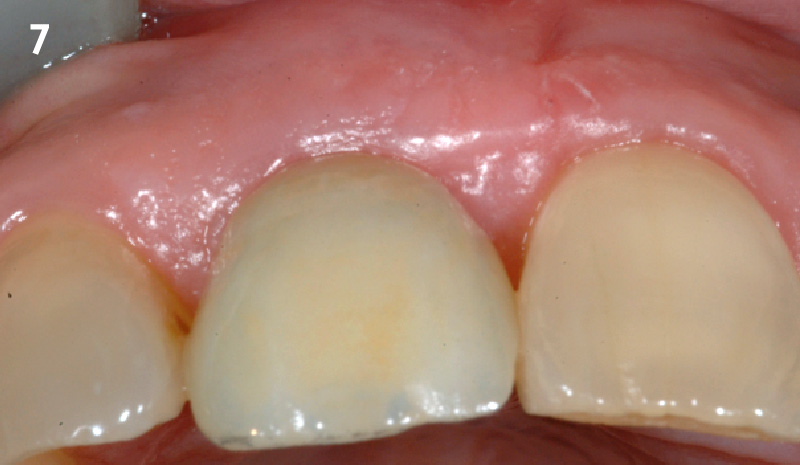
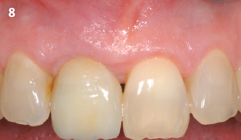
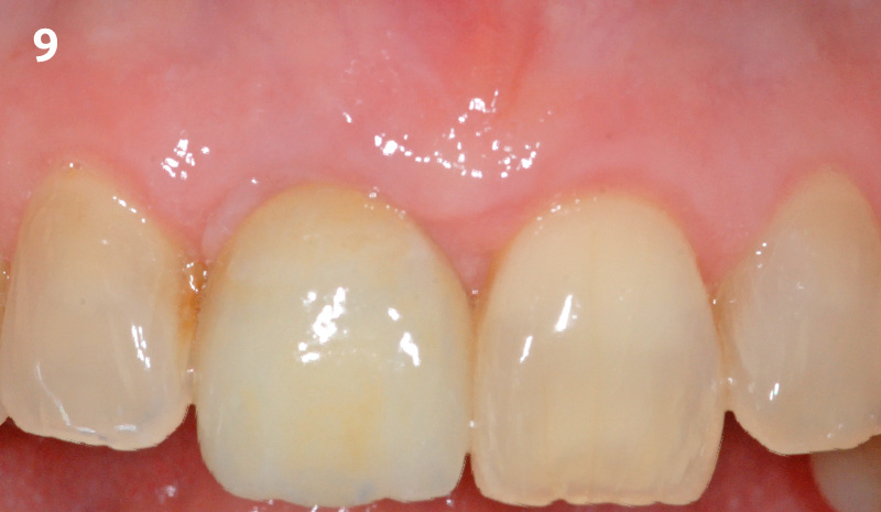
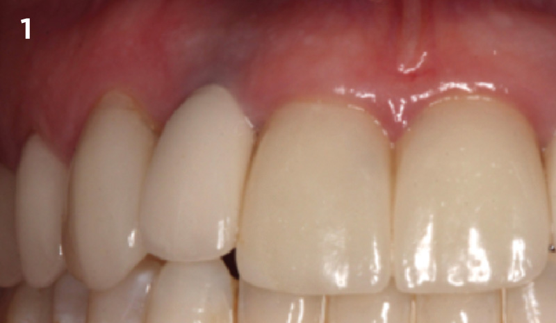
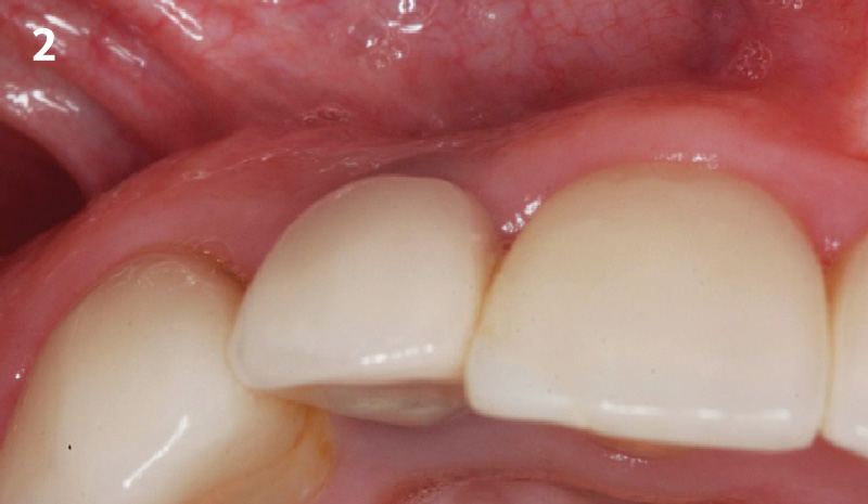
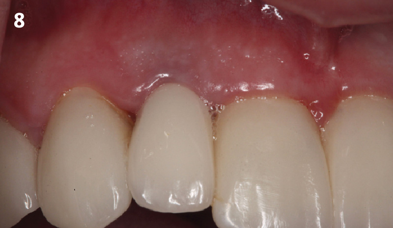
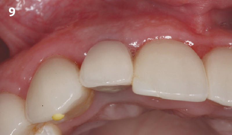
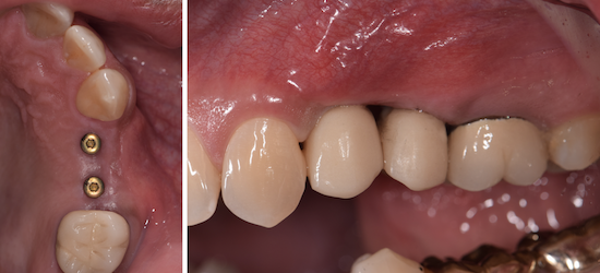
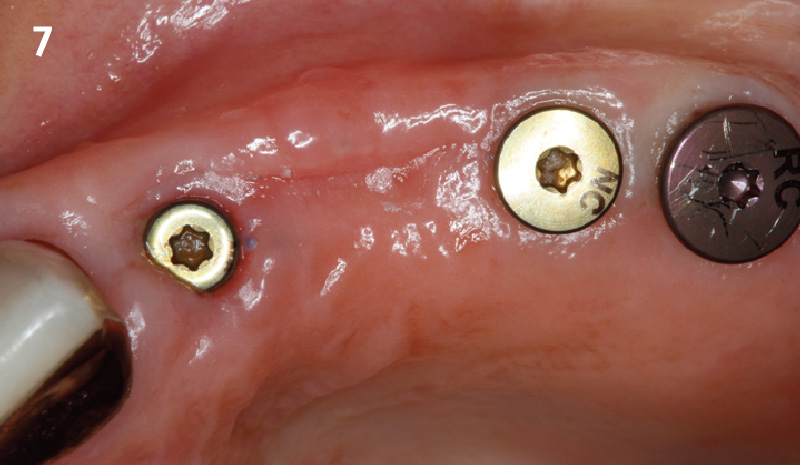
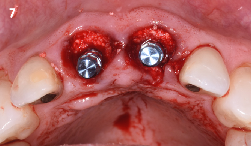
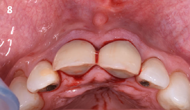
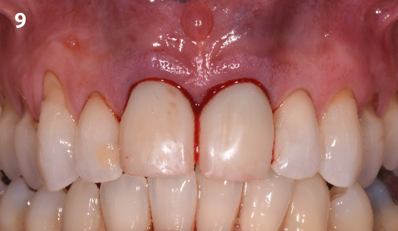
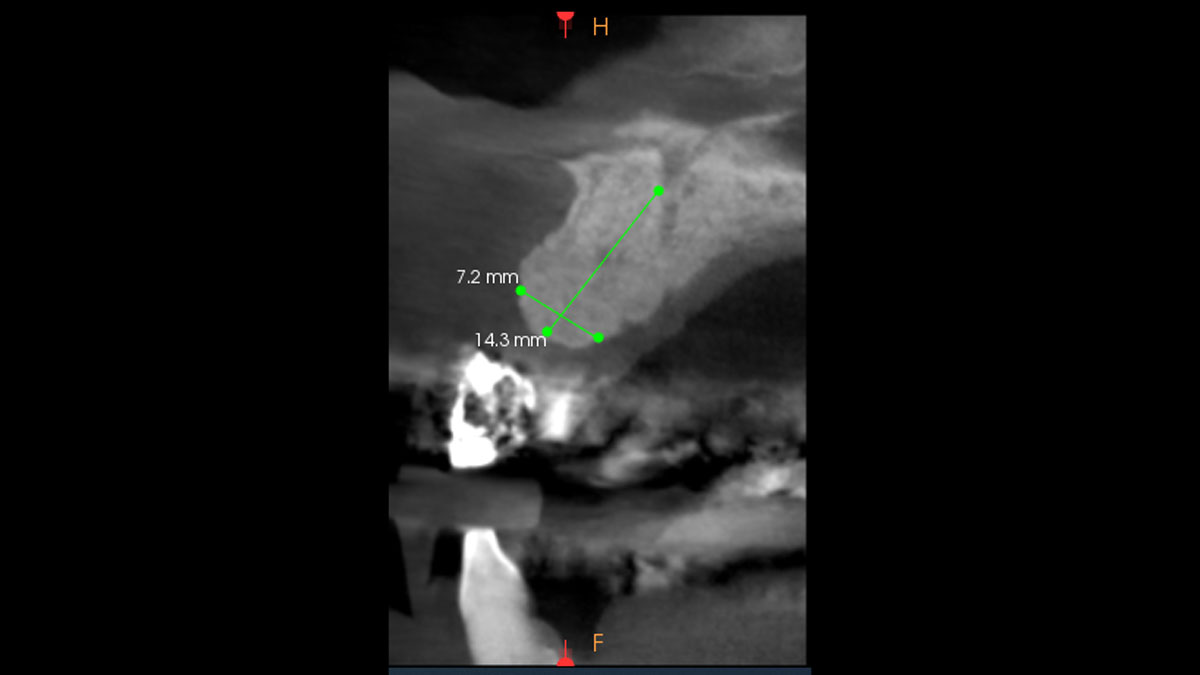
At 6 months, healing was complete with a bone fill of 10x10x6.5 mm. Tooth #7, affected by the combined defect, was fully regenerated. The adjacent ridge defect was also restored, enabling the placement of a regular-sized implant without the need for additional grafting.

CLINICAL CASE

THE APPROACH

Considering the patient’s age, condition, and good compliance, a regenerative approach was chosen to preserve all teeth. Due to the extent and severity of the defect, a bone graft, biologics, and a membrane were utilized. The goal was to restore lost attachment and stabilize the teeth through splinted therapy.

THE OUTCOME

At the 7-month follow-up, probing depths decreased from 7 mm (MB, DB) to 3 mm, with radiographic bone fill indicating a 90% success. Attachment gain of 3 mm was achieved, improving from 7 mm to 4 mm. The patient reported satisfactory outcomes, and splinting effectively maintained tooth stability with no mobility. 

CLINICAL CASE

THE APPROACH

Following flap elevation and thorough defect debridement, a xenograft bone substitute mixed with PDGF (GEM 21S®) was used to fill the defect. The graft was left uncovered without a membrane.

THE OUTCOME

The 1-year follow-up radiograph demonstrated near-complete bone fill in a previously deep defect. Clinically, shallow probing depths and healthy tissue were also observed at the 1-year follow-up.

CLINICAL CASE

THE APPROACH

The crown was biologically shaped, and the root surface was detoxified using Ellman burs. Following flap elevation and thorough defect debridement, an allograft mixed with GEM 21S® was used to fill the defect. The graft was then covered with Geistlich Bio-Gide®.

THE OUTCOME

At the 10-month follow-up, radiographs revealed evidence of bone fill, and clinically, the interdental tissue showed signs of maturation. At the 9-year follow-up, clinical photos demonstrated long-term stability, with maintained bone levels, minimal interproximal recession, and lack of facial recession. Radiographic analysis further confirmed the sustained stability of the bone.

CLINICAL CASE

CLINICAL CASE

CLINICAL CASE

CLINICAL CASE

CLINICAL CASE

CLINICAL CASE

CLINICAL CASE

CLINICAL CASE

CLINICAL CASE

CLINICAL CASE

CLINICAL CASE

CLINICAL CASE

CLINICAL CHALLENGE:

The upper premolar had to be removed due to advanced periodontal disease and severe bone loss around the infected tooth. The bone defect was an intra-alveolar defect without dehiscence or fenestration.

AIM/APPROACH:

An early implant placement approach with a healing time of six weeks before implant placement was chosen. The bone augmentation with Geistlich Bio-Oss Collagen® was conducted simultaneously with implant placement. As this patient was treated in 1991, the case is one of the very first clinical applications of Geistlich Bio-Oss Collagen®

CONCLUSION:

A premolar grafted with Geistlich Bio-Oss Collagen® during implant placement showed good long-term result after 25 years. Satisfactory hard and soft-tissue contour are present 25 years after implantation.

CLINICAL CASE

CLINICAL SITUATION:

A 60-year-old female presented to the periodontics clinic at UTHSA for implant placement at sites #18 and #19. Upon clinical and radiographic examinations, the lower left edentulous ridge was diagnosed as a Siebert class III due to the presence of bucco-lingual and apico-coronal tissue defects. The treatment proposed included soft tissue grafting for increase of keratinized tissue followed by ridge augmentation using Yxoss CBR®mesh and a mix of autograft, vallos fibers, and platelet-rich plasma (PRP)

OUTCOME:

The vallos fibers combined with autogenous bone and the PRP created a stable fibrin bone graft that could be easily molded and contained within the mesh. Hydration with PRP was progressive until the graft reached the desired consistency. Wound healing following ridge augmentation was uneventful. There were no signs of infection or membrane exposure at the site. Mesh removal and implant placement is planned at 6-months following ridge augmentation.

CLINICAL CASE

CLINICAL CASE

CLINICAL CASE

CLINICAL CHALLENGE:

  • The planning of the patient’s case takes local and general patient-specific risk factors into consideration according to the principles of backward planning for implant positioning.

AIM/APPROACH:

  • Highlights step-by-step the important procedures to regenerate the bone (horizontal and vertical) with the 3-D printing technology, Yxoss CBR®.

CLINICAL CASE

CLINICAL CASE

CLINICAL CASE

CLINICAL CASE

CLINICAL CASE

CLINICAL CASE

CLINICAL CASE

CLINICAL CASE

CLINICAL CASE

CLINICAL CHALLENGE:

  • Insufficient alveolar ridge height for implant placement and proximity to the alveolar nerve
  • Autologous bone harvesting is associated with patient discomfort

AIM/APPROACH:

  • Interpositional grafting with Geistlich Bio-Oss® Block for vertical augmentation
  • Alveolar ridge volume preservation and minimizing patient morbidity

CLINICAL CASE

CLINICAL CHALLENGE:

  • Severely atrophied alveolar ridge with insufficient bone volume for implant placement
  • ­­­­High complication rates and patient discomfort associated with large augmentations when using autologous bone grafts

AIM/APPROACH:

  • 3-dimensional augmentation of alveolar ridge by the fence technique for implant placement
  • At the same time reducing complication rates and patient discomfort

CLINICAL CASE

CLINICAL CHALLENGE:

  • Insufficient alveolar ridge width for implant placement
  • Autologous bone is subject to resorption and may lead to loss of volume

AIM/APPROACH:

  • Ridge Split procedure in combination with Geistlich Bio-Oss® and Geistlich Bio-Gide® for horizontal augmentation
  • Preservation of the alveolar ridge volume

CLINICAL CASE

CLINICAL CHALLENGE:

  • Insufficient alveolar ridge width for implant placement
  • Donor site morbidity after autologous bone block harvesting and resorption of autologous bone

AIM/APPROACH:

  • Horizontal alveolar ridge augmentation with Geistlich Bio-Oss® and Geistlich Bio-Gide®
  • Minimizing autologous bone harvesting and resorption protection

CLINICAL CASE

CLINICAL CASE

CLINICAL CASE

CLINICAL CASE

CLINICAL CASE

CLINICAL CASE

CLINICAL CASE

CLINICAL CASE

CLINICAL CASE

CLINICAL CASE

CLINICAL CASE

CLINICAL CASE

CONCLUSIONS:

  • Geistlich Mucograft® with a keratinized tissue strip was utilized to increase vestibular depth and gain additional keratinized tissue.
  • Augmentation of severely atrophied alveolar ridge provided sufficient bone for implant placement 8 months following augmentation.

CLINICAL CASE

CLINICAL CASE

CLINICAL CASE

CLINICAL CASE

CLINICAL CASE