BIOBRIEF

Selecting Biomaterials for Combined Complex Defects

Irina F. Dragan, DDS, DMD, MS, eMBA

THE SITUATION

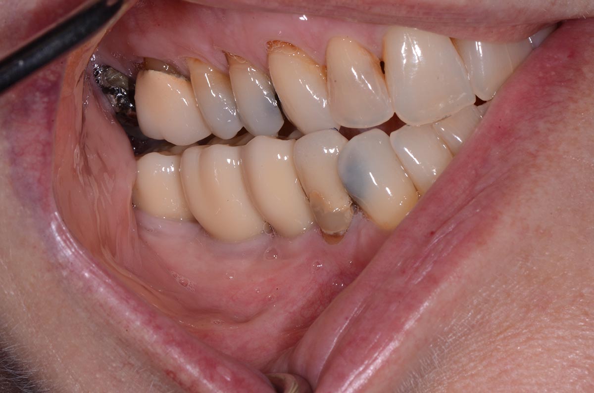
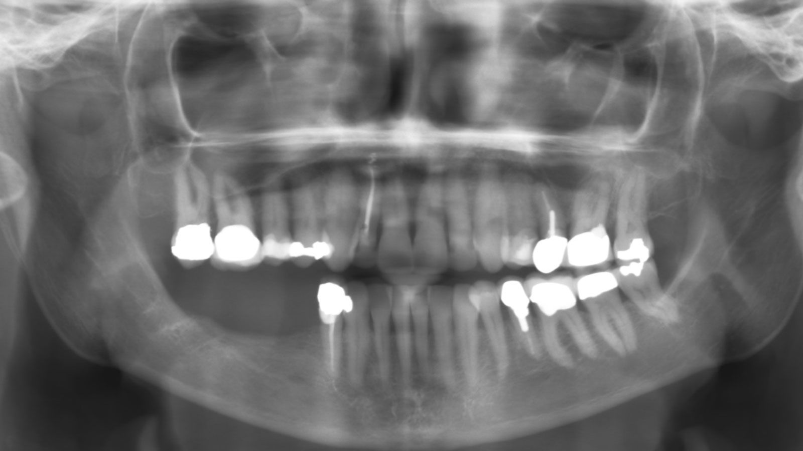
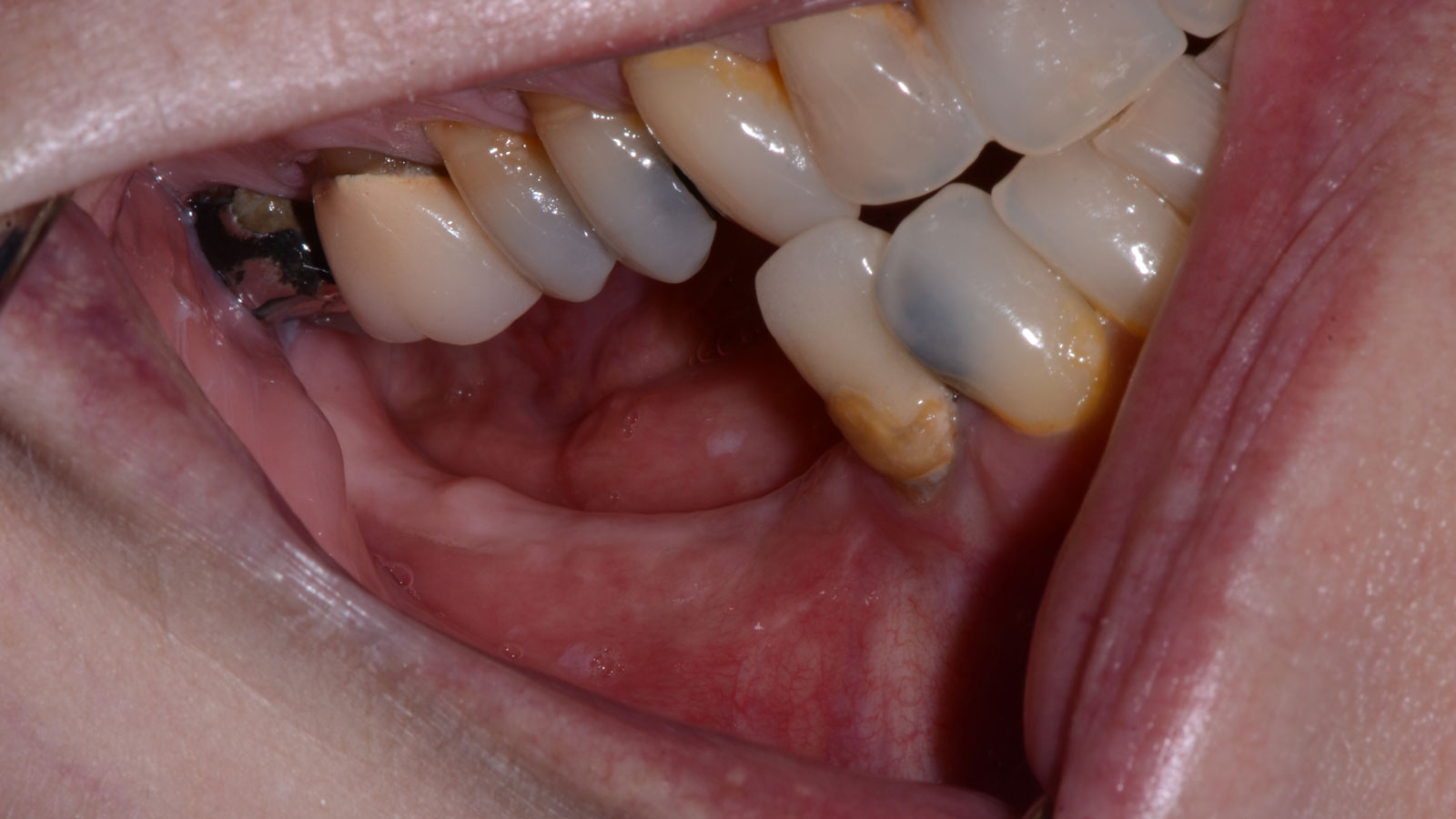
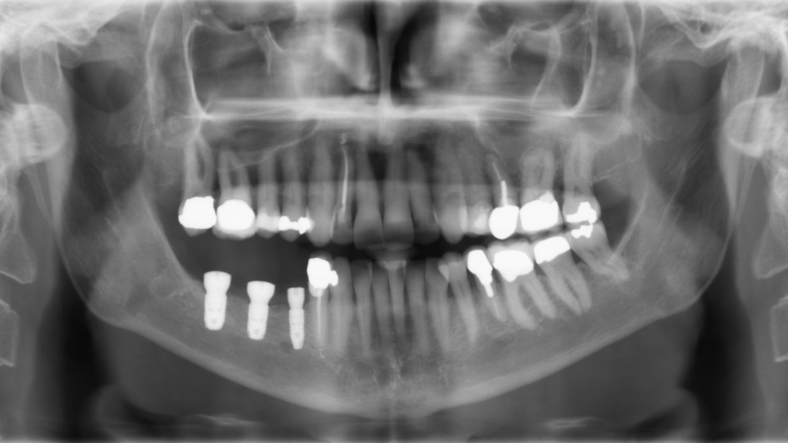
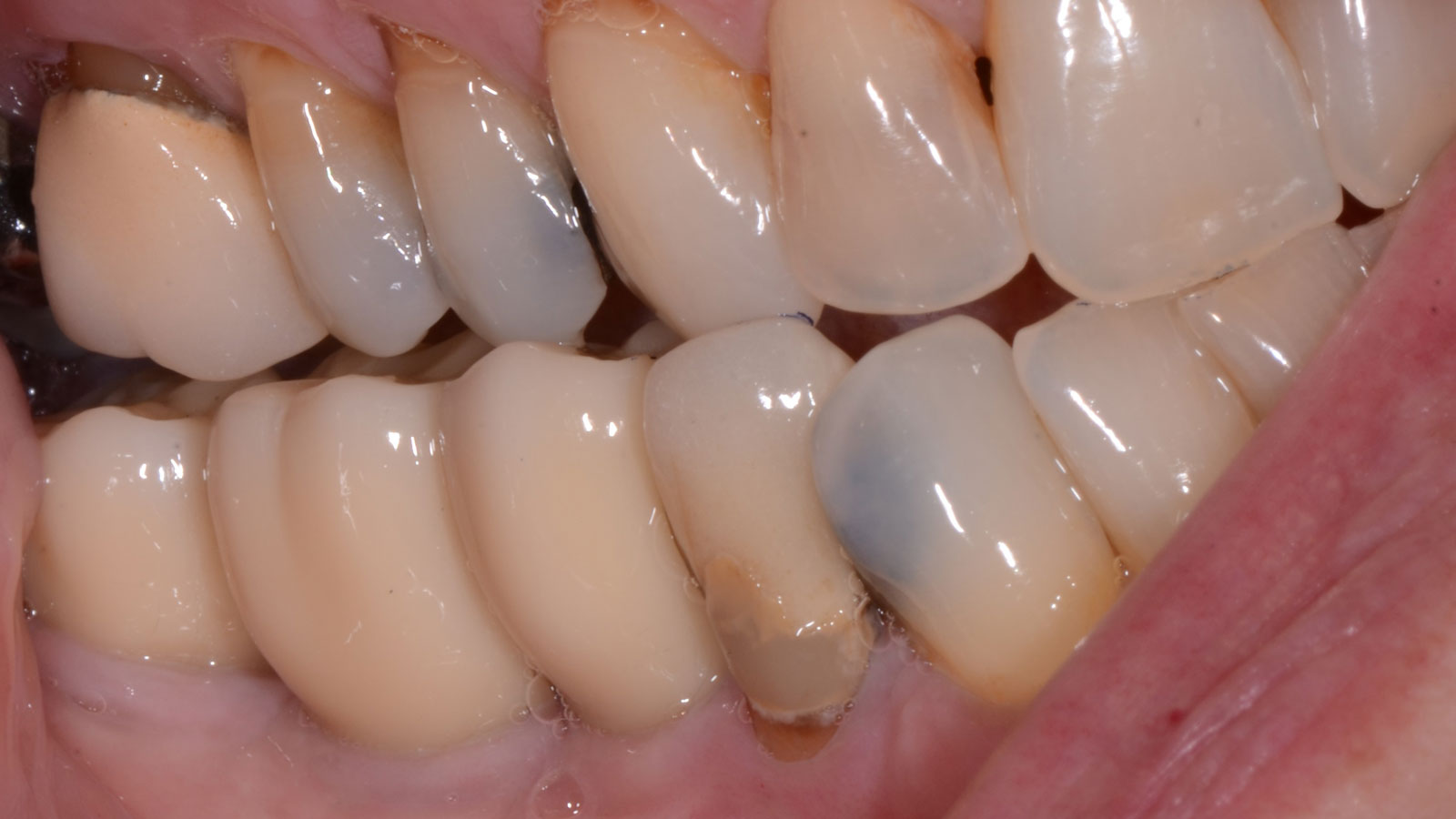
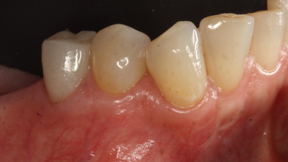
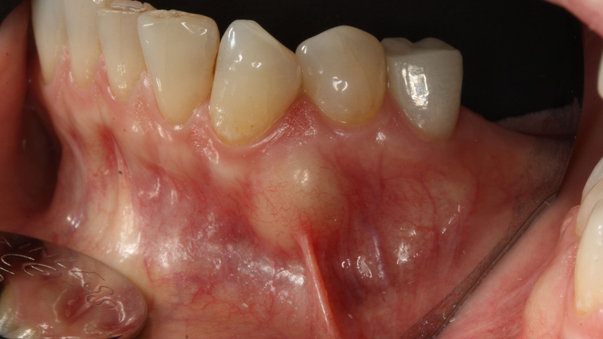
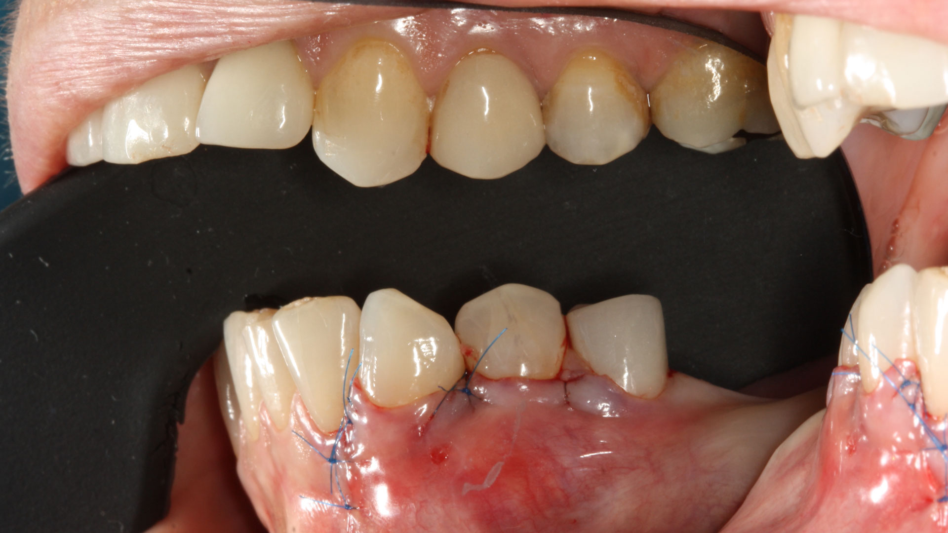
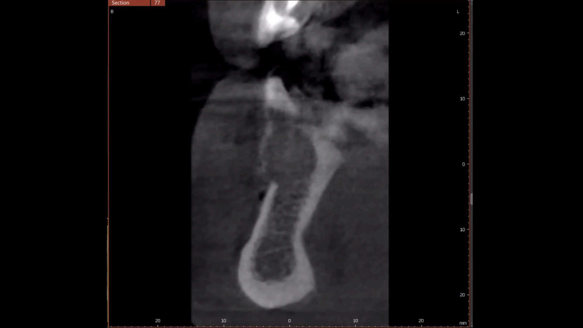
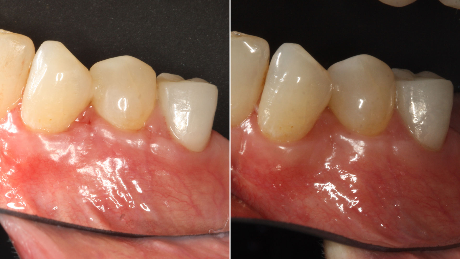
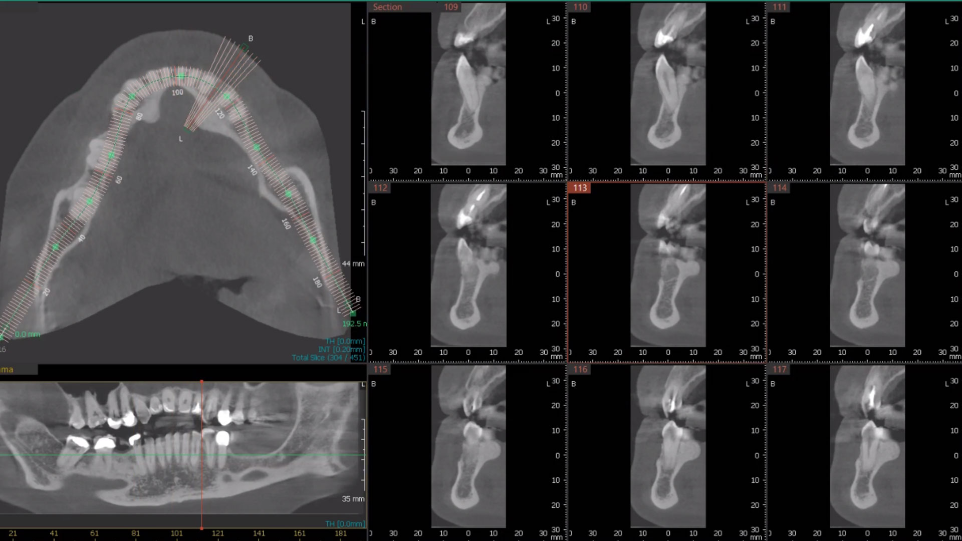
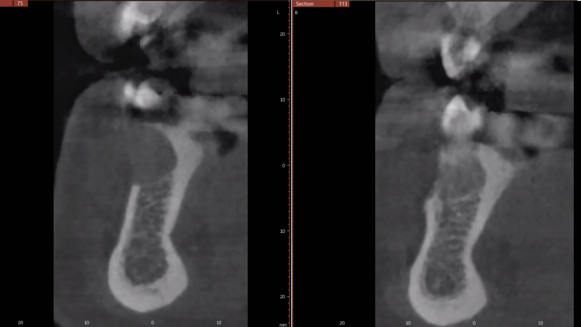
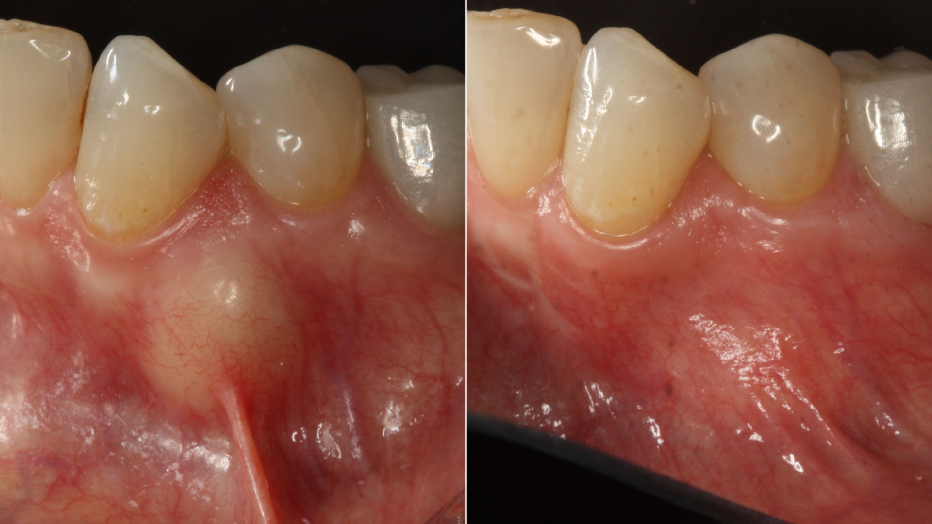
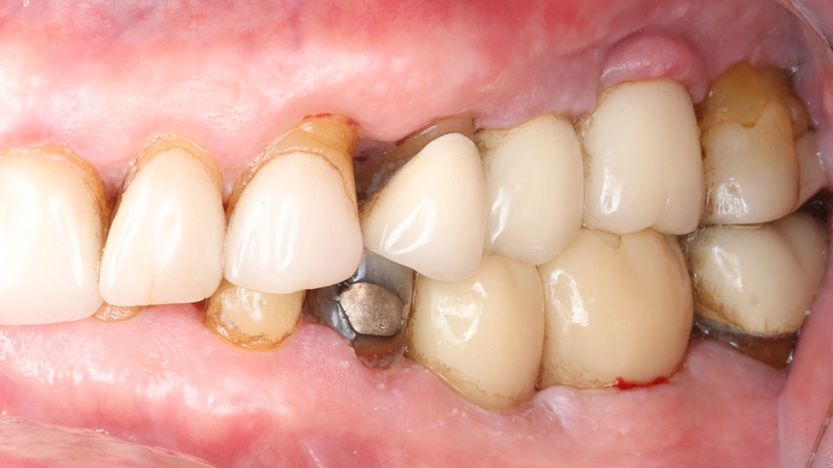
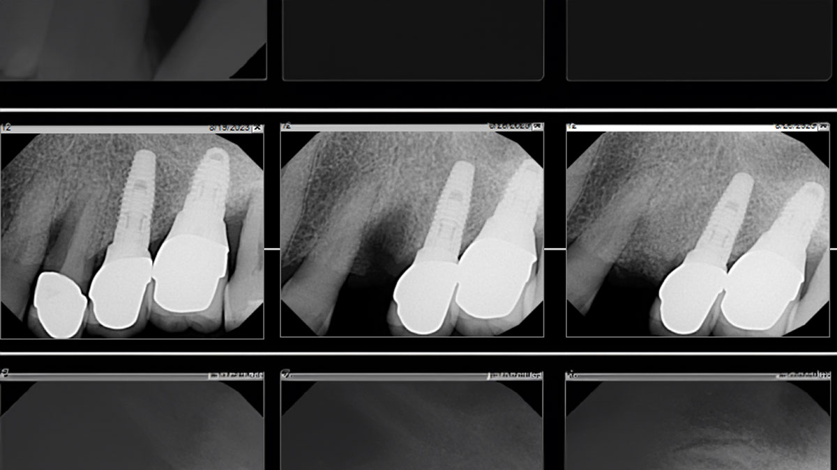
The patient called the office complaining of sensitivity and swelling in the maxillary left quadrant. He was seen and prescribed an antibiotic. Tooth #12 was deemed hopeless, and the peri-apical and radicular lesion presented on the radiograph extended significantly on the mesial aspect, impacting the interproximal bone level for tooth #11. Patient presents with implant supported restorations distal to the affected area and was concerned about the infection spreading to that area as well. The area was treated successfully, and the patient was pleased with the outcome, allowing him to preserve the tooth, on the mesial aspect of the lesion and the implant distally.

THE RISK PROFILE

Low RiskMedium RiskHigh Risk
Patient’s healthIntact immune system/Non-smoker Light smokerImpaired immune system 
Patient’s esthetic requirementsLowMediumHigh
Height of smile lineLowMediumHigh
Gingival biotypeThick – “low scalloped”Medium – “medium scalloped”Thin – “high scalloped”
Shape of dental crownsRectangularTriangular
Infection at implant sightNoneChronicAcute
Bone height at adjacent tooth site≤ 5 mm from contact point5.5 – 6.5 mm from contact point≥ 7 mm from contact point
Restorative status of adjacent toothIntactRestored
Width of tooth gap1 tooth (≥ 7 mm)1 tooth (≤ 7 mm)2 teeth or more
Soft-tissue anatomyIntactCompromised
Bone anatomy of the alveolar ridgeNo defectHorizontal defectVertical defect

THE APPROACH

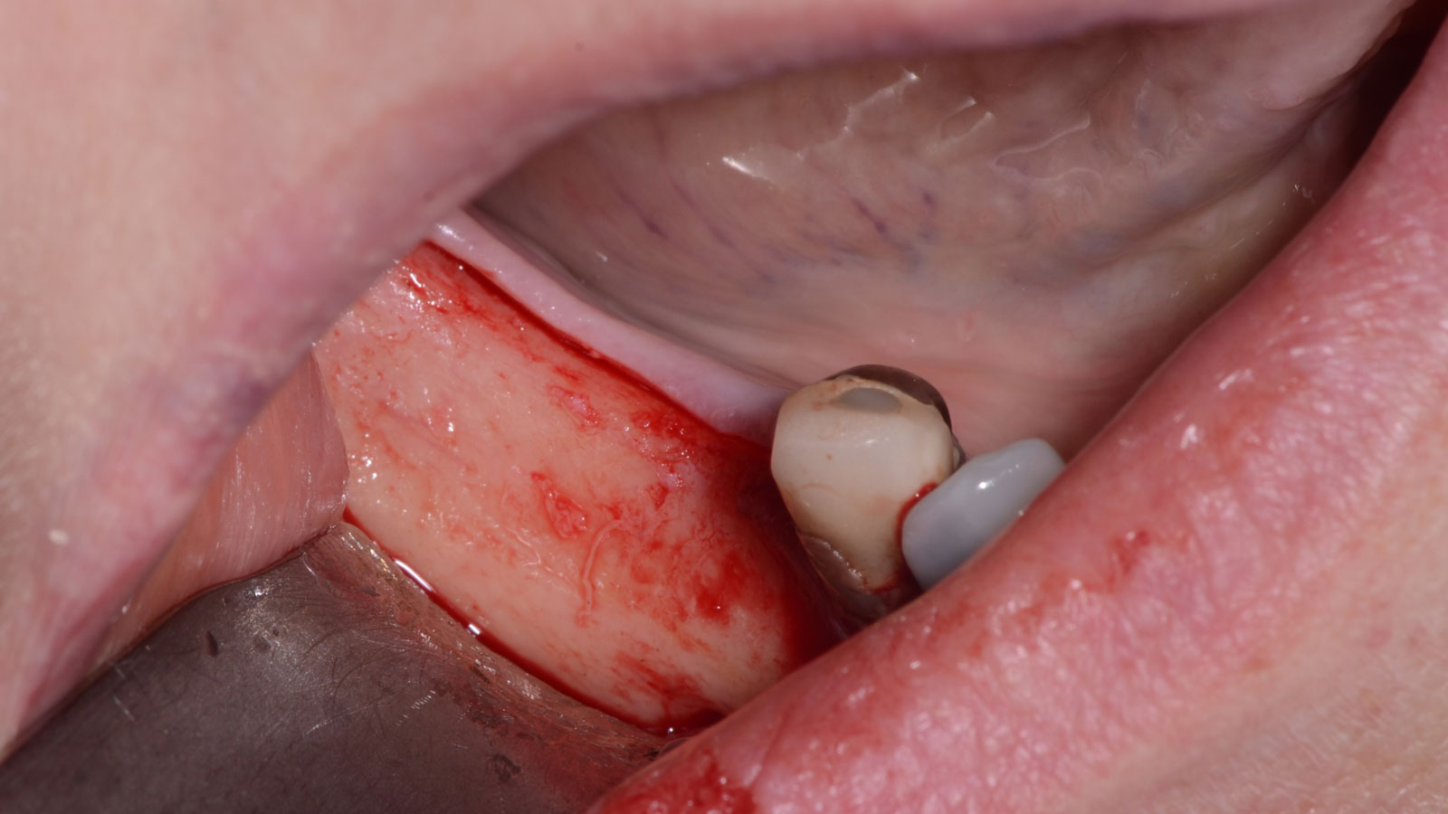
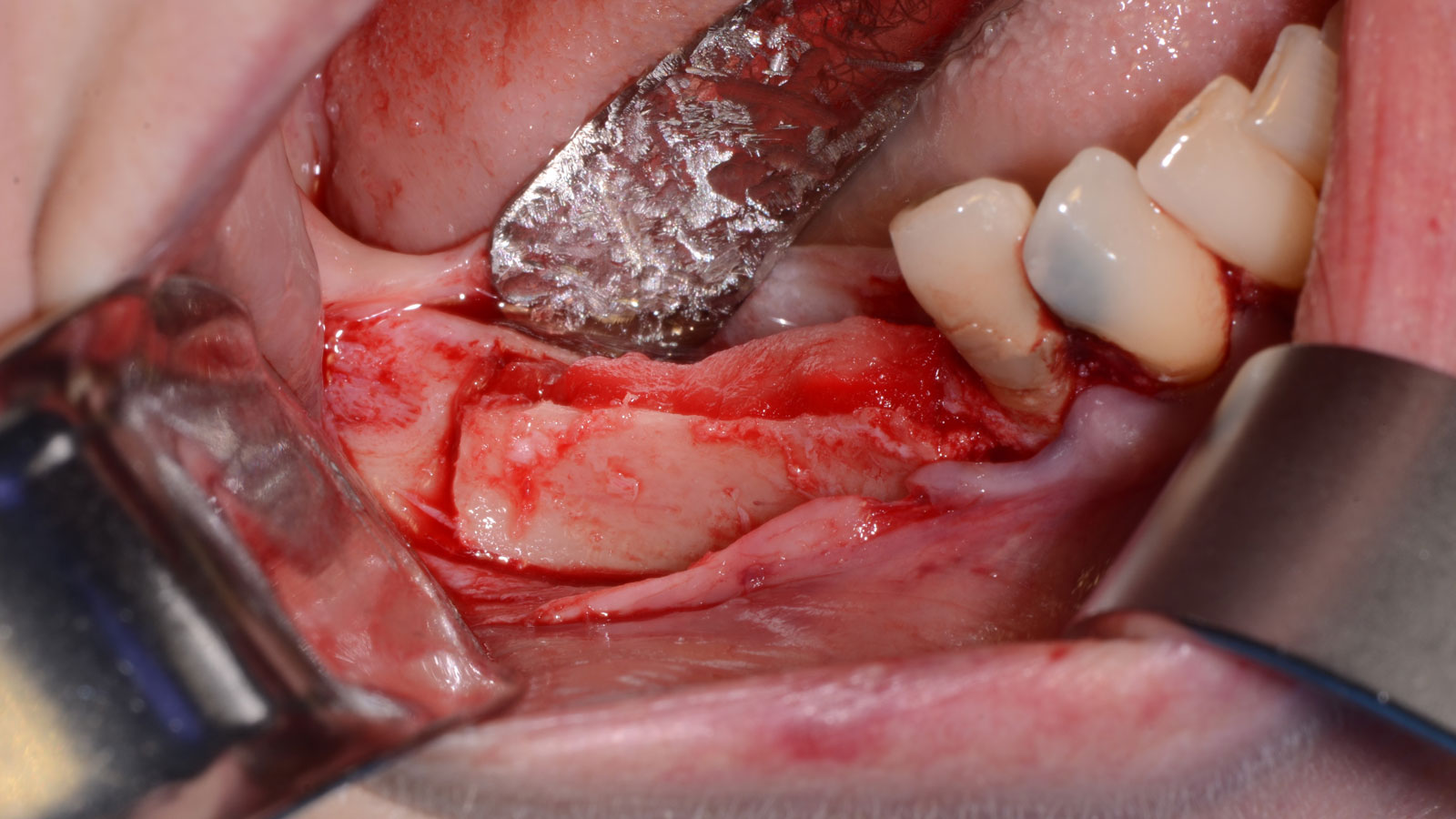
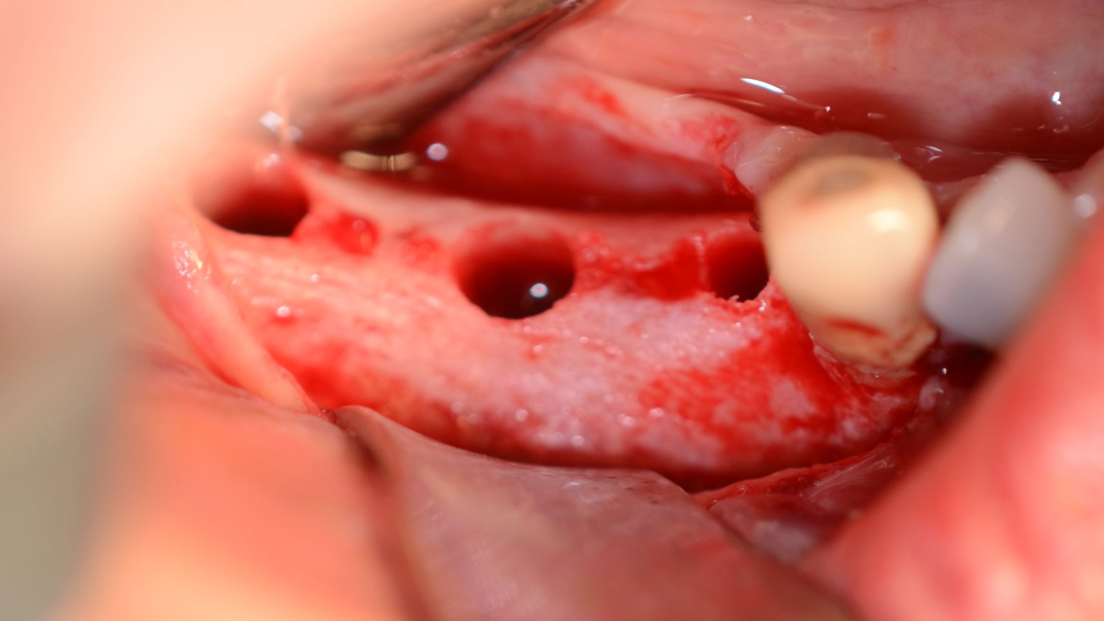
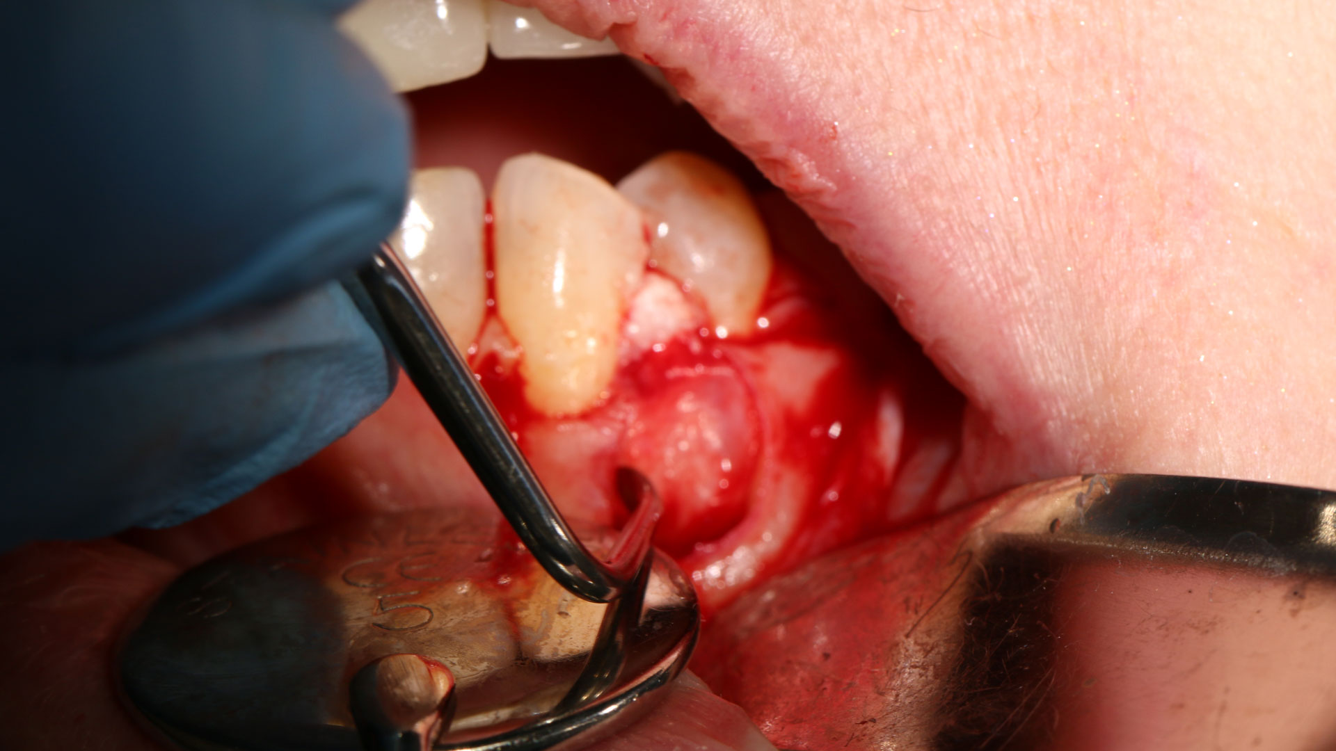
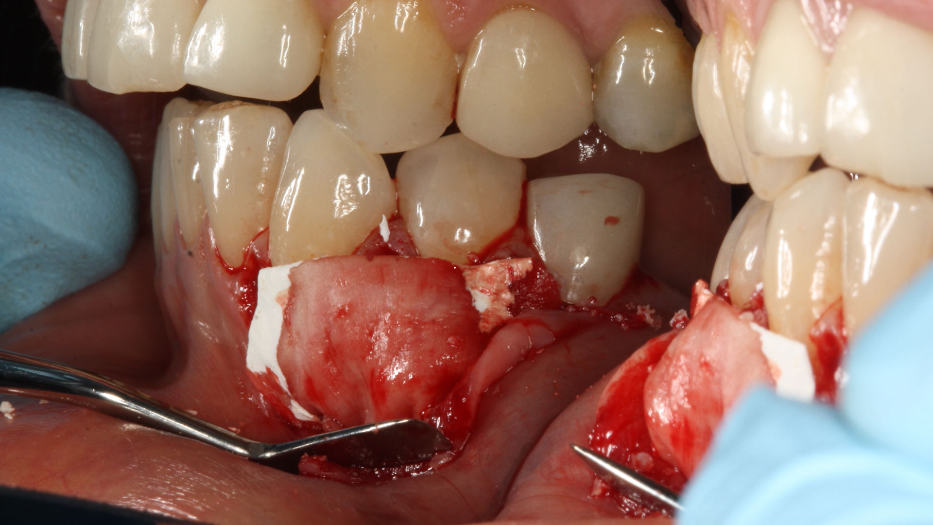
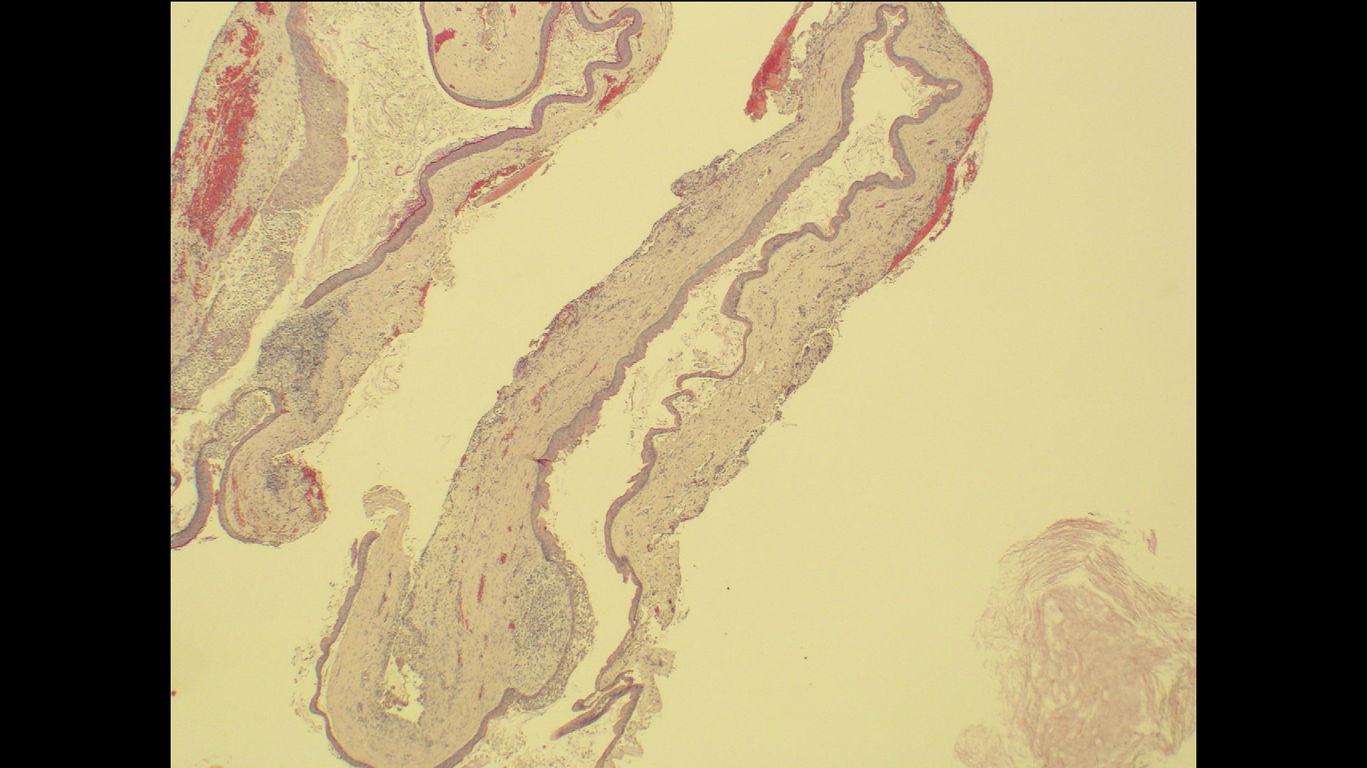
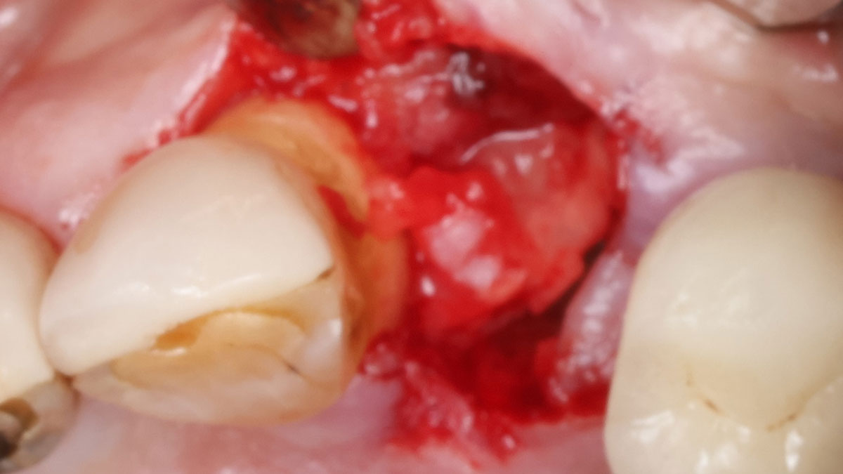
The goals of the procedure were to eliminate infection, the source of pain, and reduce periodontal problems to the adjacent tooth and implant. Full thickness flap was reflected, #12 was removed and the socket was debrided and irrigated. A peri-radicular lesion was removed and submitted for histopathological exam.

Initial presentation – buccal view.
Full thickness flap elevation exposing the complex clinical situation.
Alveolar socket after the tooth removal exposing the loss of bone on the distal of tooth #11, prior to the debridement of the granulation tissue and root preparation.
Adaptation on the buccal defect prior to placement of bone grafting with vallos® mineralized cortical cancellous mix granules (bottom) followed Geistlich Bio-Oss® (top).
Post adaptation with Geistlich Bio-Gide® for alveolar ridge preservation and guided tissue regeneration, followed by final suturing of the site using ePTFE material.
Radiographic overview of the clinical procedure: initial presentation with the bony defect impacting distal of #11 and #12 – mesial and inter-radicular, site after the tooth #12 was extracted, radiographic bone fill of the defect post-operative.
Post-operative healing of the site, 4 weeks after the procedure was completed.

“A localized infection can easily spread and impact adjacent teeth and implants. It is critical for clinicians to intervene as soon as possible to prevent further complications. Patient education and motivation is key to successfully treat these types of clinical situations encountered in a daily practice.”

— Dr. Irina Dragan

THE OUTCOME

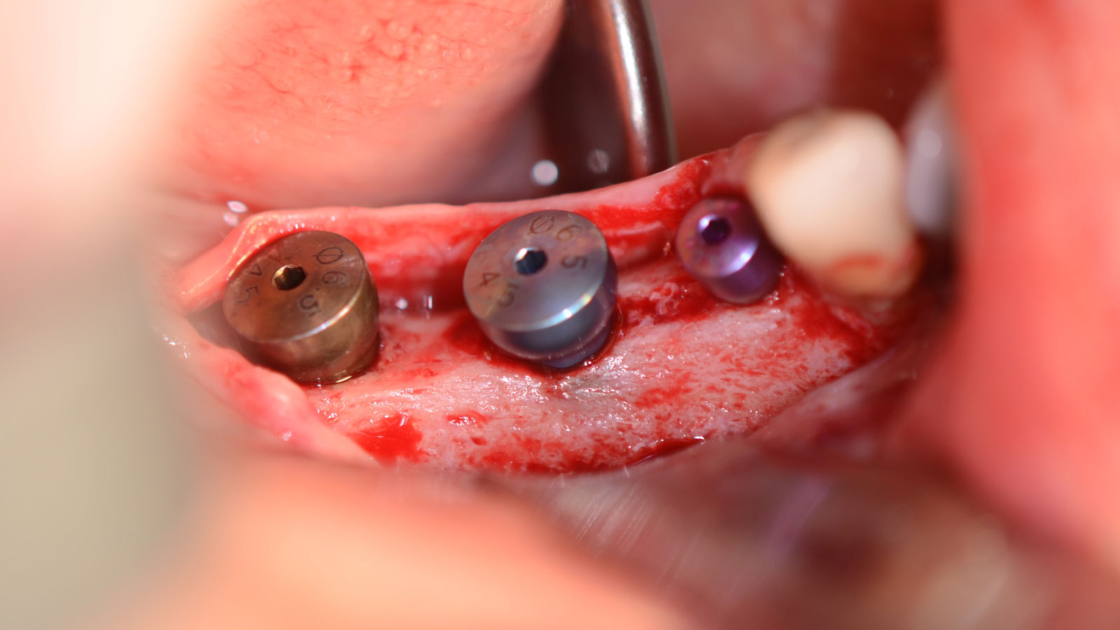
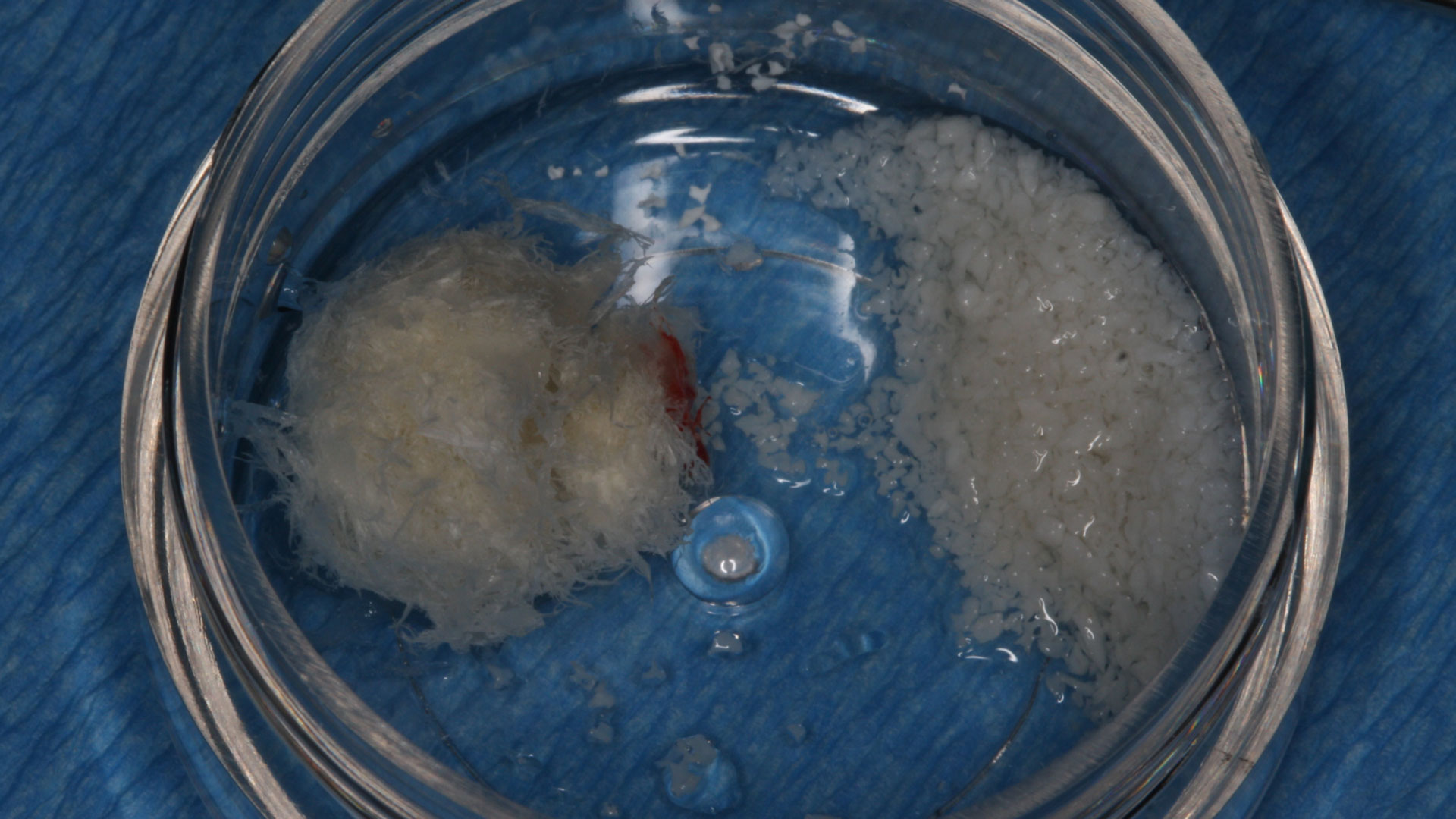
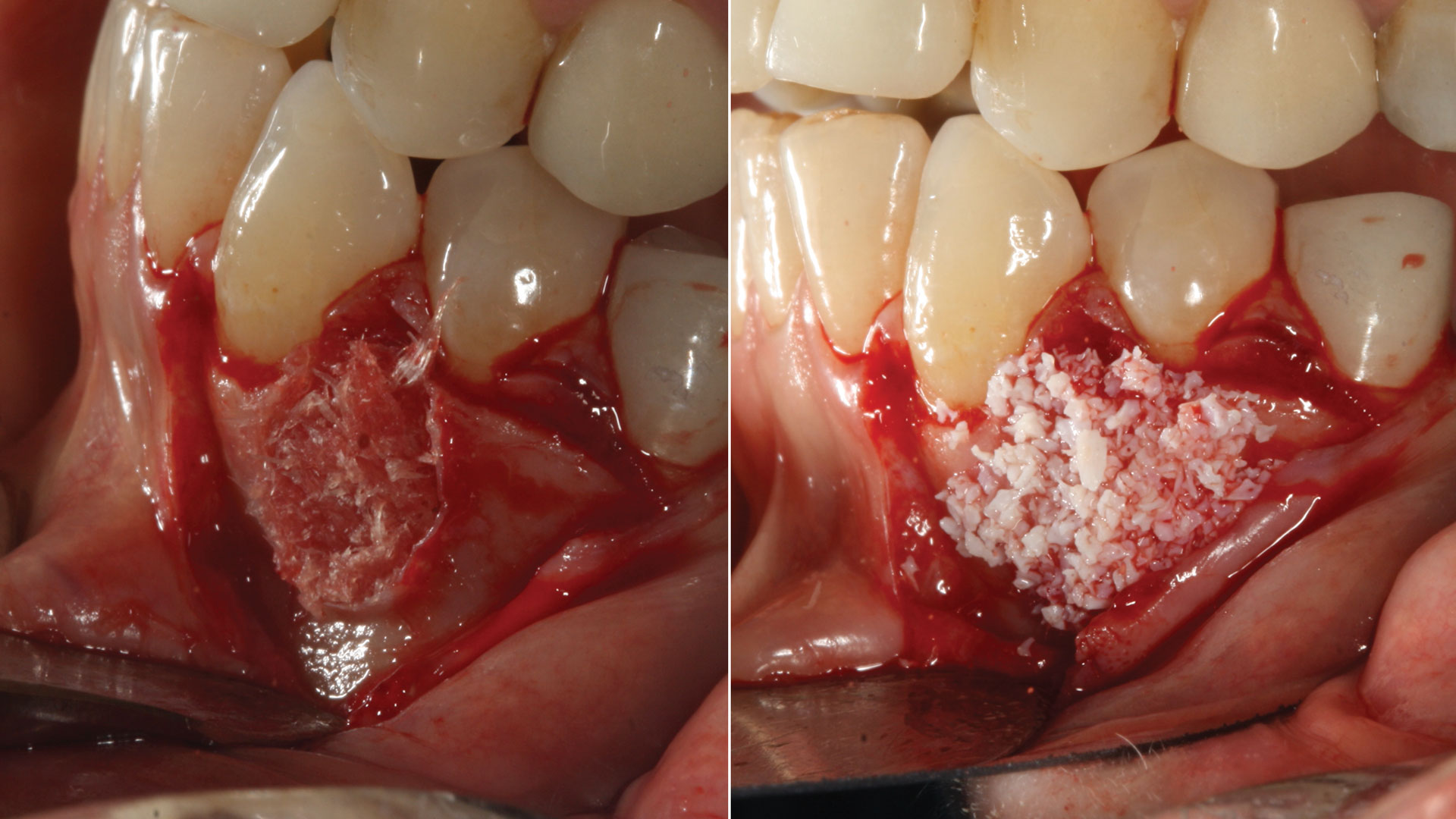
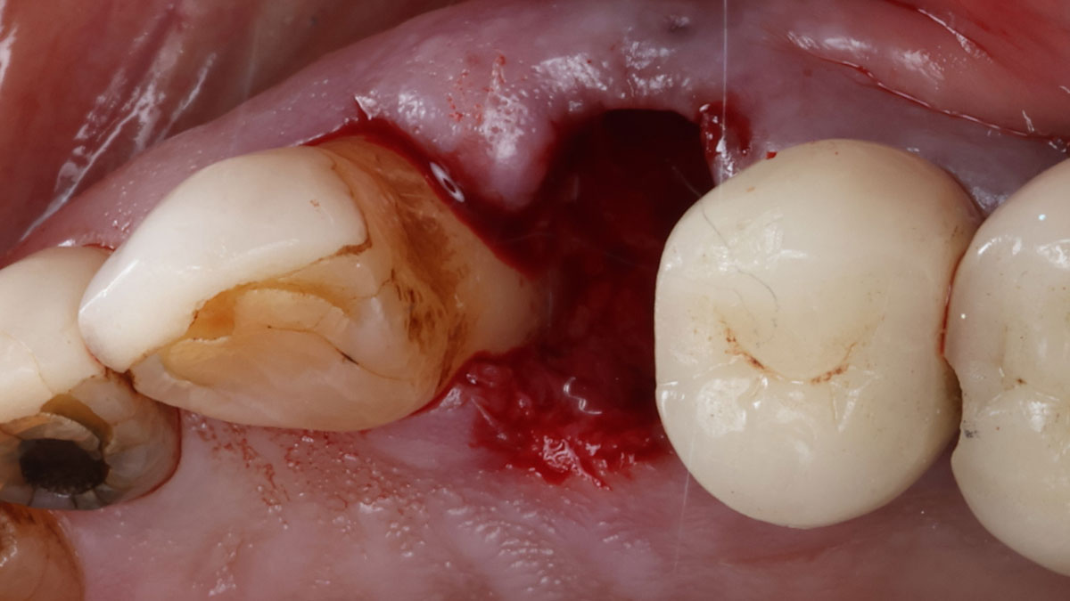
The combined defect: #11 distal guided tissue regeneration and #12 alveolar ridge preservation for #12. This area was treated with vallos®, Geistlich Bio-Oss Collagen®, and Geistlich Bio-Gide®. The xenograft was placed in the apical portion of the socket and the allograft towards the coronal surface.

Healing of the site at 4 weeks post-operative.

Considering today’s advancements in regeneration we are able to successfully treat complex clinical scenarios that involve combined therapeutic applications, such as guided tissue regeneration and alveolar ridge preservation.”

Dr. Irina Dragan

Periotomes were able to support with an atraumatic extraction of tooth #12 and maintaining as much as possible the soft and hard tissue present in this compromised area.”

Dr. Irina Dragan

Irina F. Dragan, DDS, DMD, MS, eMBA

Periodontology and Implant Dentistry
Dr. Irina Dragan is board certified and an examiner for the American Board of Periodontology and Implant Dentistry. She is part-time faculty in postgraduate periodontics at Harvard School of Dental Medicine and an adjunct associate professor of periodontology at Tufts University School of Dental Medicine. She is a periodontist and clinical researcher at The Perio Studio, a practice limited to periodontology and implant dentistry in Boston, MA.

BIOBRIEF

Mandibular Alveolar Ridge Split with Delayed Implant Placement

Gregory A. Santarelli, DDS

THE SITUATION

A healthy (ASA 1) non-smoker 63-year-old female presented to my office with Kennedy Class II partial edentulism in the mandibular right posterior quadrant for several years.  She denied removable options and wanted dental implants to individually replace her missing teeth.  The clinical and radiographic evaluation revealed atrophic mandibular bone height and width at site #’s 29, 30 & 31.  The edentulous site required engineering prior to the placement of conventional dental implants and prosthetics.

THE RISK PROFILE

Low RiskMedium RiskHigh Risk
Patient’s healthIntact immune system/Non-smoker Light smokerImpaired immune system 
Patient’s esthetic requirementsLowMediumHigh
Height of smile lineLowMediumHigh
Gingival biotypeThick – “low scalloped”Medium – “medium scalloped”Thin – “high scalloped”
Shape of dental crownsRectangularTriangular
Infection at implant sightNoneChronicAcute
Bone height at adjacent tooth site≤ 5 mm from contact point5.5 – 6.5 mm from contact point≥ 7 mm from contact point
Restorative status of adjacent toothIntactRestored
Width of tooth gap1 tooth (≥ 7 mm)1 tooth (≤ 7 mm)2 teeth or more
Soft-tissue anatomyIntactCompromised
Bone anatomy of the alveolar ridgeNo defectHorizontal defectVertical defect

THE APPROACH

The goal is to provide adequate soft and hard tissue at edentulous site #’s 29, 30 & 31 in order to place dental implants and restore a stable balanced occlusion.

Initial panoramic radiograph.
Atrophic edentulous alveolar ridge.
Alveolar ridge split with Geistlich Bio-Oss® graft in place (subsequently applied Geistlich Mucograft®).
6 months post grafting with Geistlich Bio-Oss® and Geistlich Mucograft®.
Implant osteotomies with adequate alveolar width.
Implants in tooth positions 29, 30 and 31.
Panoramic radiograph of implants.
Final restorations.

“The hard and soft tissue of the edentulous posterior mandible were inadequate to rehabilitate with dental implants.”

— Dr. Gregory Santarelli

THE OUTCOME

The patient summarized this challenging case very well – “I never imagined I would have fixed teeth again.” Geistlich Bio-Oss® and Geistlich Mucograft®  allowed for retention of the hard and soft tissue volume to achieve our final result and for maintenance of the final prosthesis.

Final restorations.

Careful patient selection, treatment planning and operative efficiency were used to provide a previously non-functional segment with fixed stable dental implant prosthetics and a balanced occlusion.”

Dr. Gregory Santarelli

Precise osteotomies along with the use of Geistlich Bio-Oss® and Geistlich Mucograft® provide adequate bone volume for dental implants.”

Dr. Gregory Santarelli

Gregory A. Santarelli, DDS

Dr. Santarelli earned his DDS degree in 1998 from the University School of Dentistry, Milwaukee, WI, after graduating with his B.S. in Biology from Arizona State University (Tempe, AZ). In 1999, he completed his General Practice Residency at the University of Iowa Hospital and Clinics, and went on to an Oral & Maxillofacial Surgery Internship at the Medical College of Virginia (Richmond, VA) as well as an Oral & Maxillofacial Surgery Residency Program, Christiana Care Health System (Wilmington, DE).

After completing his formal training in 2004, Dr. Santarelli’s work experience includes the Bankor Hospital for Children, Cambodia (2003), Adjunct Clinical Professor, University of Marquette, School of Dentistry, Department of Oral Sugery, Marquette, WI (2005), and Oral Surgery Associates of Milwaukee, Milwaukee, WI (2004-2005). He now maintains a private practice in Kenosha, WI with his partner Dr. Deno Tiboris.

Dr. Santarelli performs numerous hard/soft tissue regeneration surgeries in preparation for dental implants and is actively involved in clinical research with The McGuire Institute (iMc).

BIOBRIEF

Odontogenic Keratocyst Management

Bassam Kinaia, DDS, MS, DICOI

THE SITUATION

A 60-year-old-heathy Caucasian female presented with the chief complaint: “I noticed a bump on my lower left teeth since last year.” An examination revealed a stable periodontium except for enlarged gingival tissue between #21-22 measuring 10x8x5mm, well-defined borders, depressible, non-painful, and vital teeth without displacement. The treatment plan included flap surgery, excisional biopsy, GTR #21-22 (Diff Dx: Lateral periodontal cyst (LPC), Odontogenic Keratocyst (OKC), Benign Fibro-Osseous lesion (BFOL).

Guided Tissue Regeneration (GTR) using Geistlich Bio-Oss® and vallos®f was performed and covered with a resorbable collagen membrane (Geistlich Bio-Gide®).

Primary closure was completed using non-resorbable sutures. Follow-up at 2, 4 weeks, 3, 6 months showed stable periodontium without re-occurrence. The pathology report indicated OKC and the area is monitored annually.

THE RISK PROFILE

Low RiskMedium RiskHigh Risk
Patient’s healthIntact immune system Light smokerImpaired immune system 
Patient’s esthetic requirementsLowMediumHigh
Height of smile lineLowMediumHigh
Gingival biotypeThick – “low scalloped”Medium – “medium scalloped”Thin – “high scalloped”
Shape of dental crownsRectangularTriangular
Infection at implant sightNoneChronicAcute
Bone height at adjacent tooth site≤ 5 mm from contact point5.5 – 6.5 mm from contact point≥ 7 mm from contact point
Restorative status of adjacent toothIntactRestored
Width of tooth gap1 tooth (≥ 7 mm)1 tooth (≤ 7 mm)2 teeth or more
Soft-tissue anatomyIntactCompromised
Bone anatomy of the alveolar ridgeNo defectHorizontal defectVertical defect
watch video download pdf

THE APPROACH

The treatment goal was to excise the lesion around #21-22 and stabilize the periodontium. Sulcular incisions #20-22 with vertical incision #22 MF were performed. Upon full thickness flap reflection, the lesion was removed (excisional biopsy). The defect extended #21M-#22D with complete facial bone loss. It was a wide 1-2 bony wall defect measuring 10x8x5mm. GTR procedure using Geistlich Bio-Oss® and vallos®f and Geistlich Bio-Gide® for the collagen membrane were employed. Primary closure was obtained using 6-0 prolene suture.

Initial clinical and radiographic presentation shows buccal soft tissue enlargement and bone loss #21-22 area.
Clinical facial view showing full thickness flap reflection with complete enucleation of cystic lesion (excisional biopsy).
Clinical view showing hydration of vallos®f and Geistlich Bio-Oss® as two separate grafts.
Clinical facial view showing placement of vallos®f internally for maximum osteogenic/osteoinductive potential and Geistlich Bio-Oss® externally for space maintenance.
Clinical facial view showing placement of Geistlich Bio-Gide® covering the defect and extending one tooth mesillay and distally.
Clinical facial view showing primary closure using 6-0 prolene sutures.
CBCT immediately post-surgery showing radiolucent allograft internally for osseoinduction and radiopaque xenograft externally for space maintenance.
After flap elevation at 4 months showing, the new buccal bone plate together with a completely filled alveolus.
Clinical facial views showing healing at 2 and 4 weeks with proper soft tissue healing.
6 months post-surgery radiographic presentation showing stable periodontium and proper bone fill #21-22 area.
Comparison of pre- and post-surgical CBCT views showing good bone formation.
Comparison of pre- and post-surgical clinical views showing stable periodontium.

“Excisional biopsy and guided tissue regeneration is indicated to treat the pathology (#21-22 area) and stabilize the periodontium.”

— Dr. Bassam Kinaia

THE OUTCOME

Complete excision of pathology and biopsy followed by GTR using vallos®f internally for maximum osteogenic/osteoinductive potential and Geistlich BioOss® externally for space maintenance showed excellent radiographic bone fill and stable periodontium.

Six-month post-surgical clinical view shows stable periodontium.

Guided tissue regeneration using vallos®f bone graft (allograft as an internal first layer), Geistlich Bio-Oss® (as an outside second layer), and collagen membrane showed predictable periodontal regeneration.

Dr. Bassam Kinaia

Bassam Kinaia, DDS, MS, DICOI

Dr. Kinaia is the Associate Director of the Graduate Periodontology Program at the University of Detroit Mercy (UDM). He is also the former Director of the Periodontology Program at UDM in Michigan and Boston University Institute for Dental Research and Education in Dubai. He is a Diplomate of the American Academy of Periodontology (AAP) and International Congress of Oral Implantology (ICOI). He received a certificate of Excellence from the AAP in recognition of teaching-research fellowship.