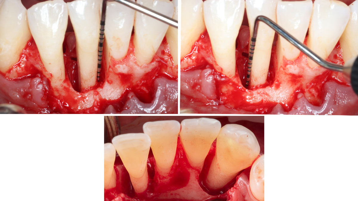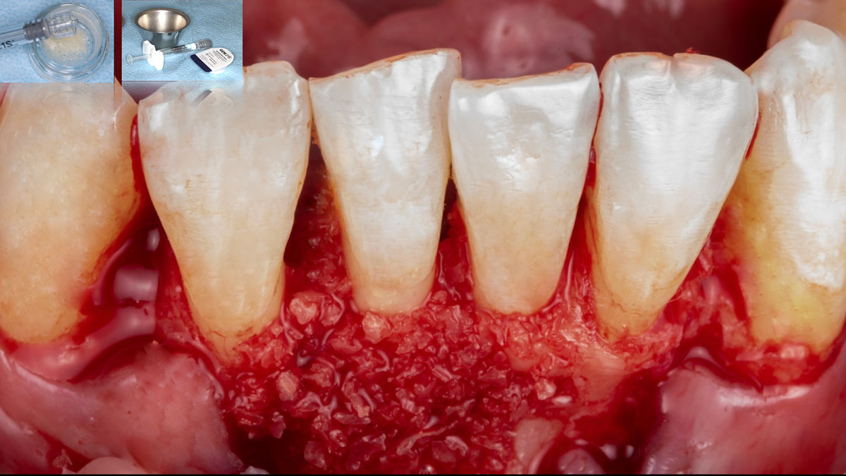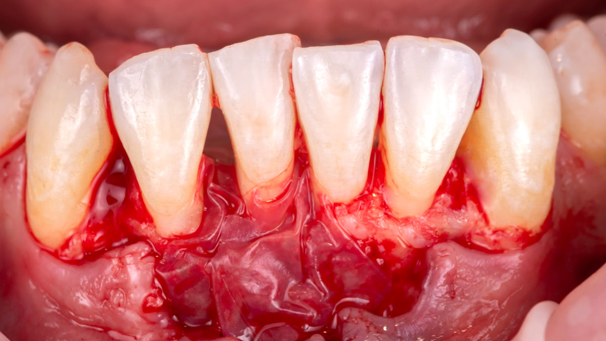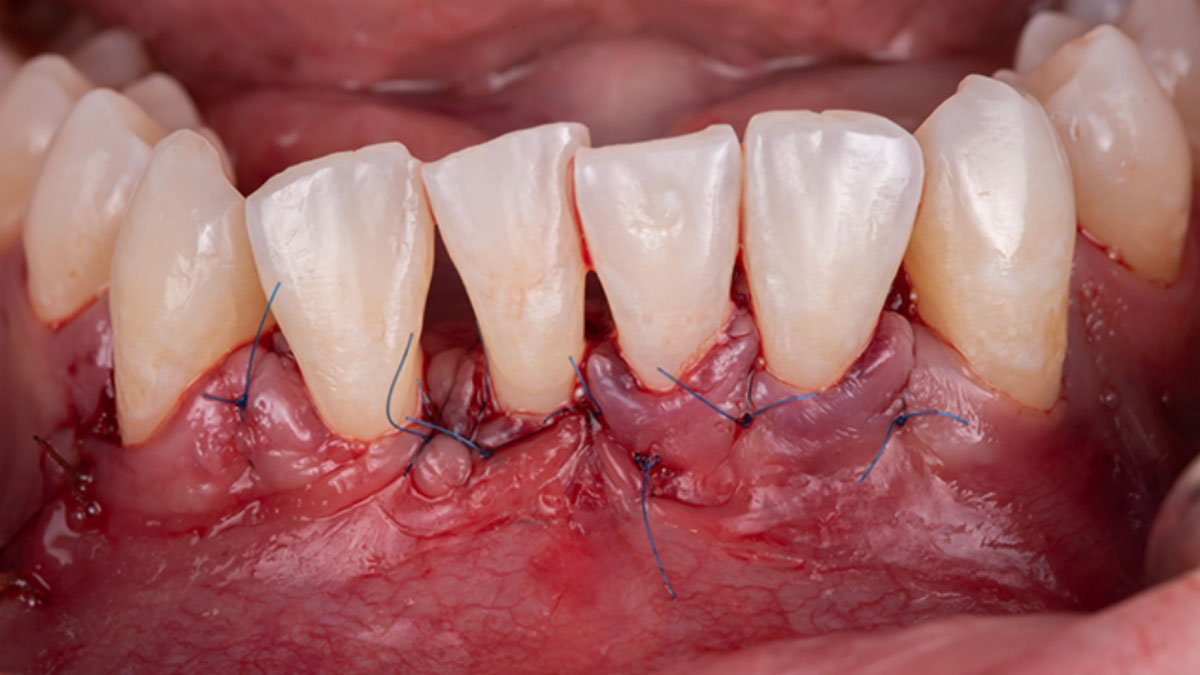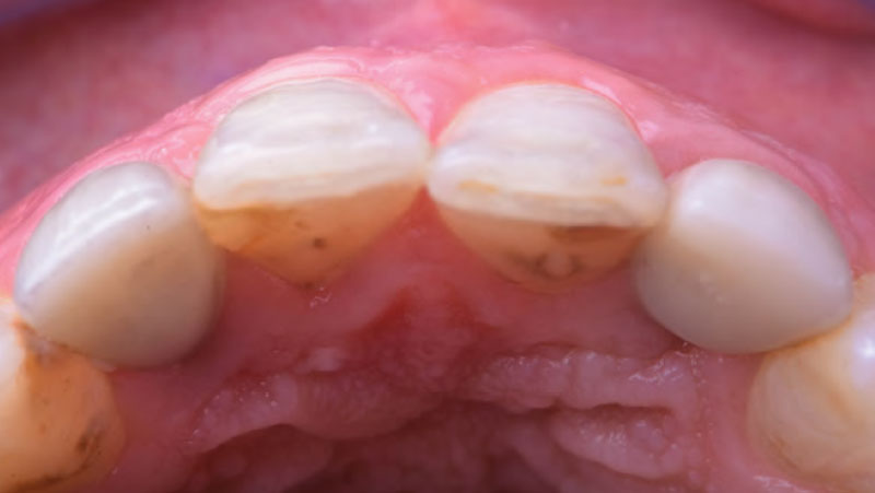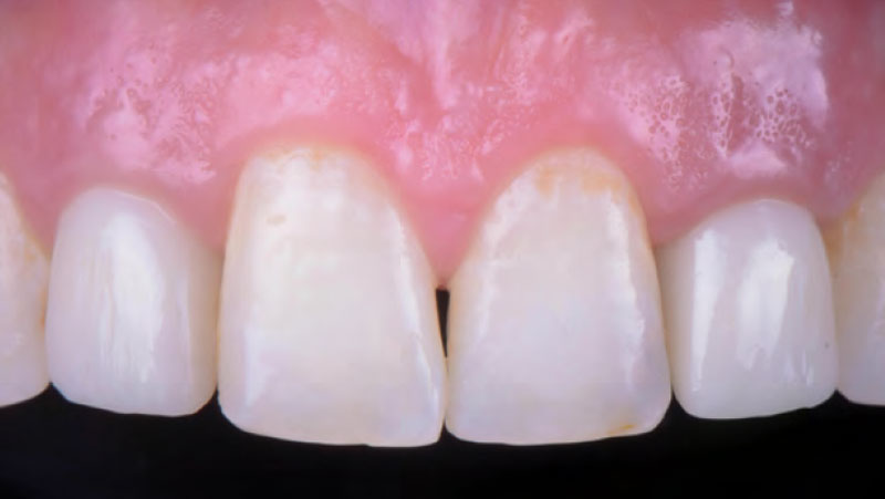


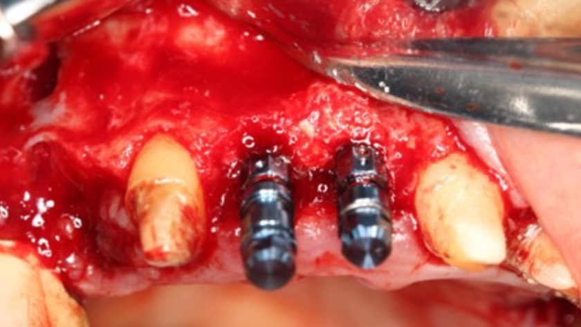
CLINICAL CASE

THE SITUATION

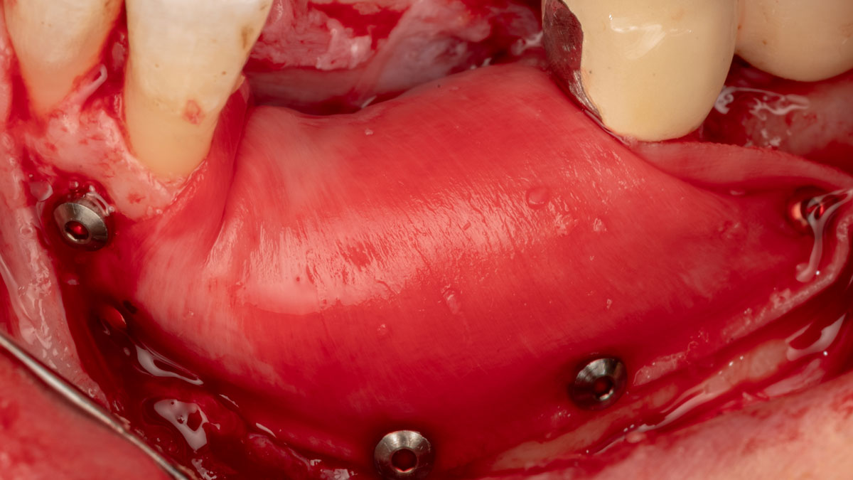
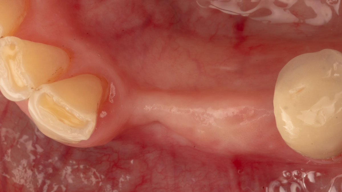
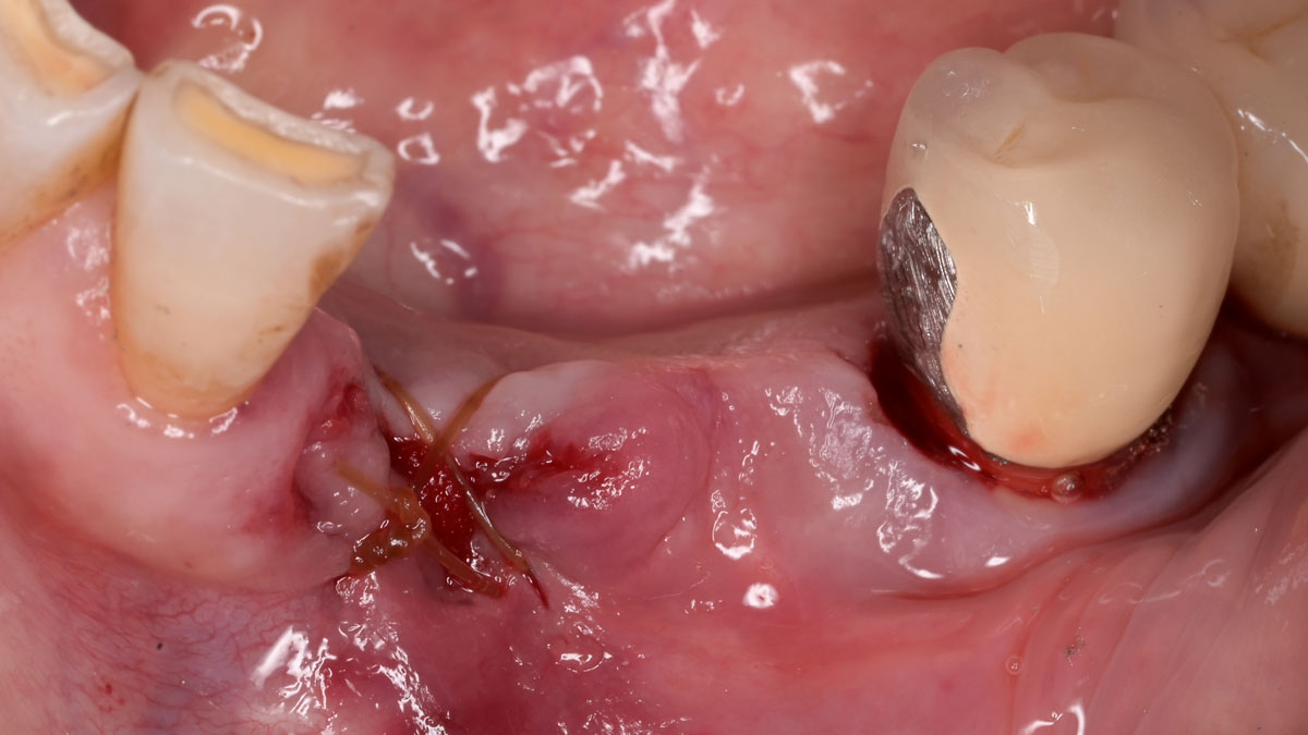
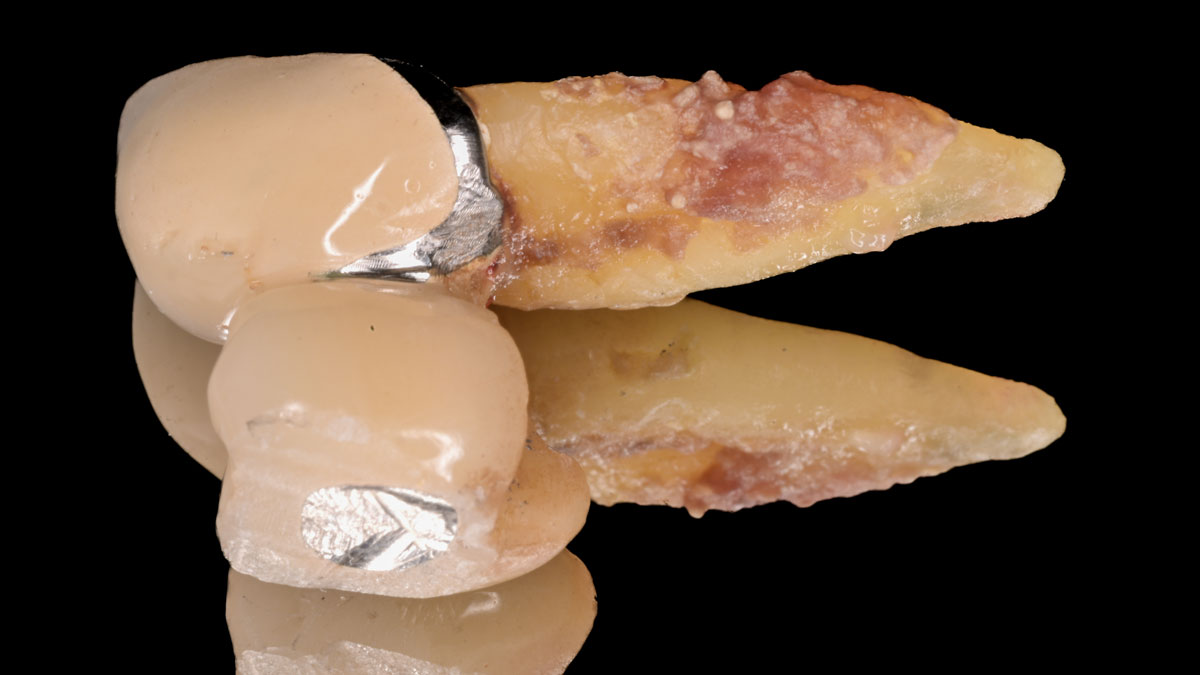
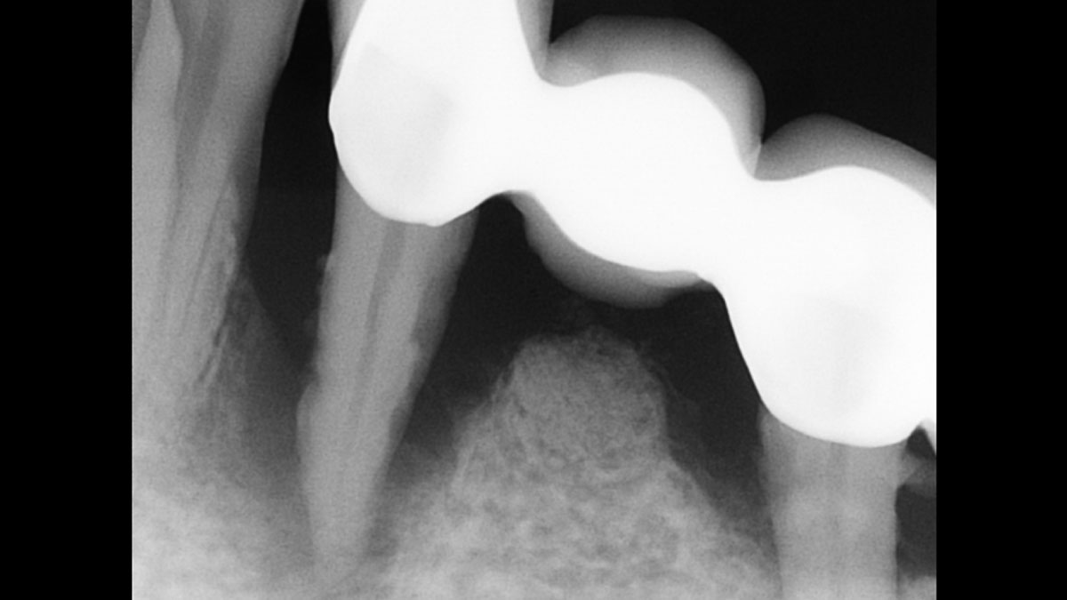
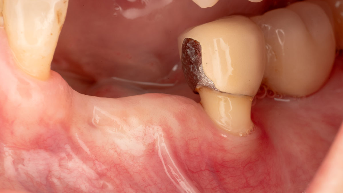
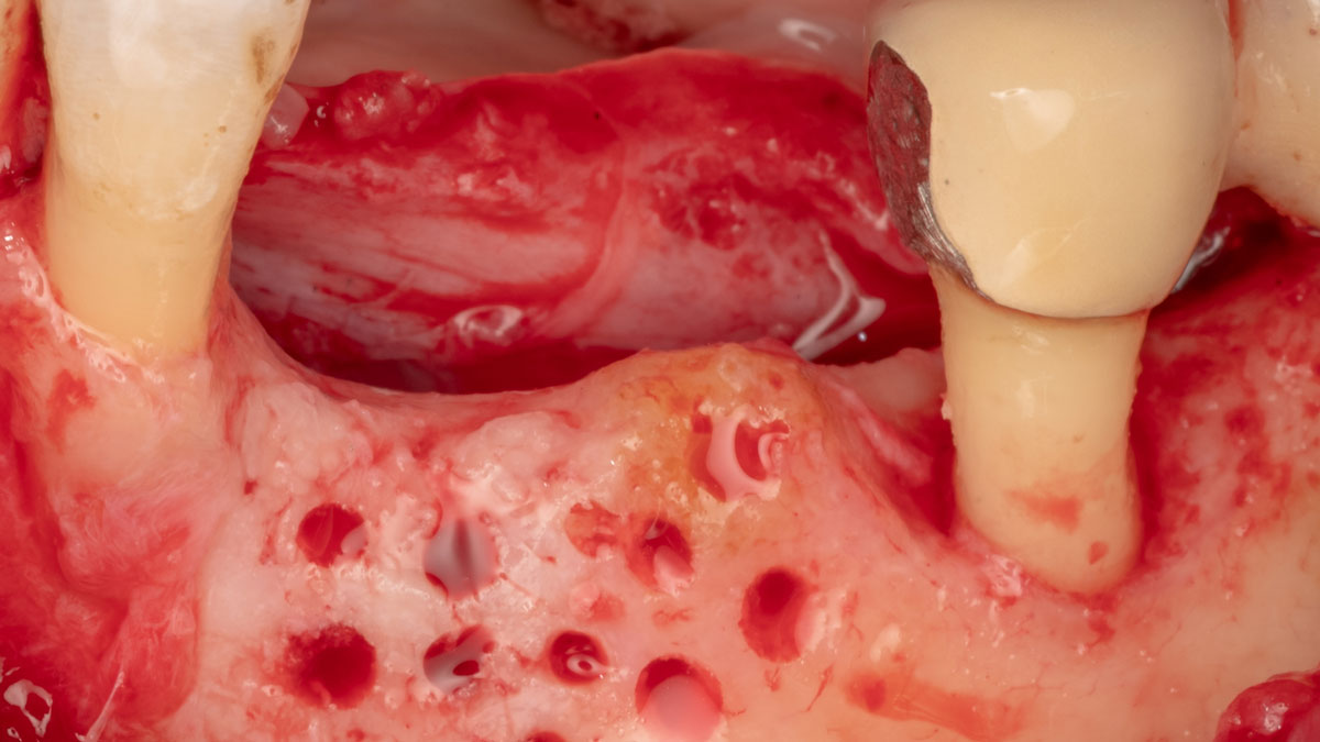
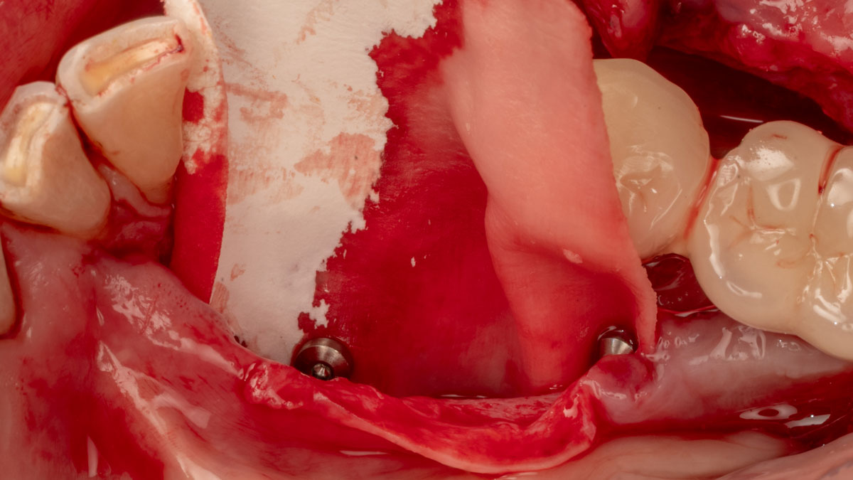
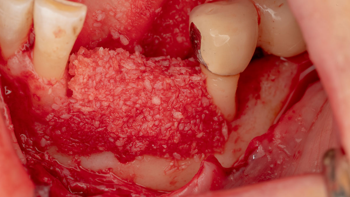
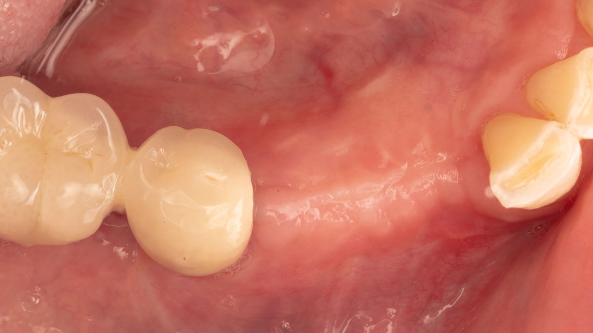
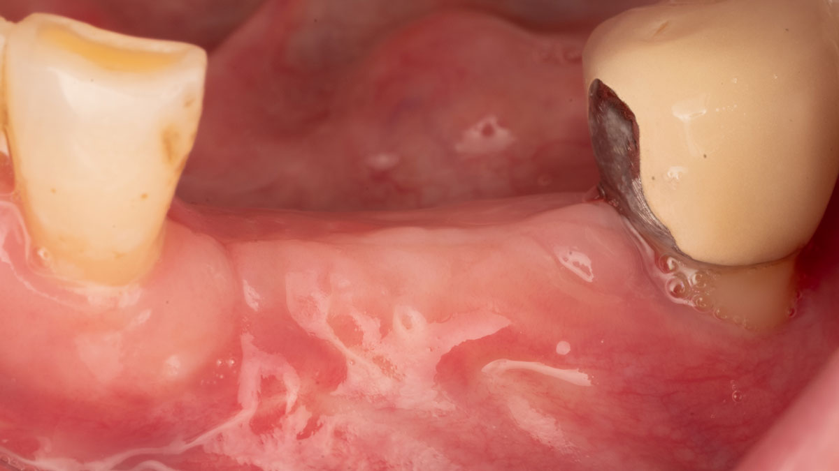
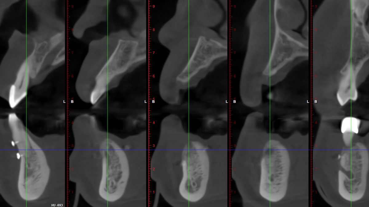
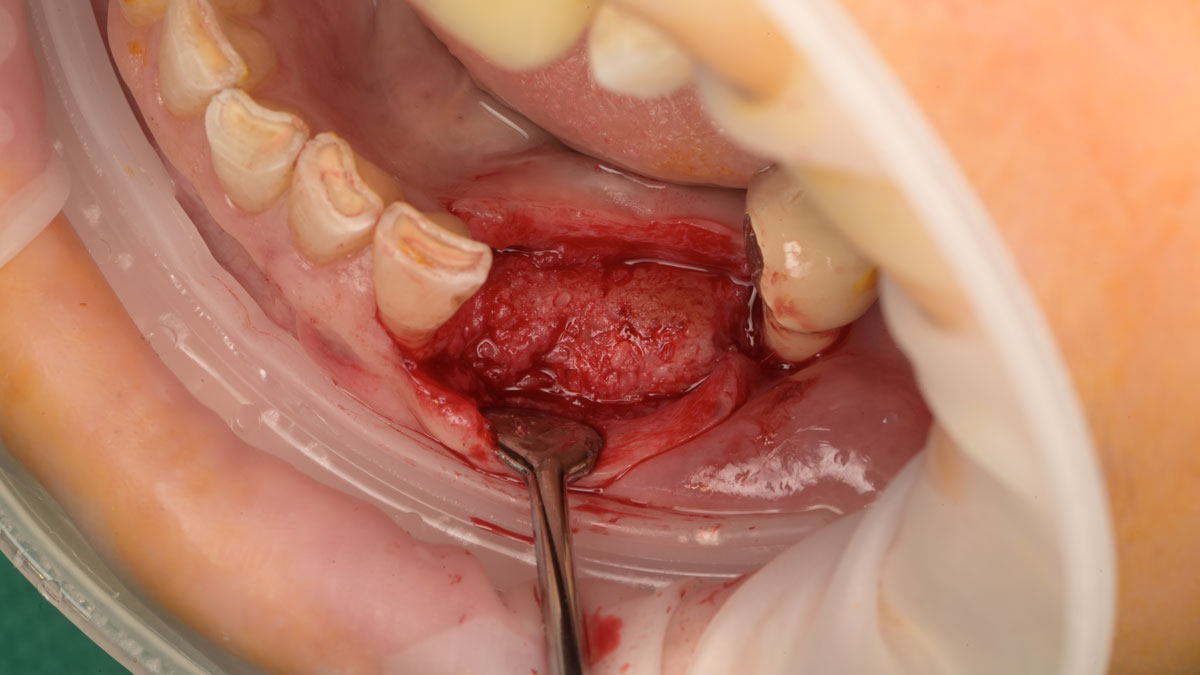
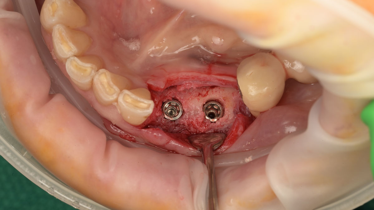
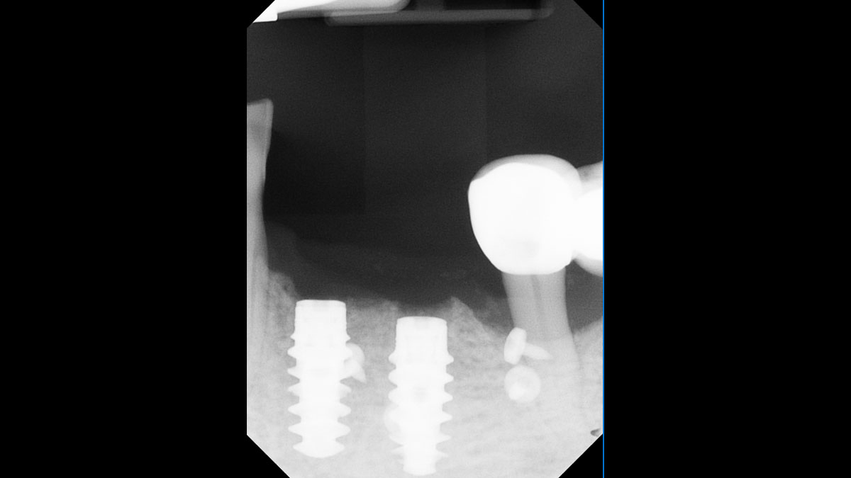
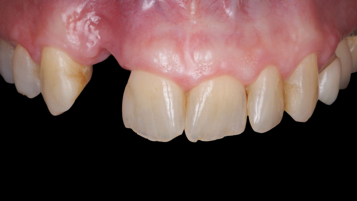
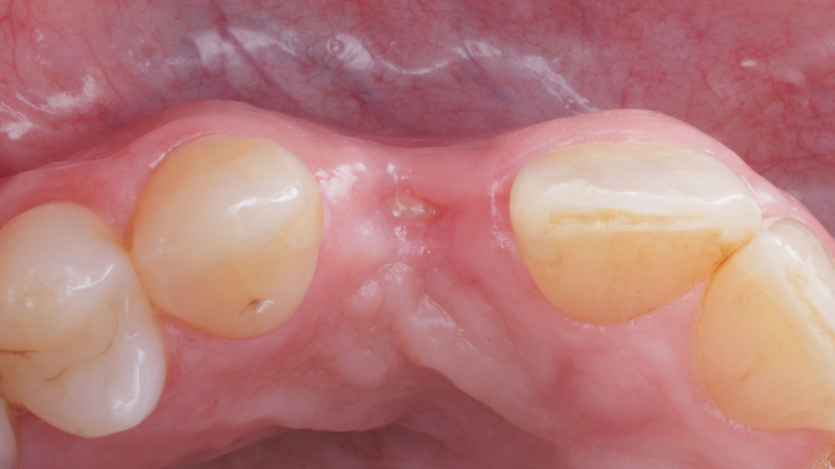
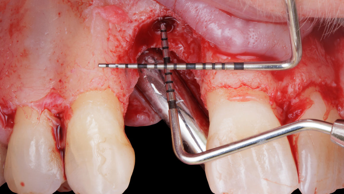
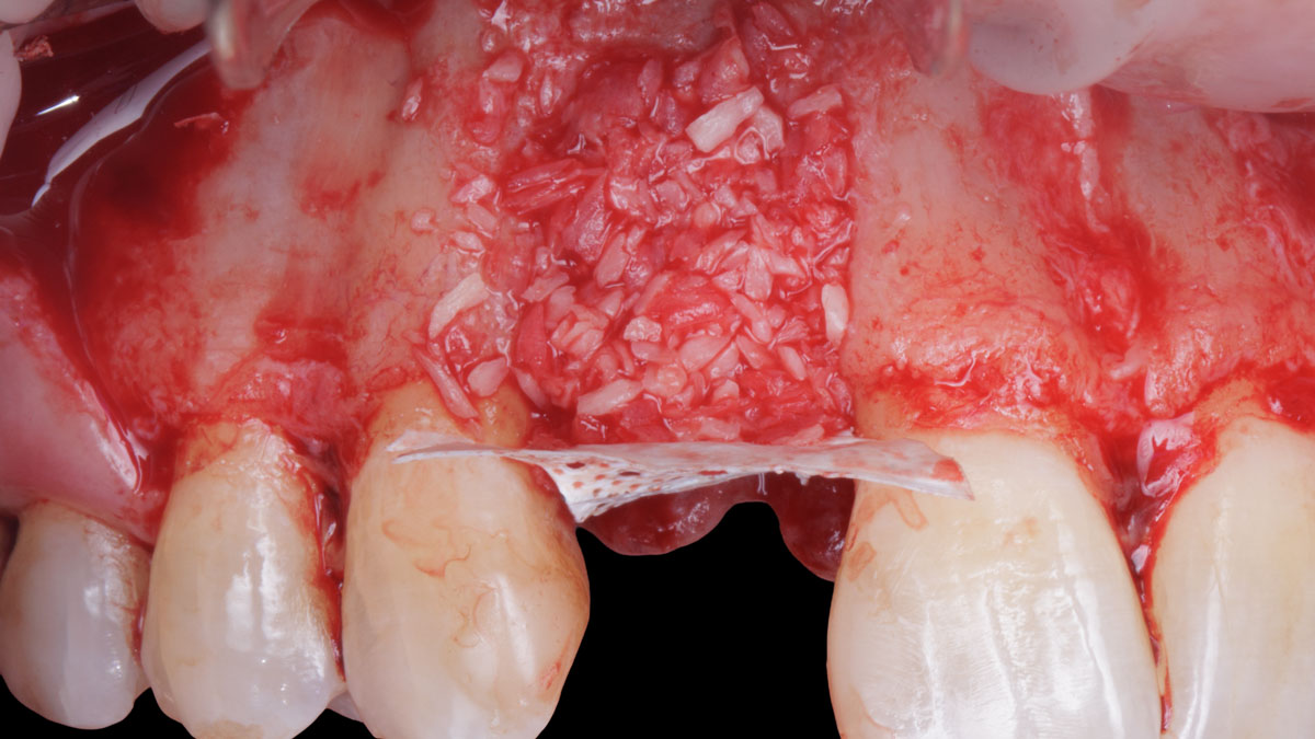
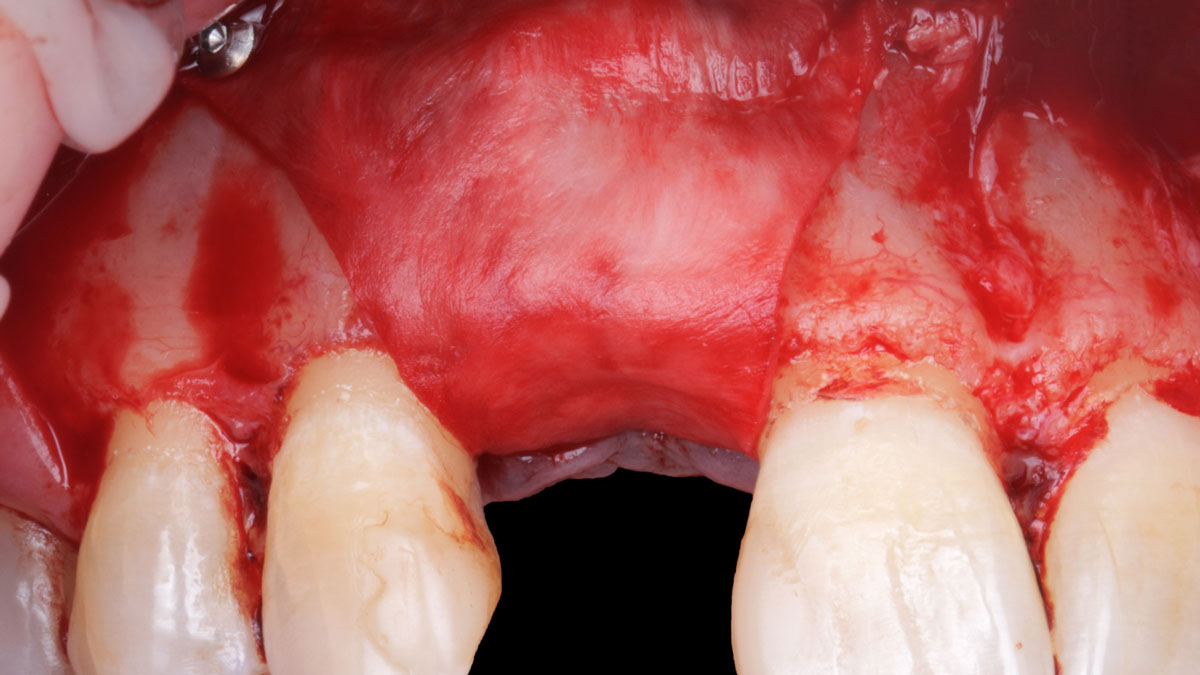
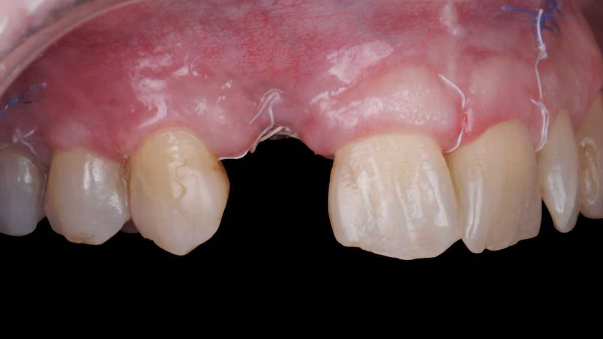
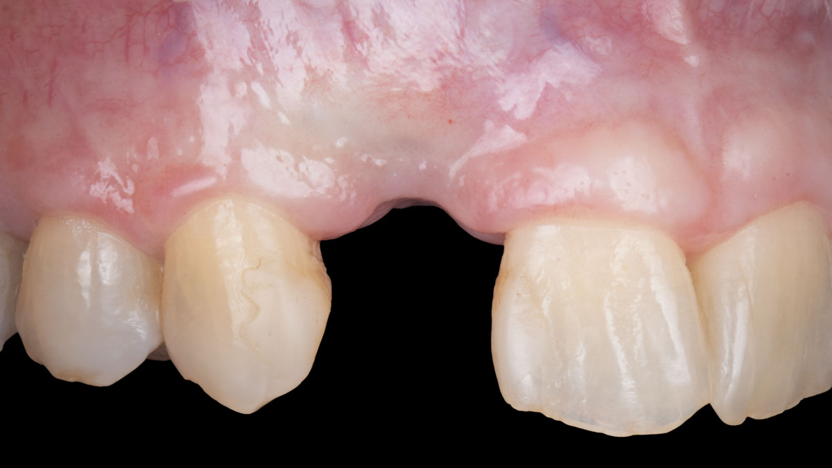
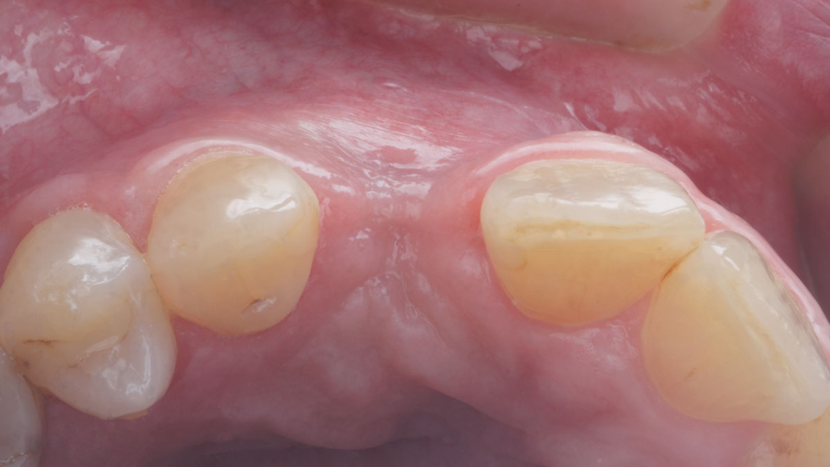
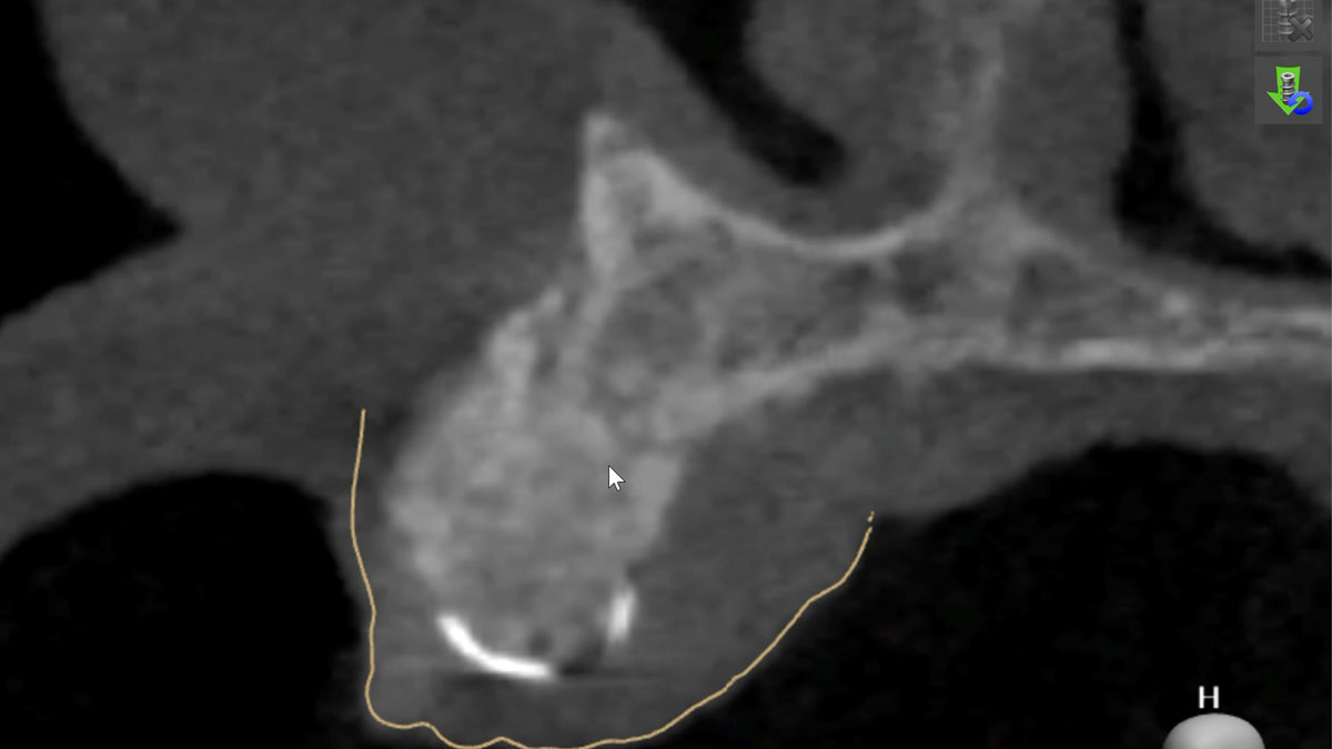
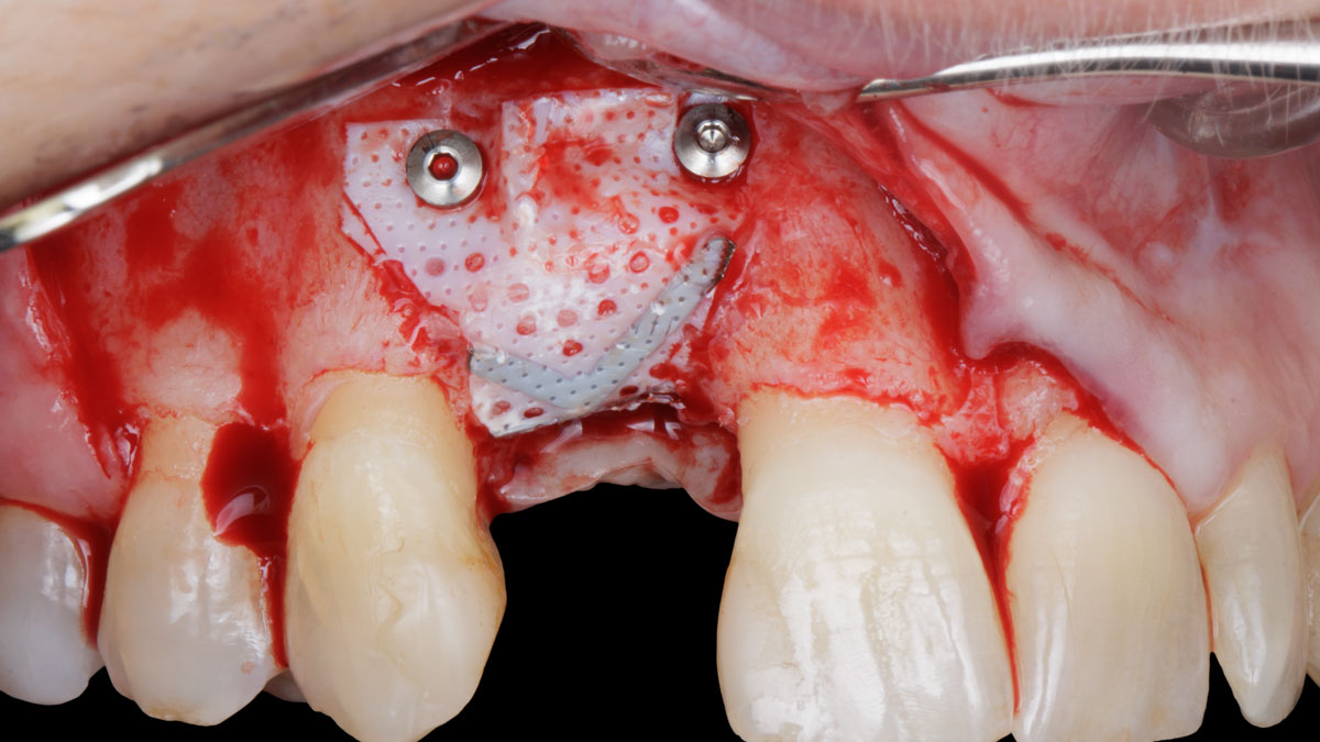
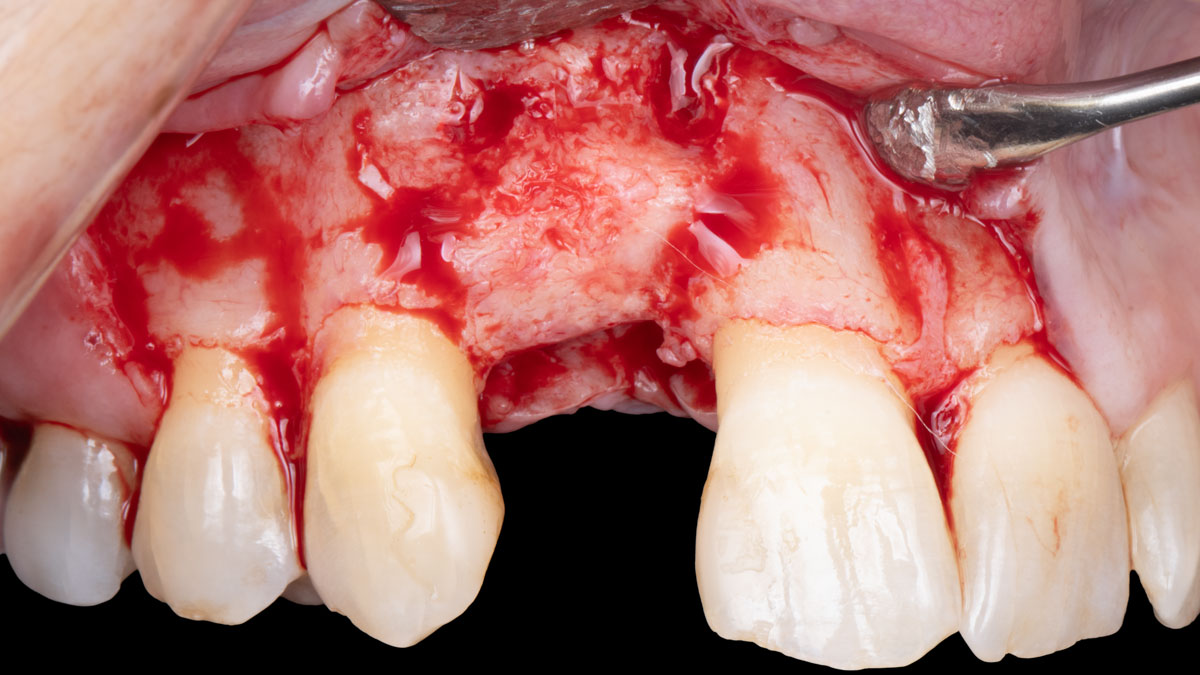
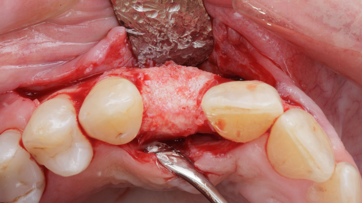
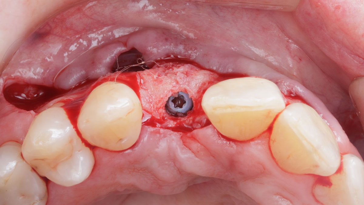
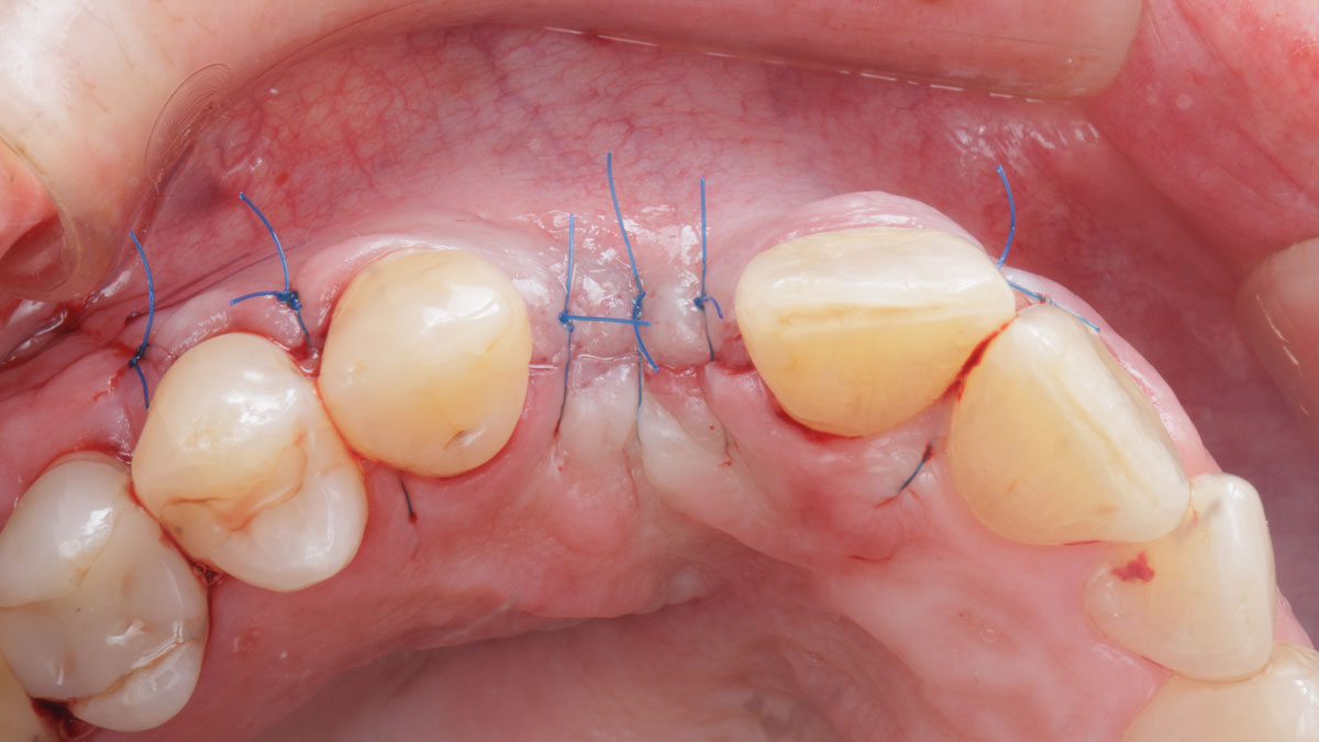
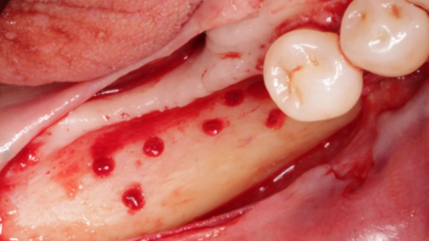
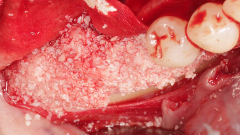
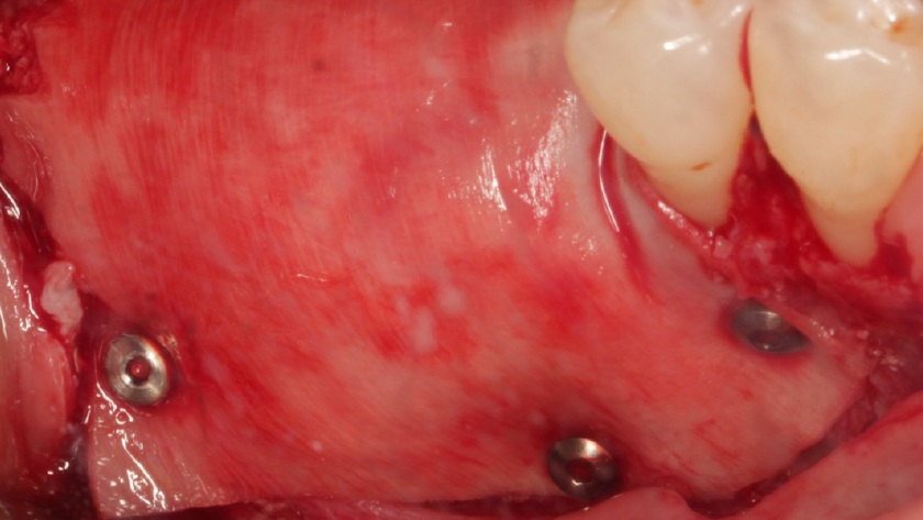
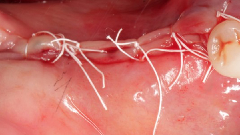
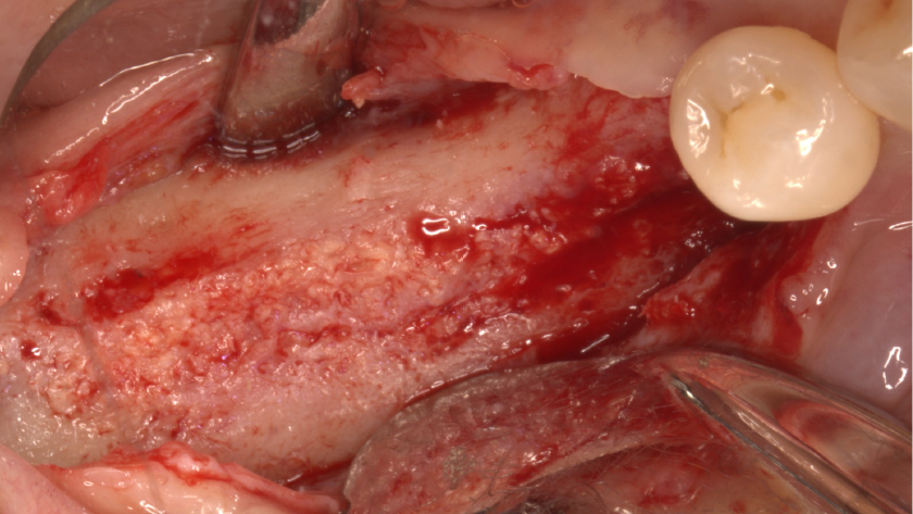
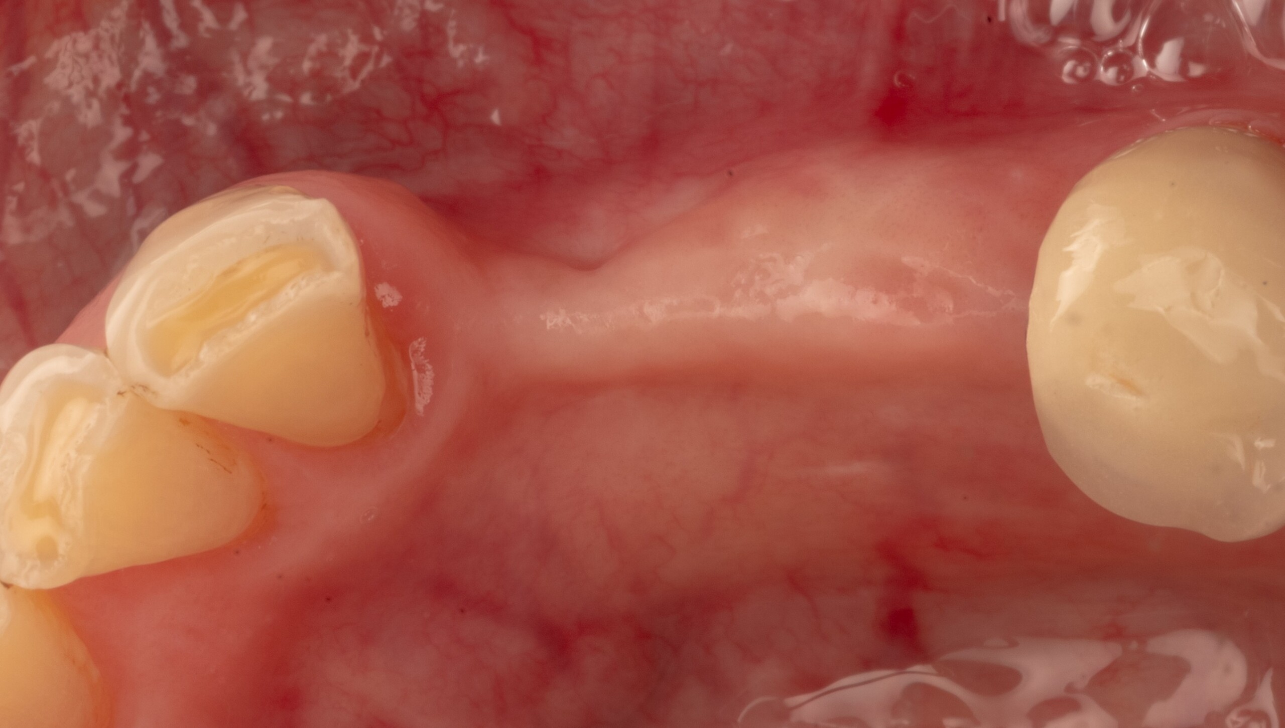
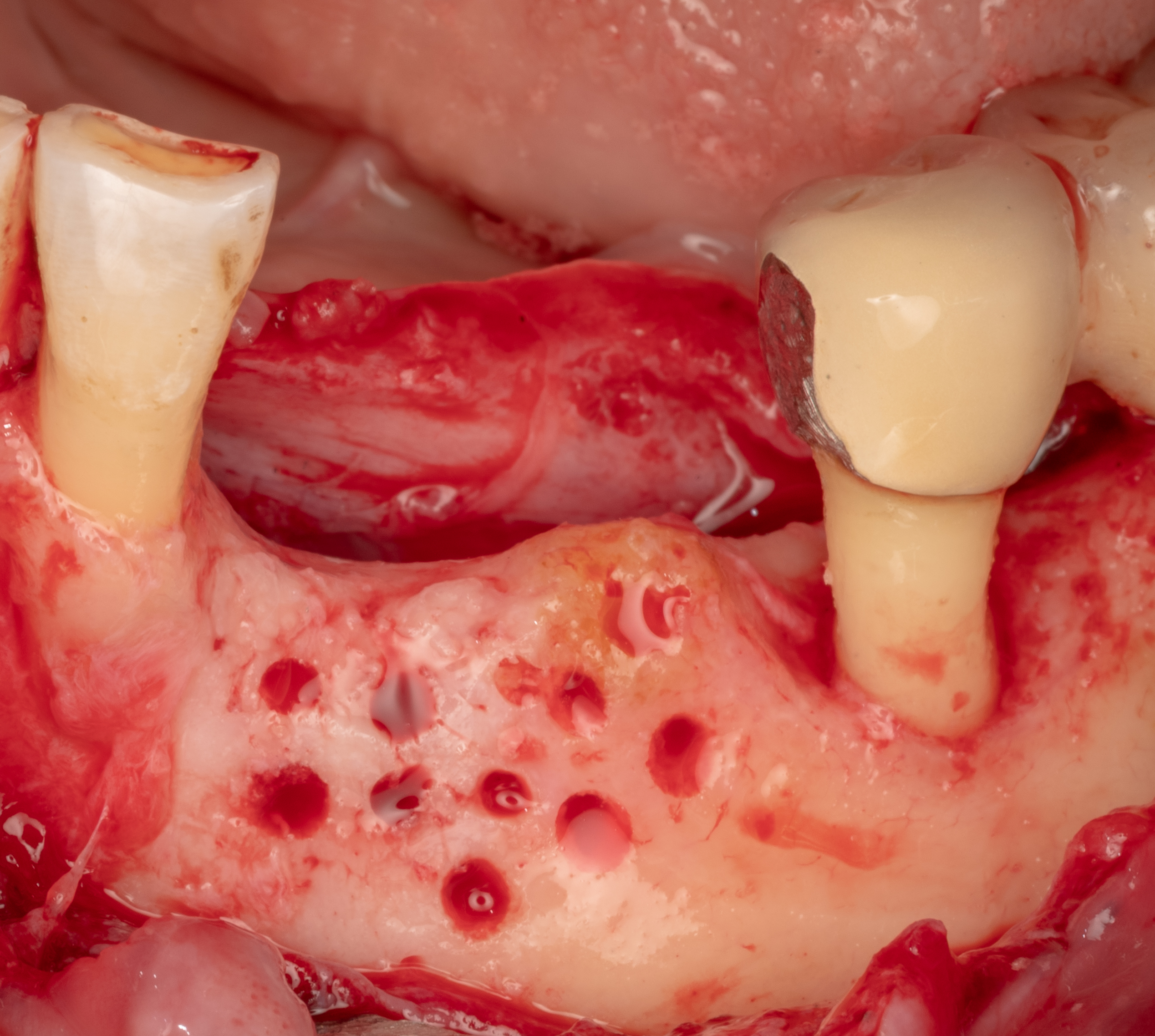
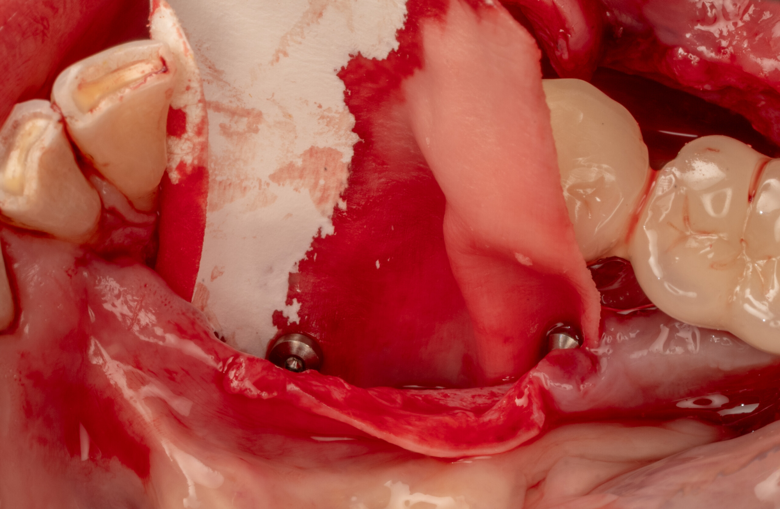
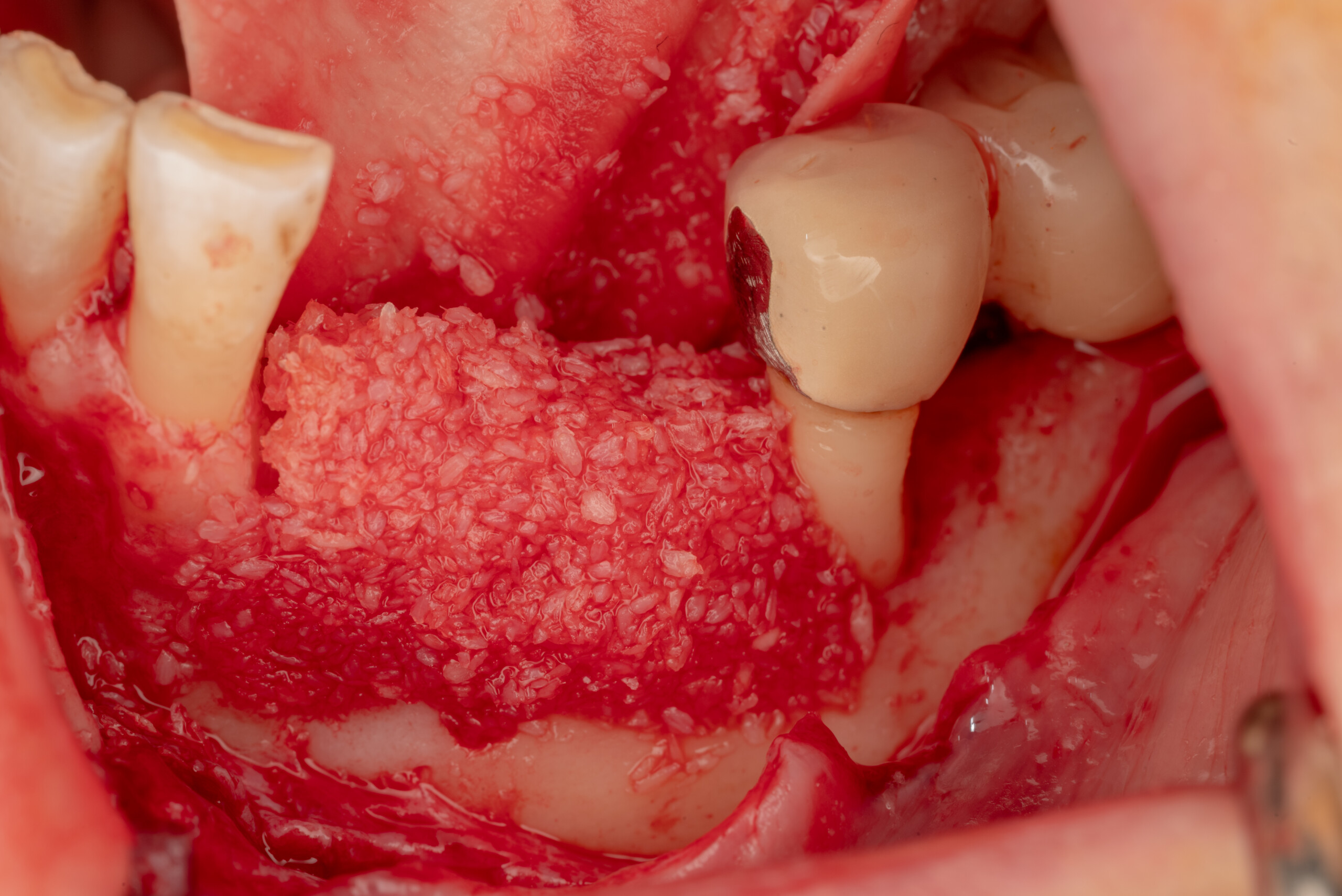
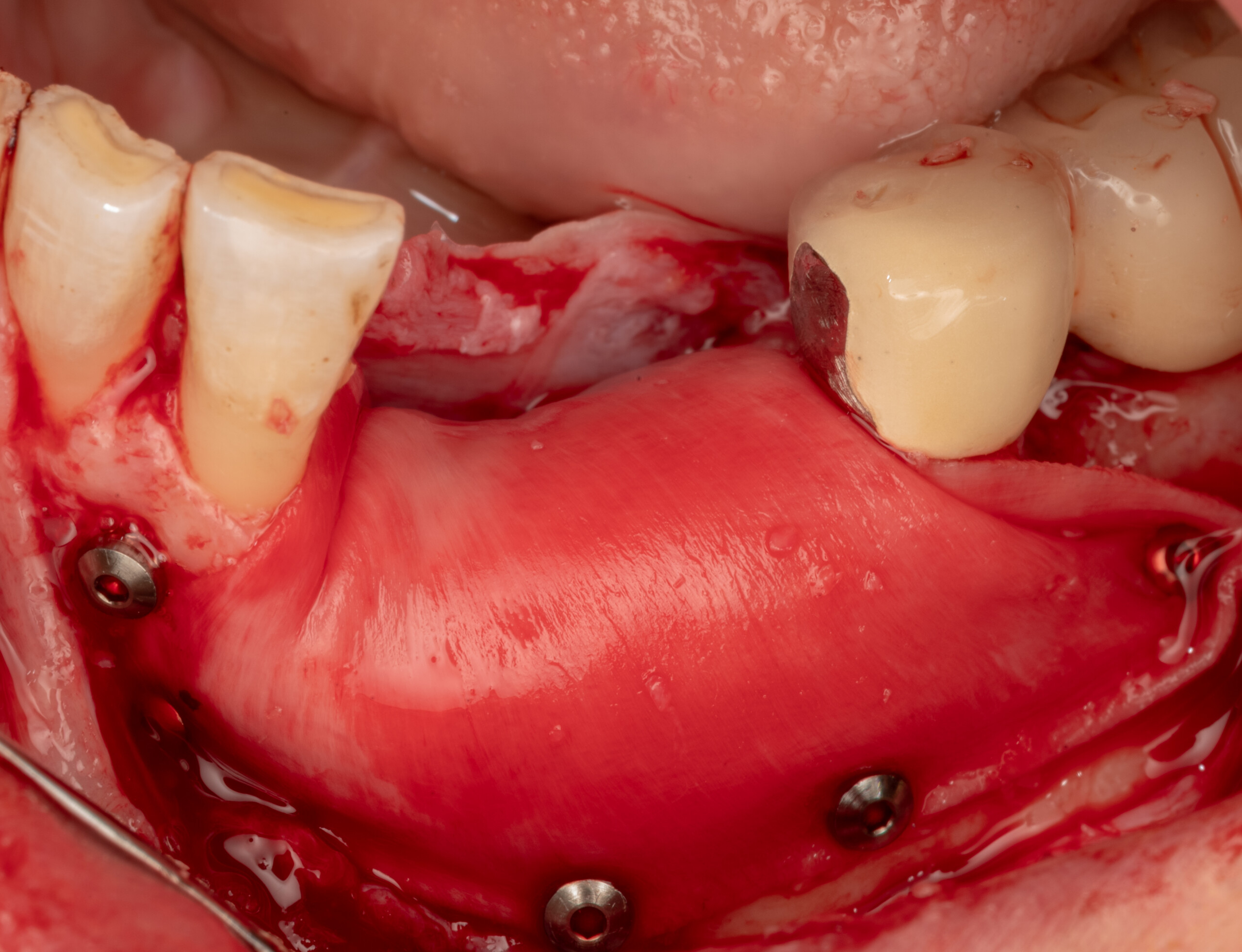
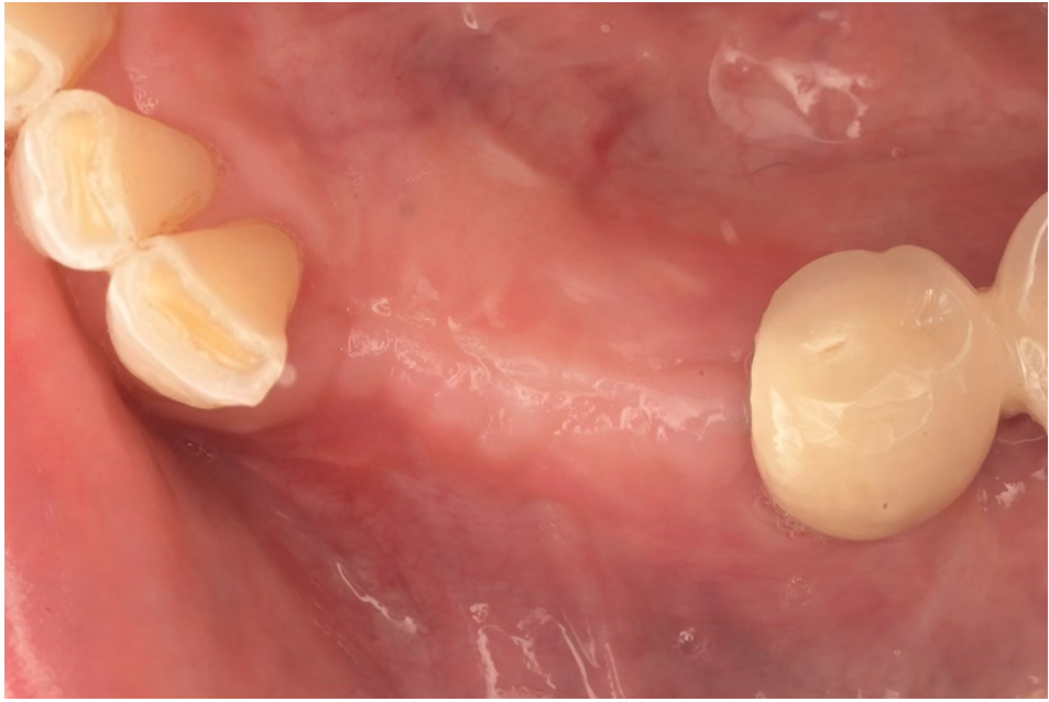
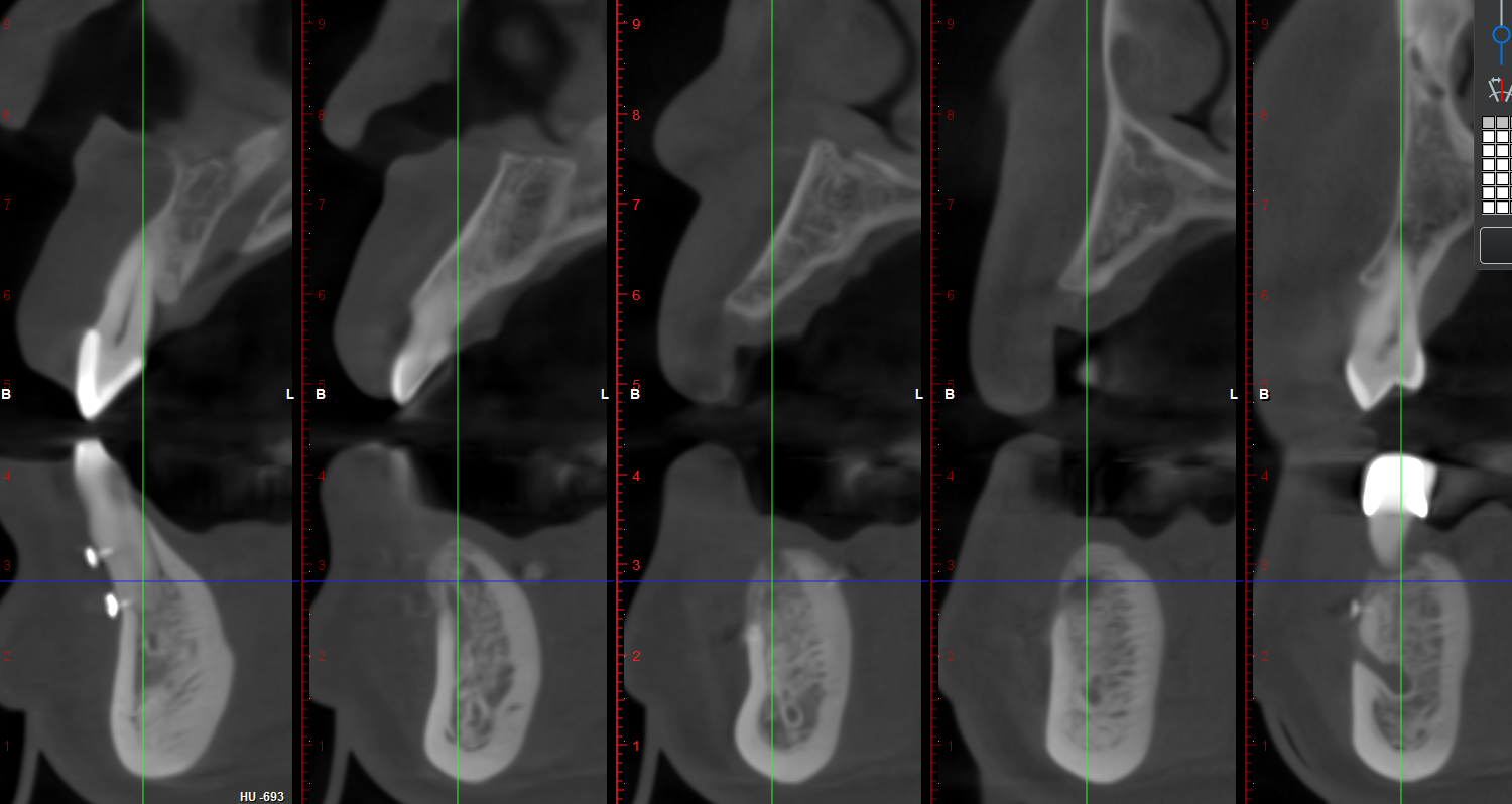
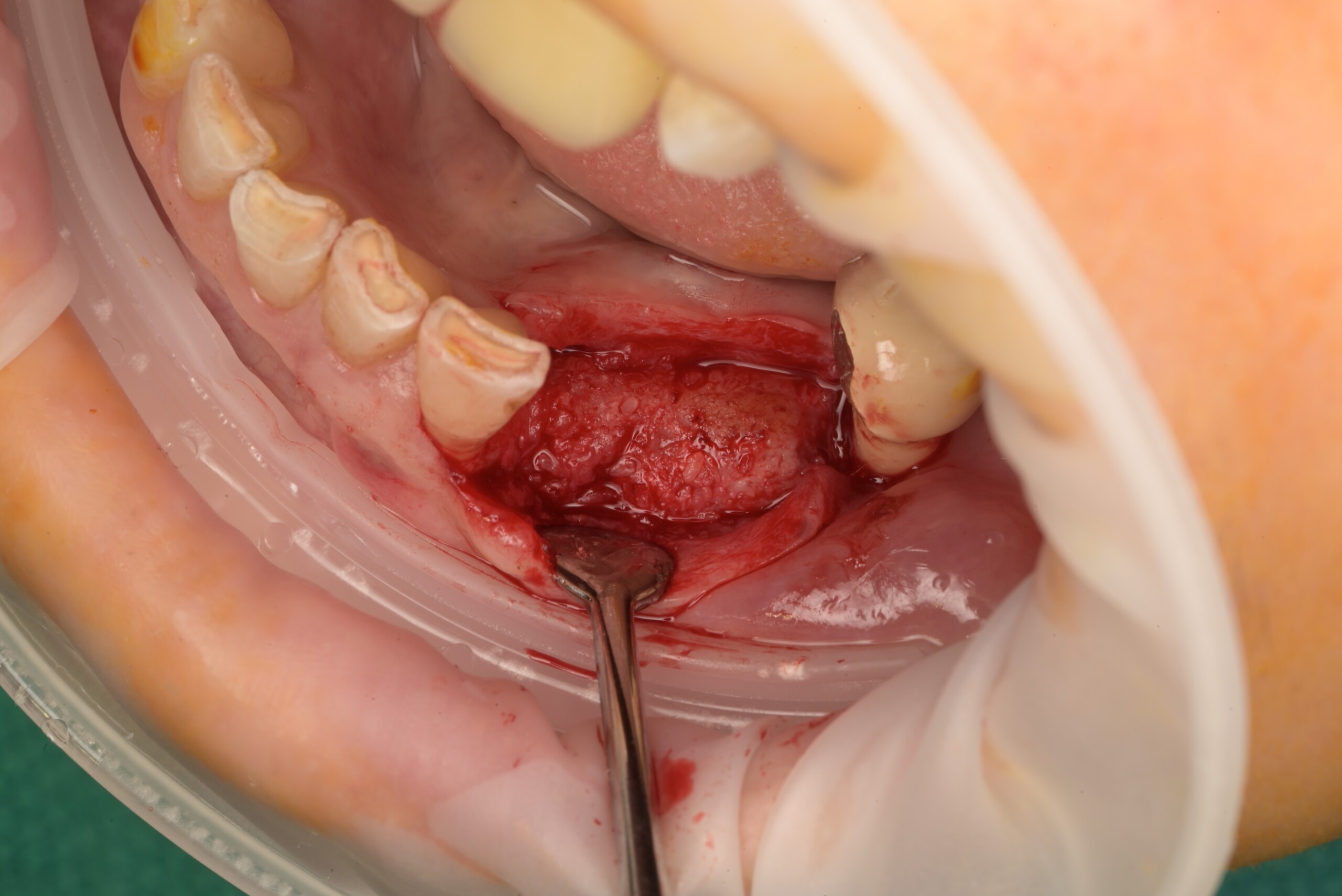
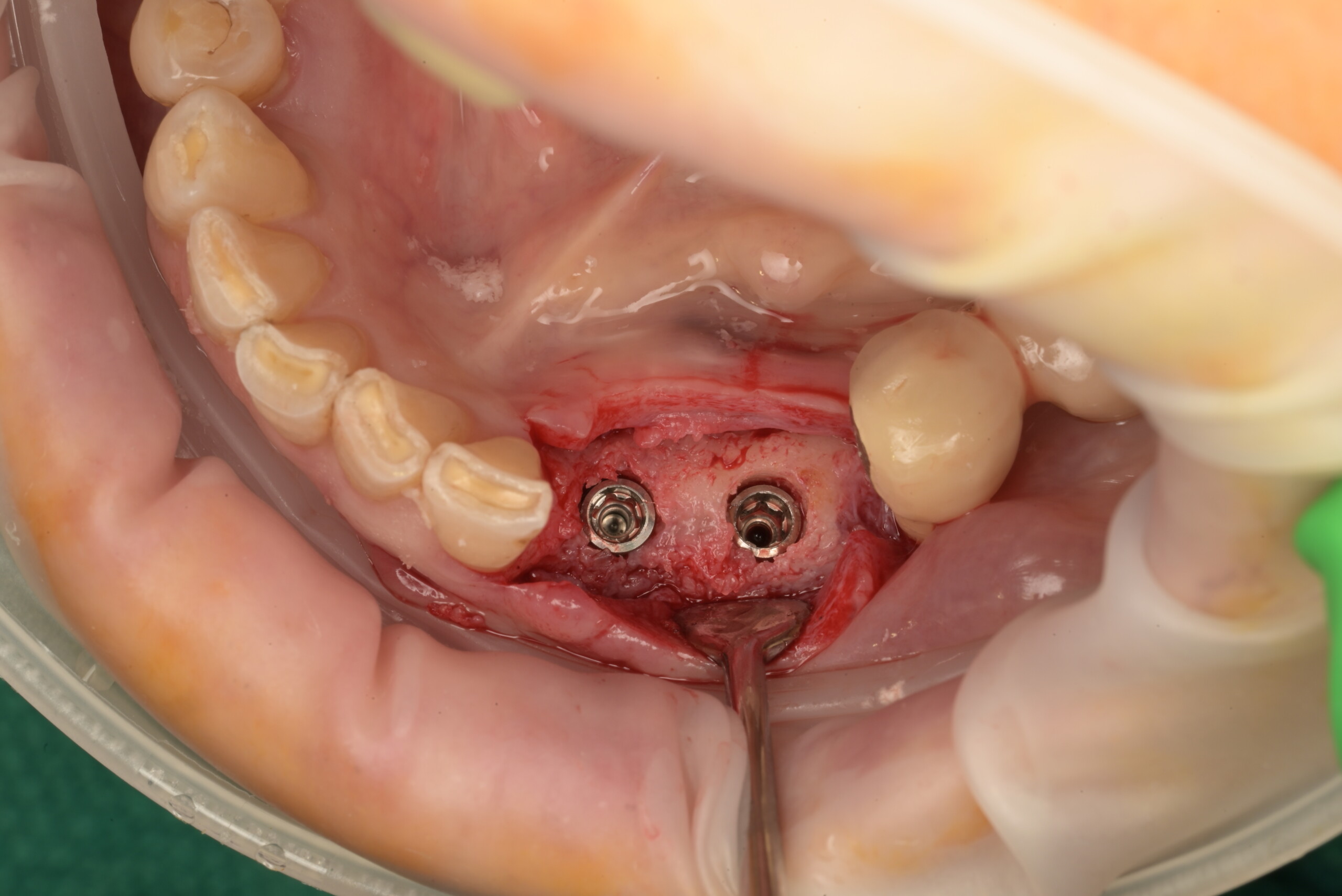
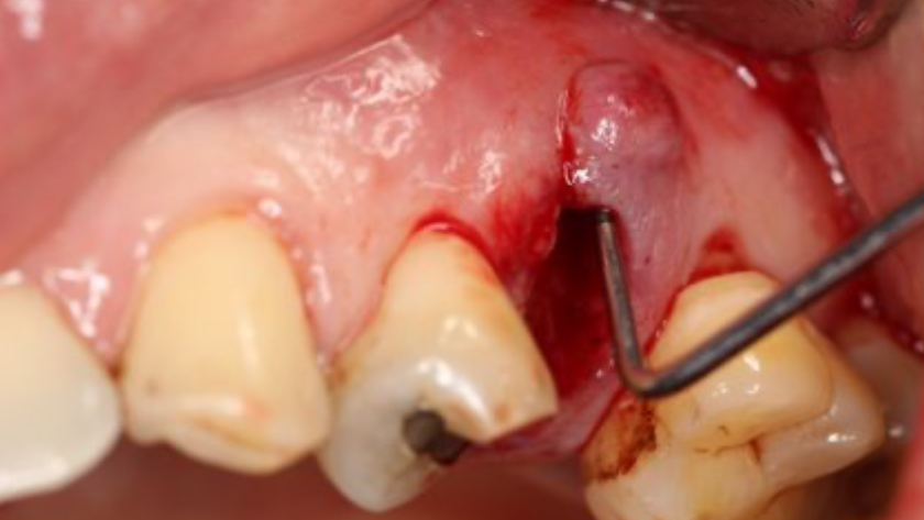
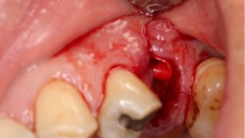
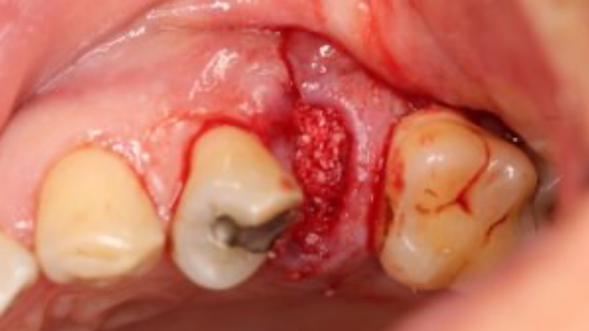
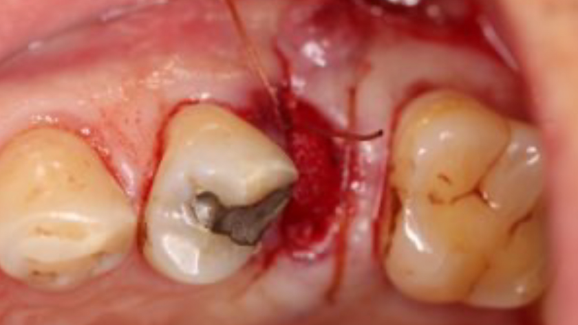
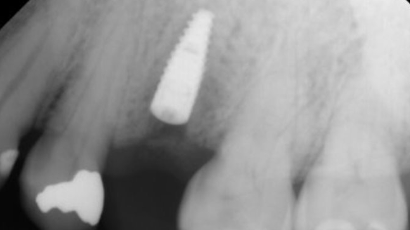
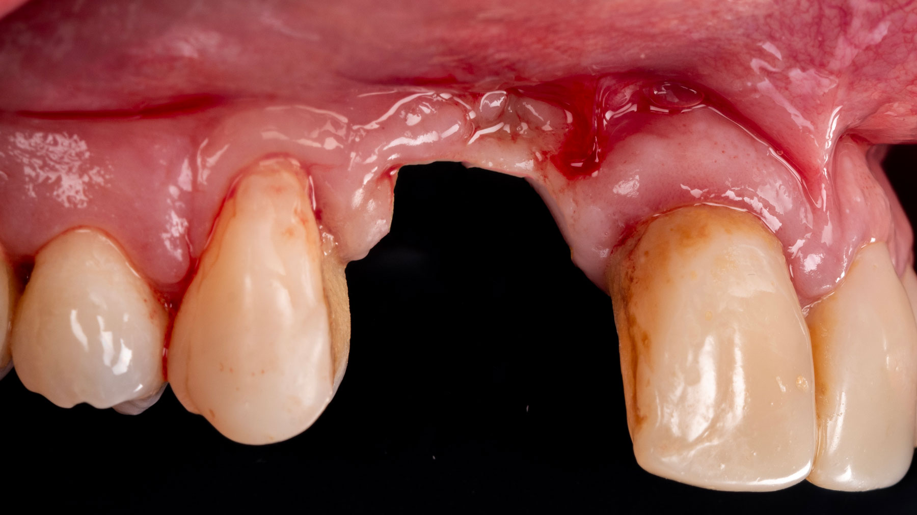
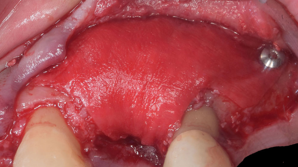
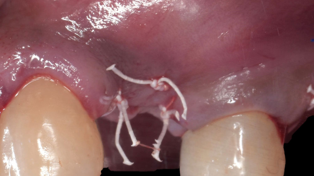
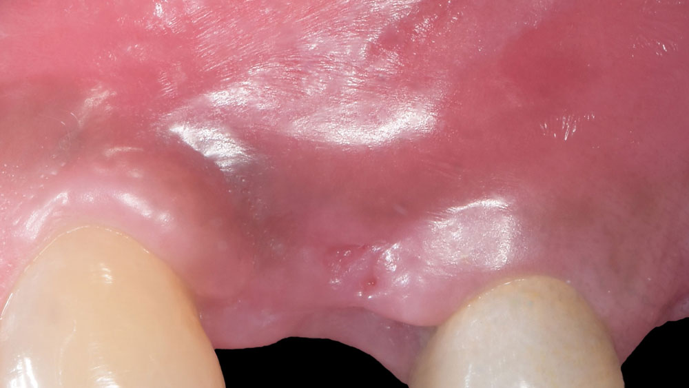
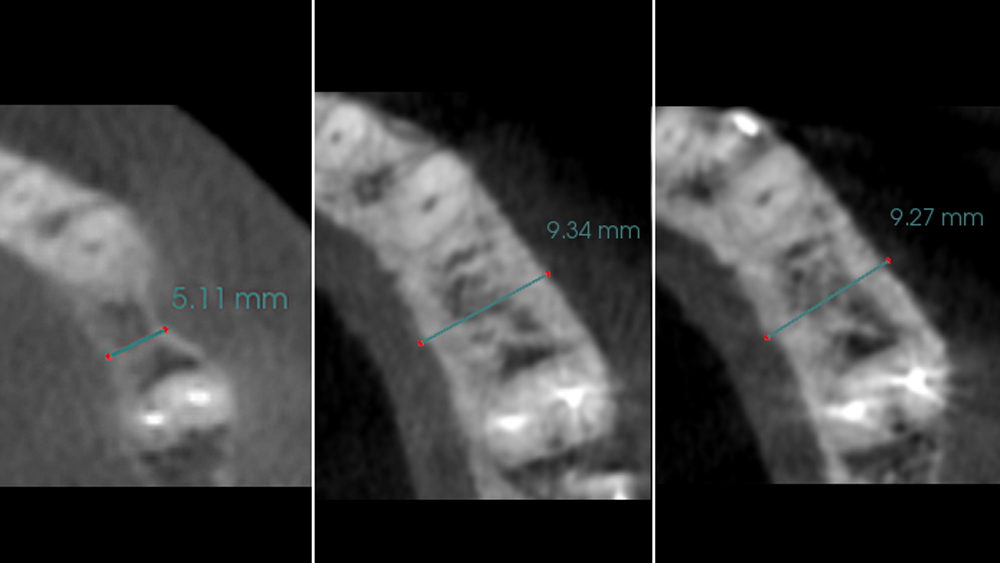
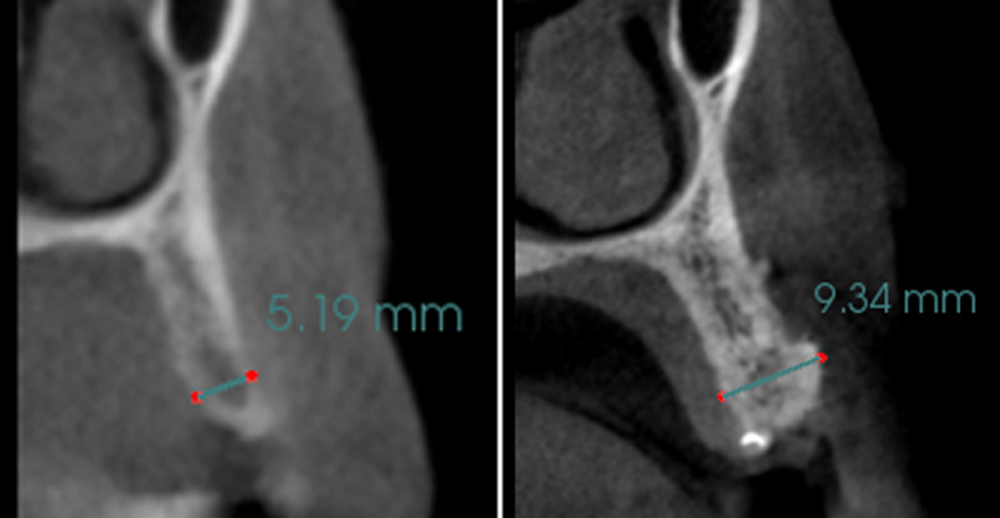
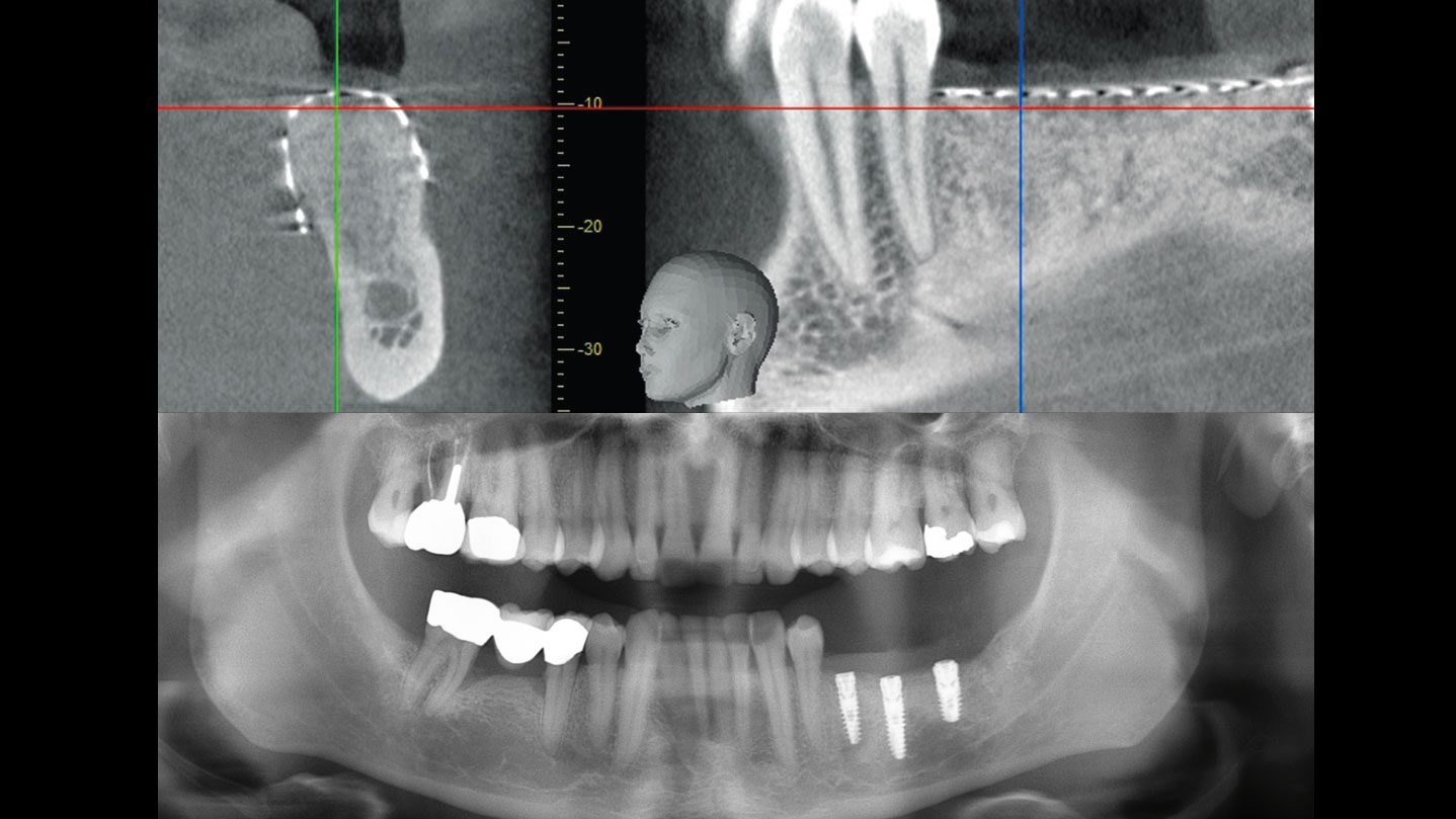
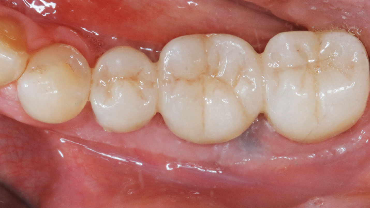
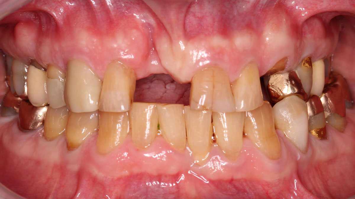
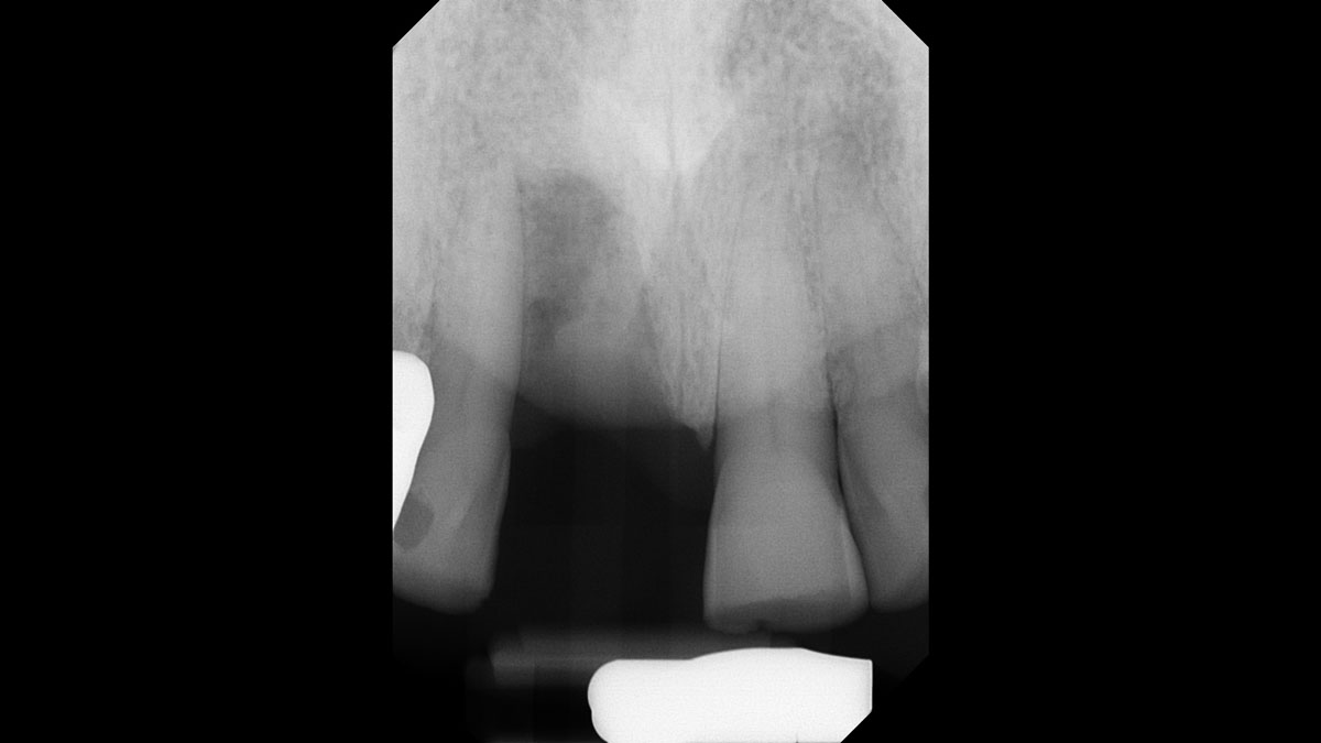
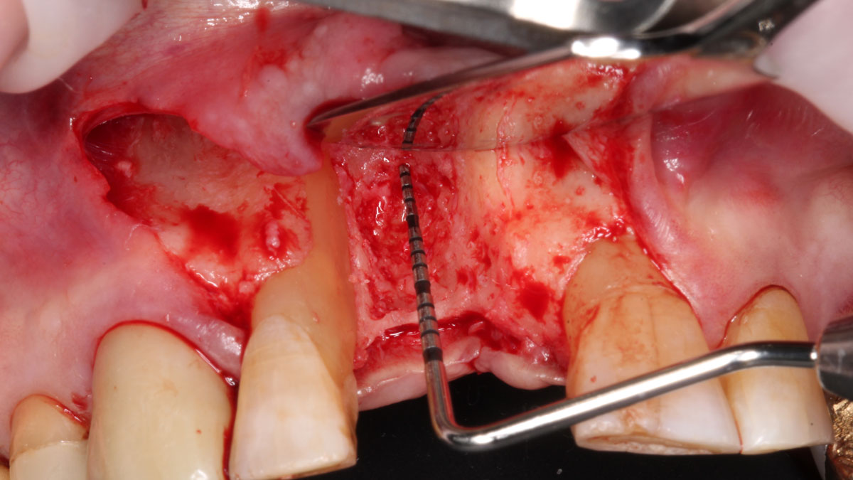
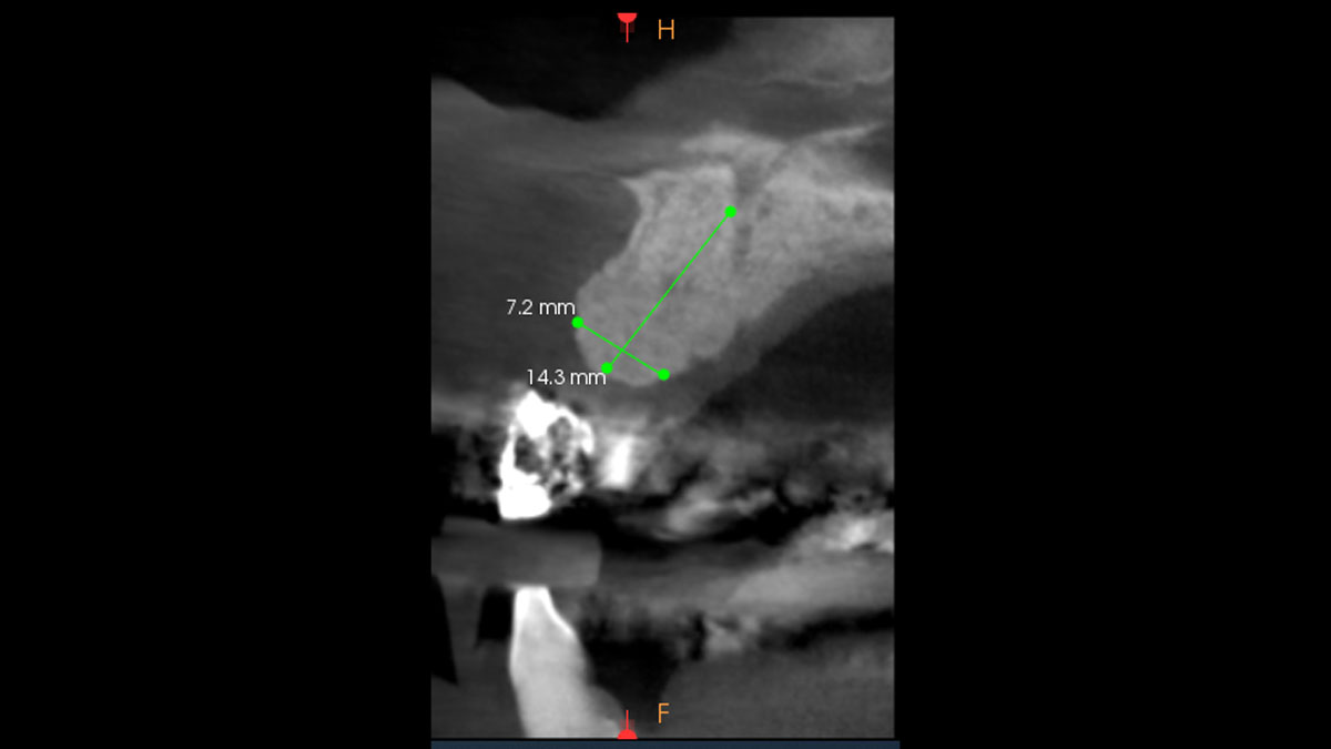
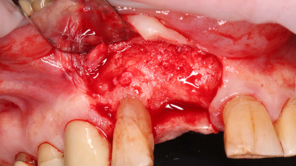
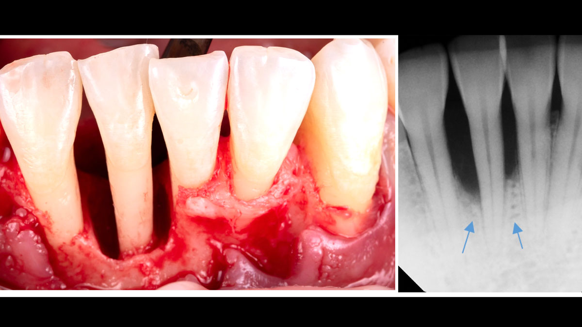
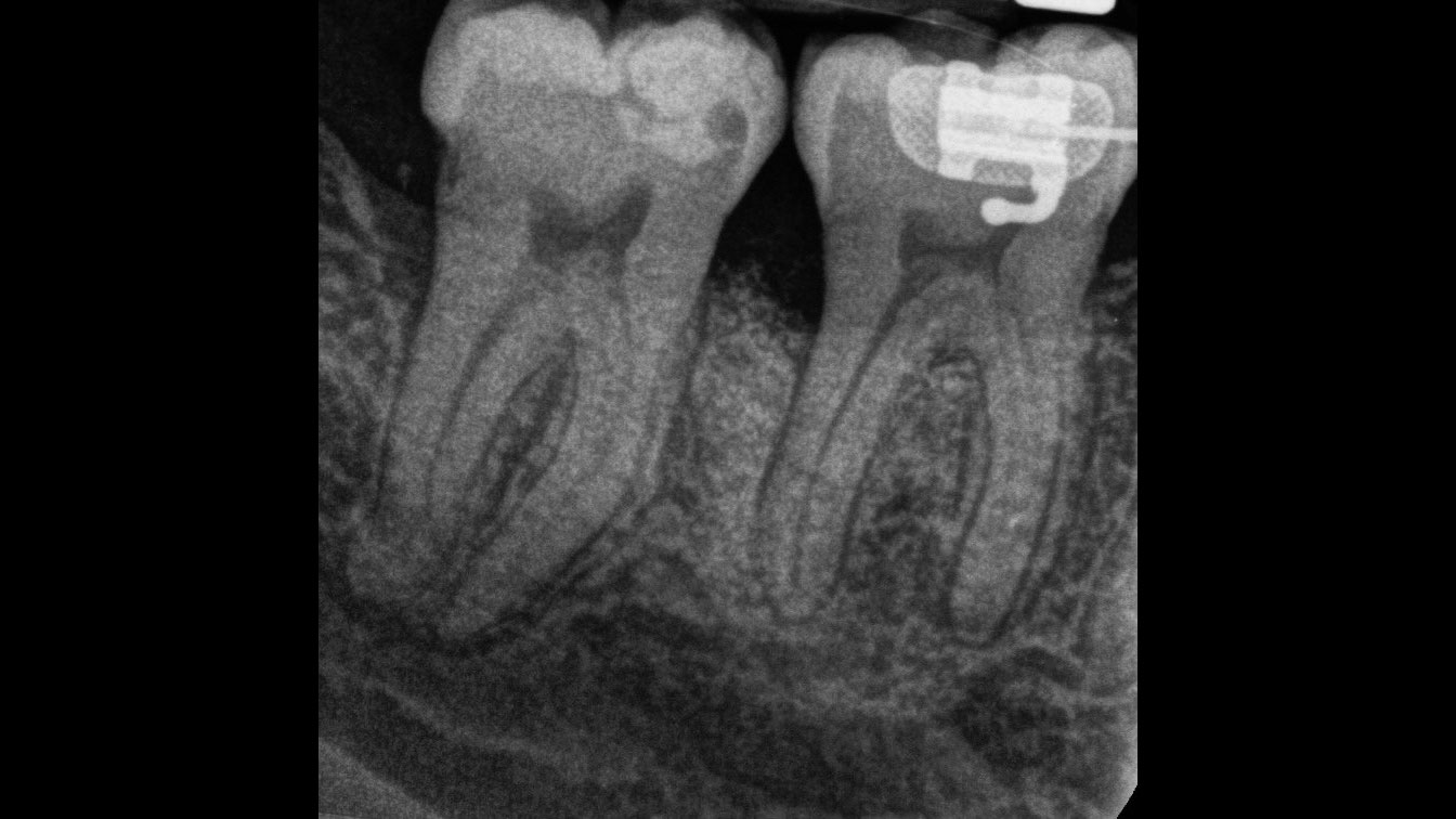
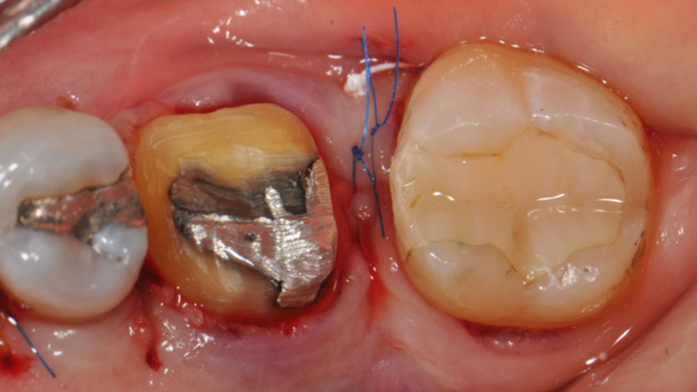
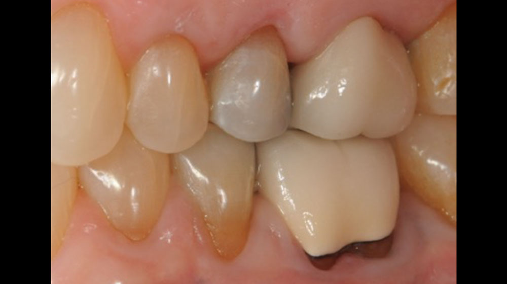
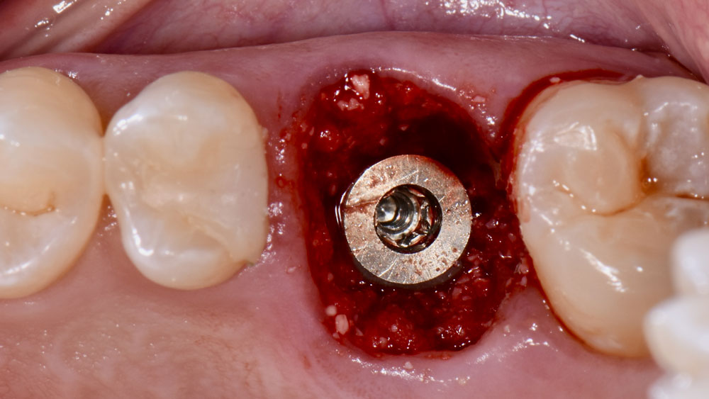
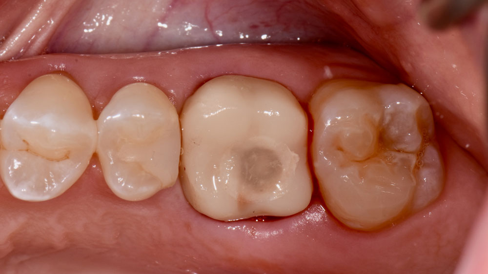
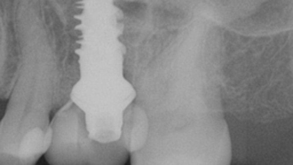
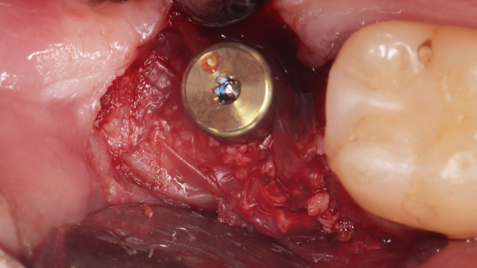
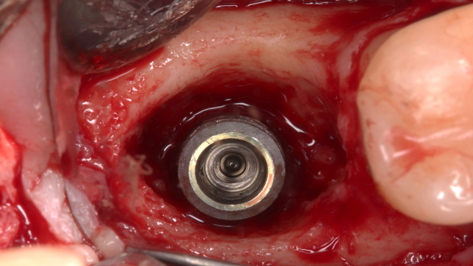
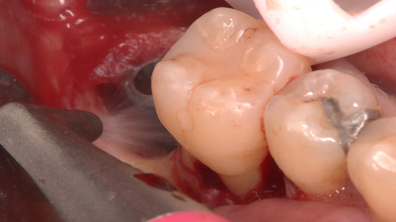
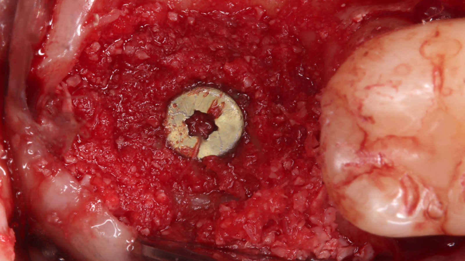
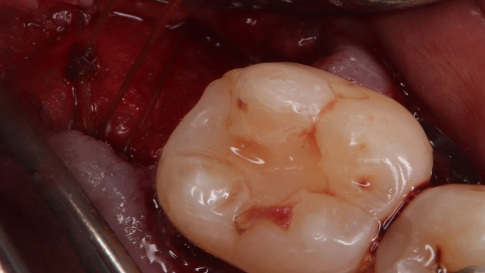
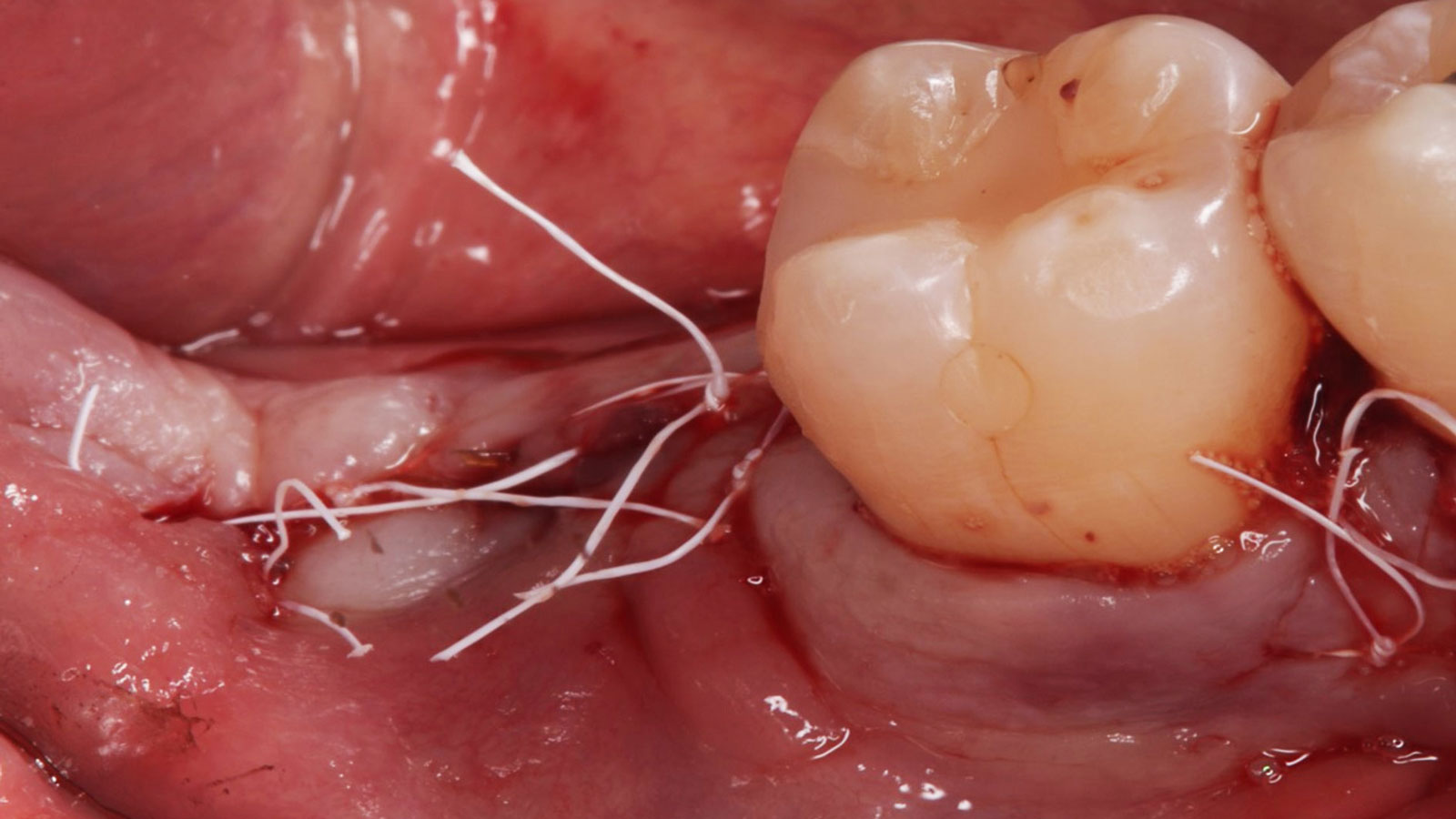
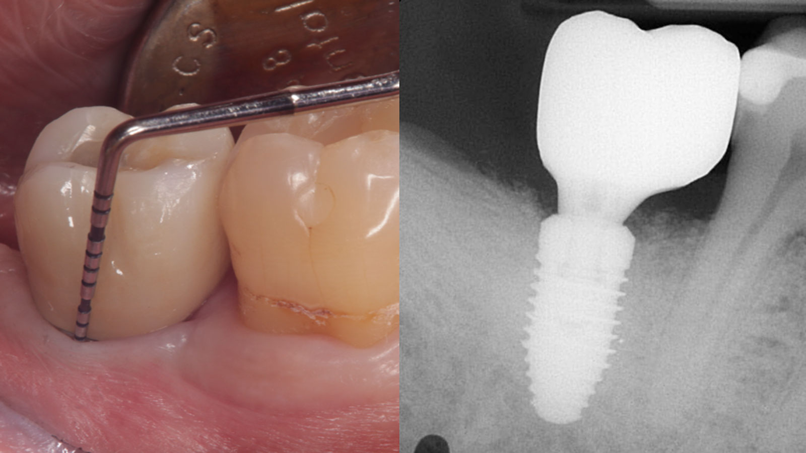
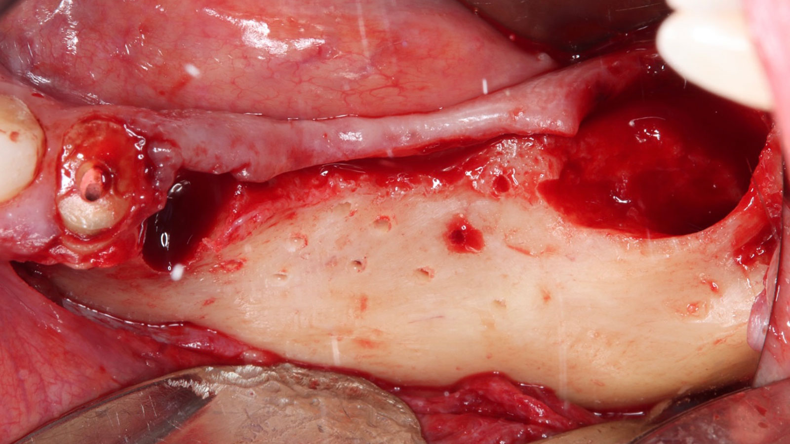
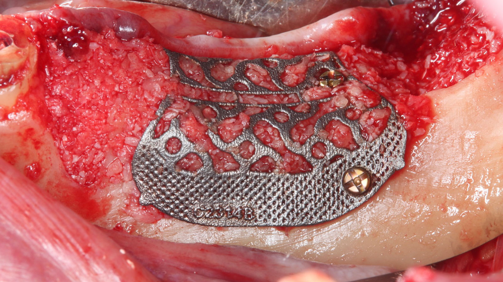
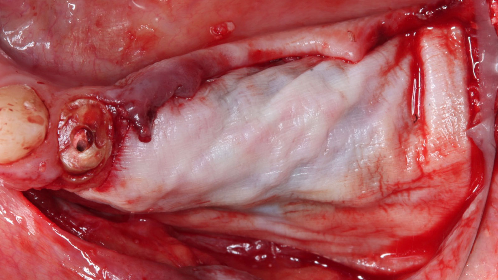
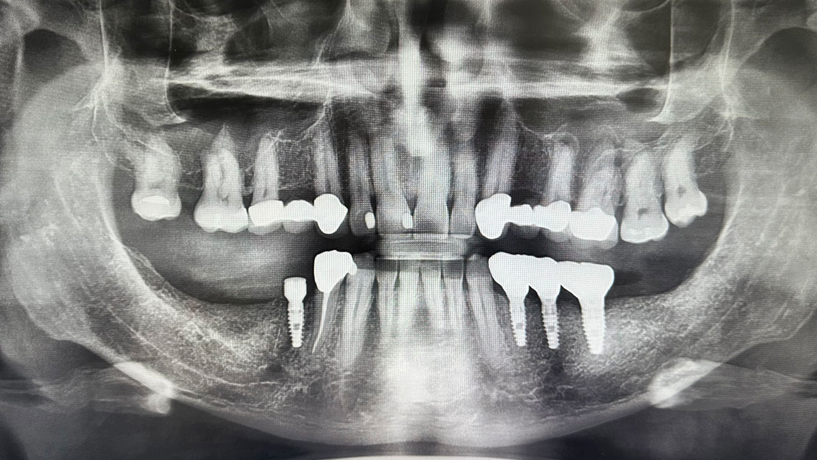
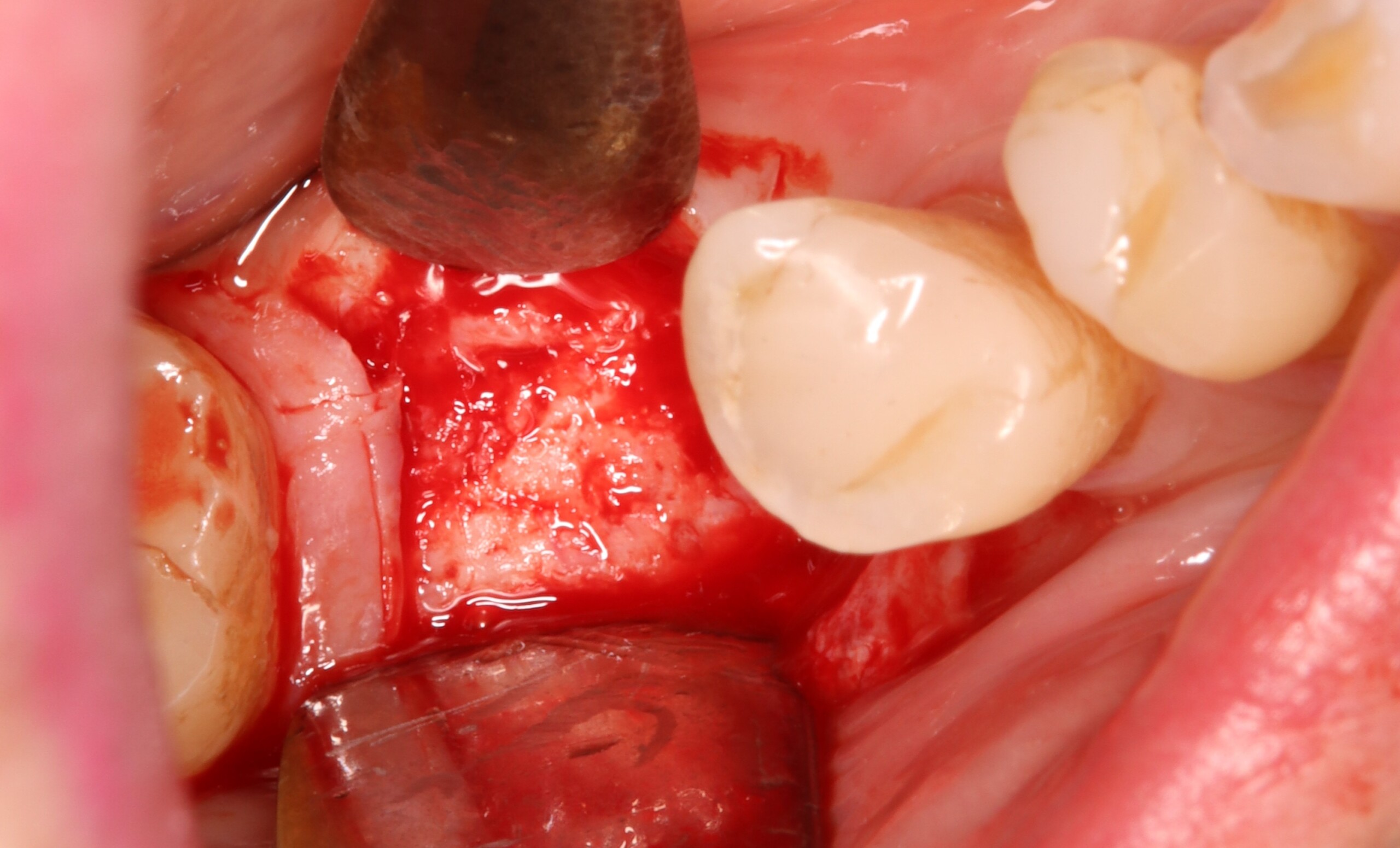
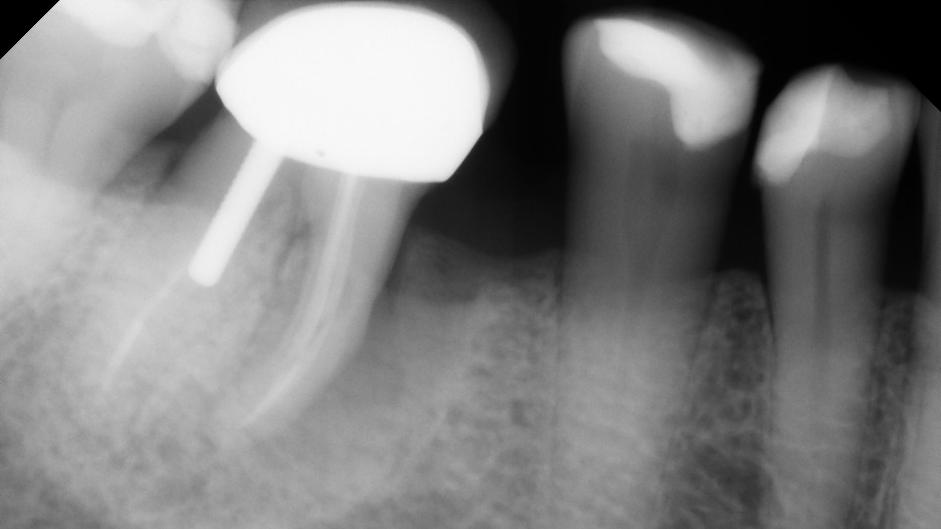
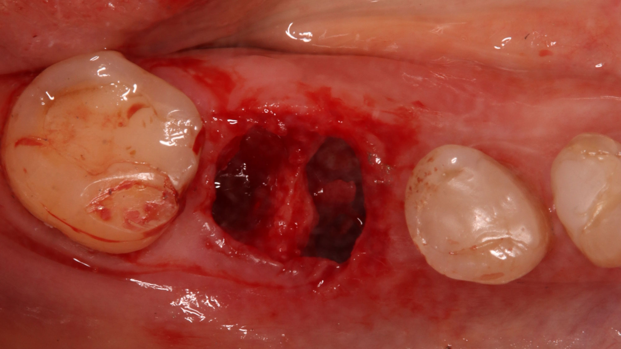
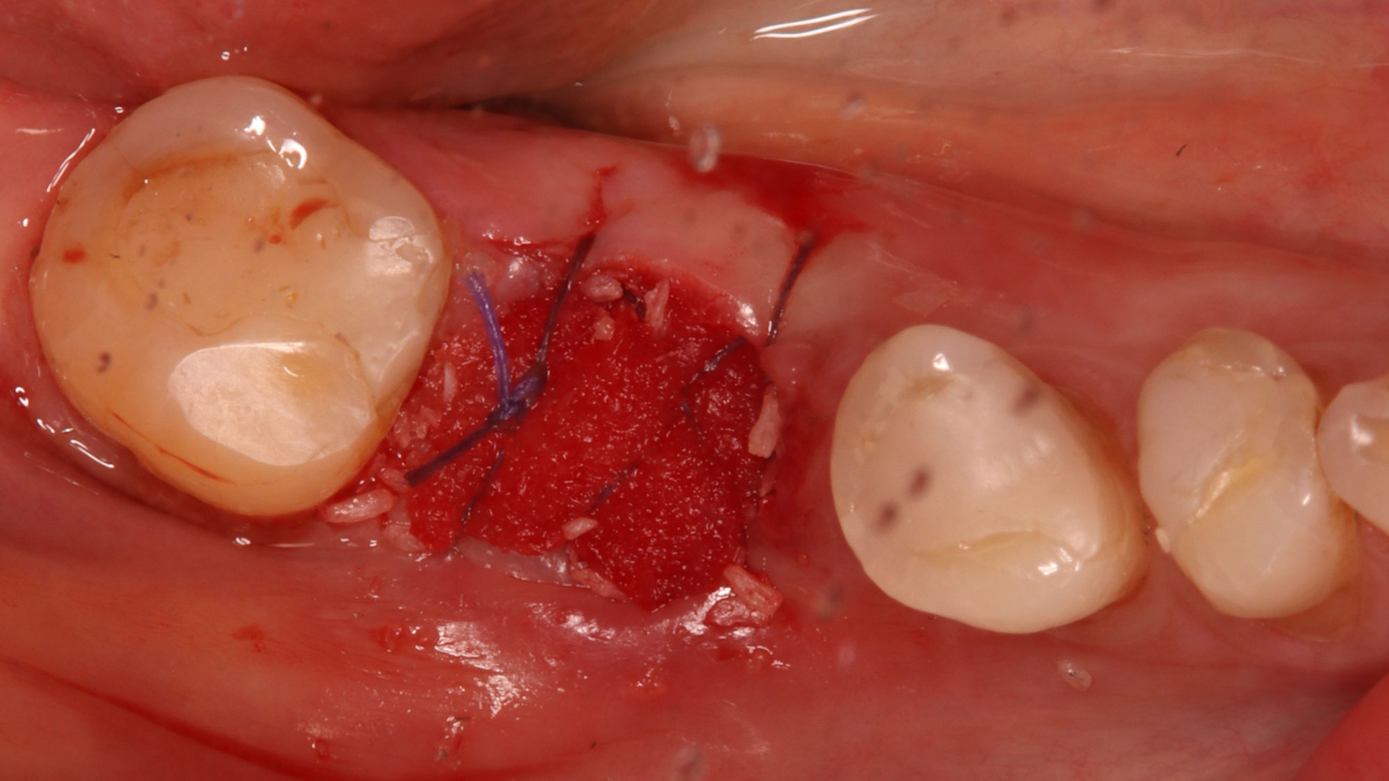
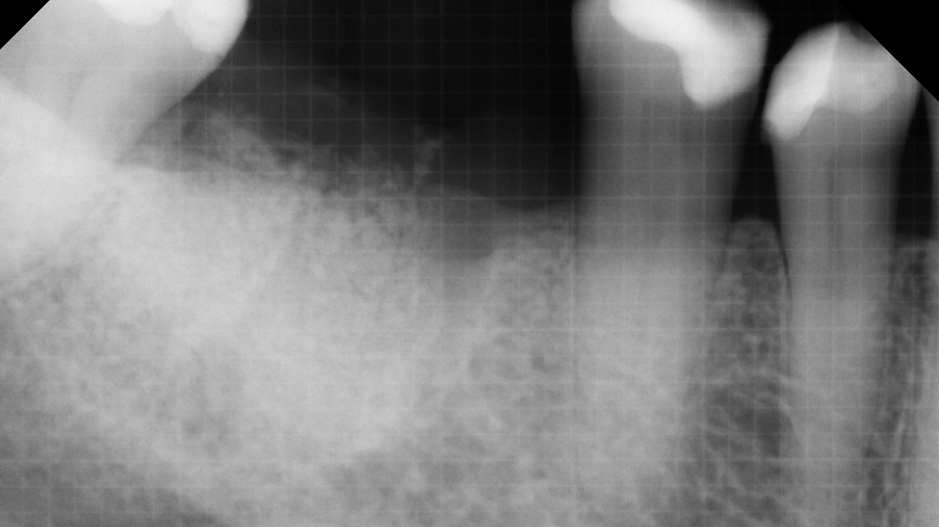
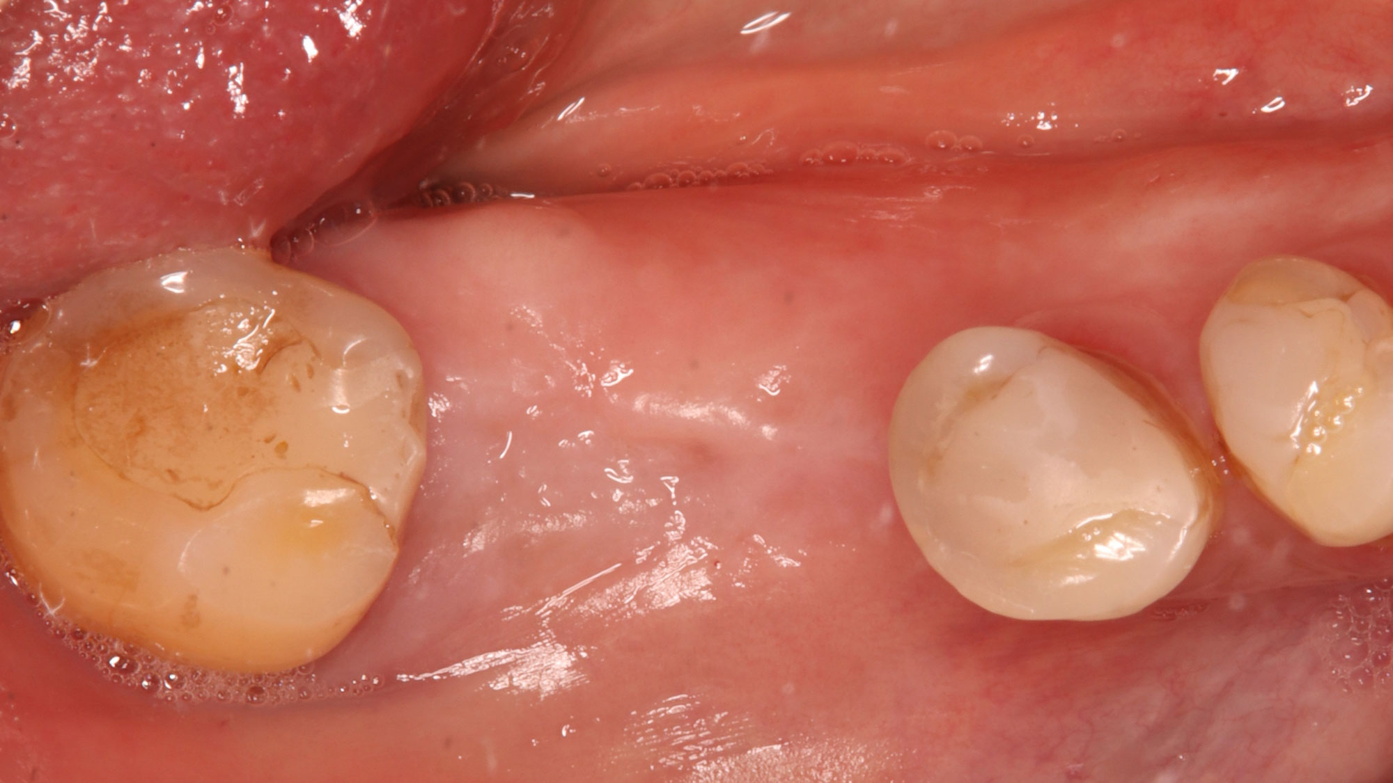
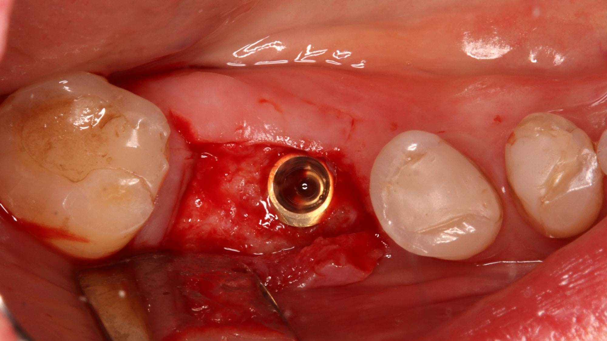
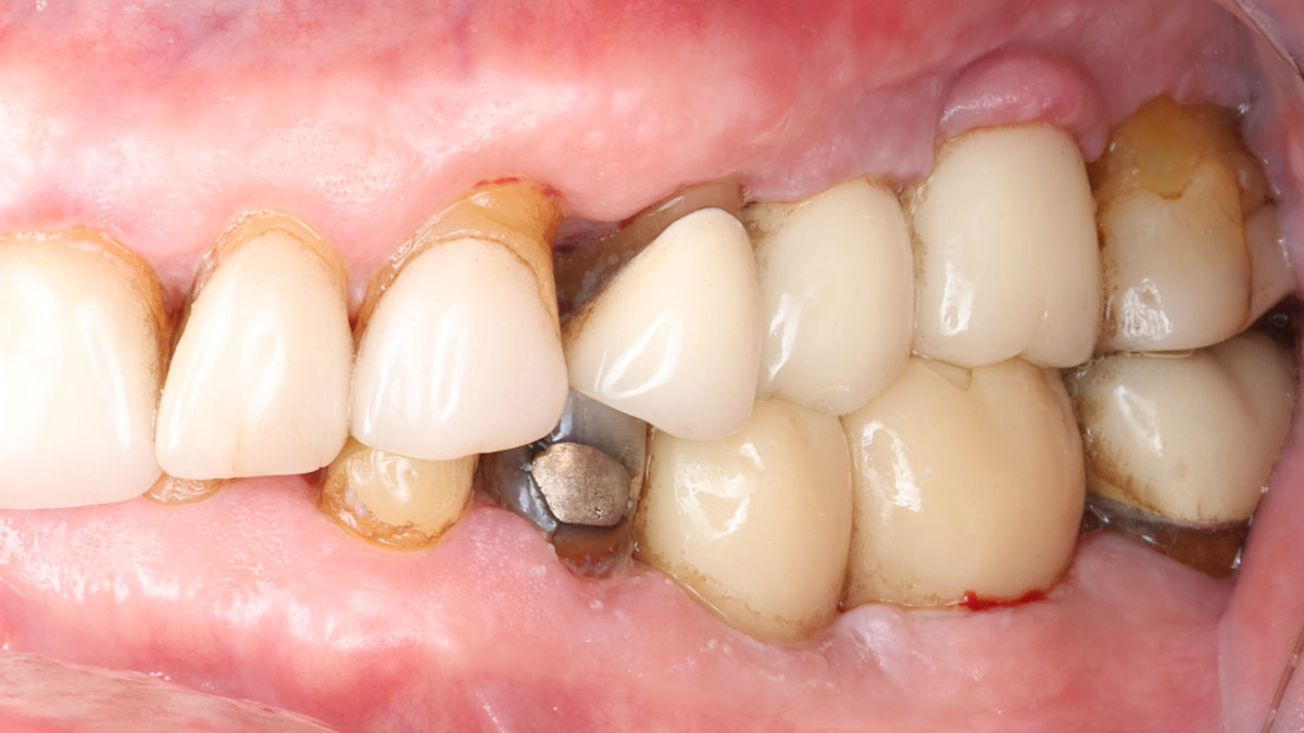
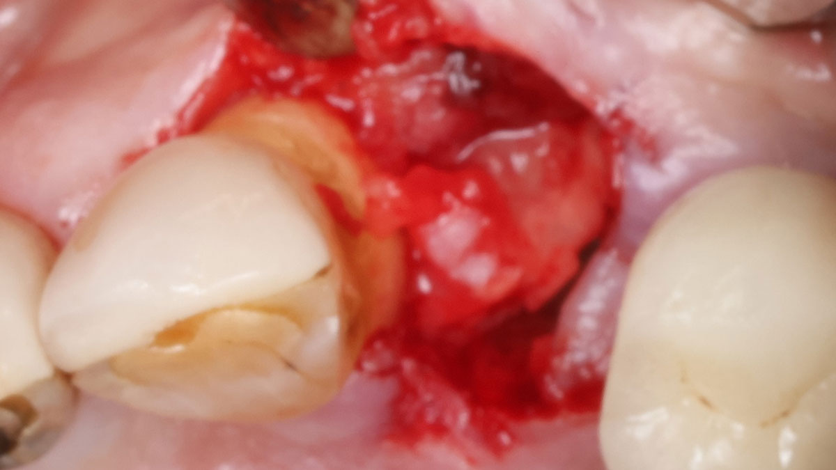
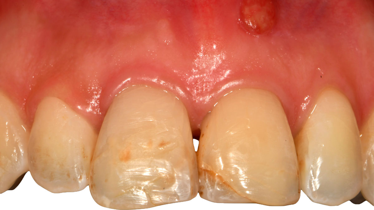
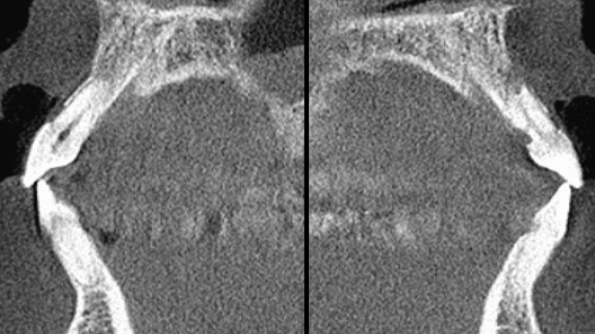
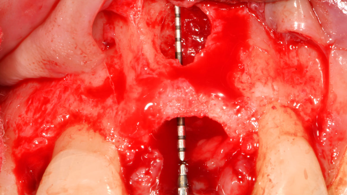
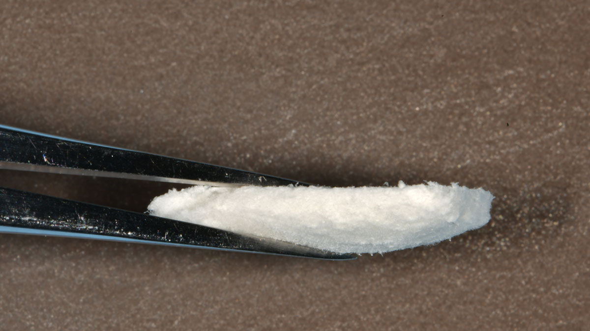
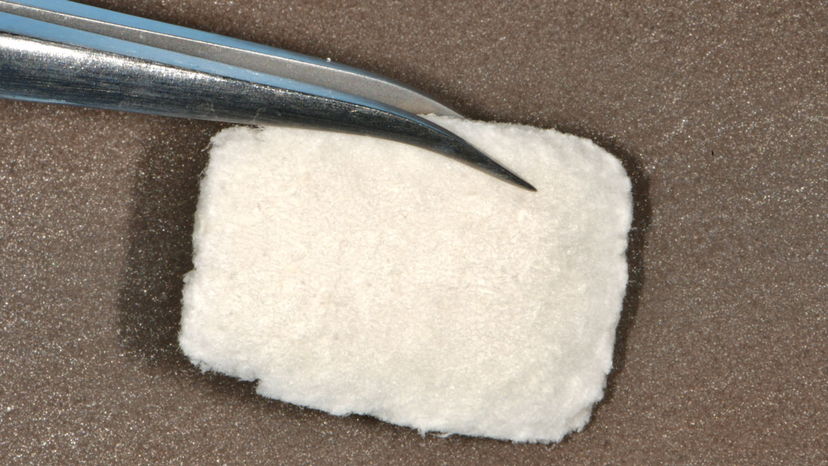
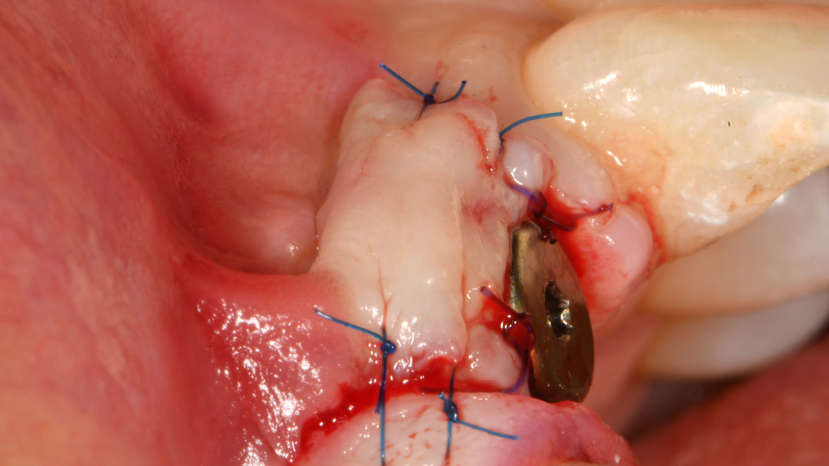
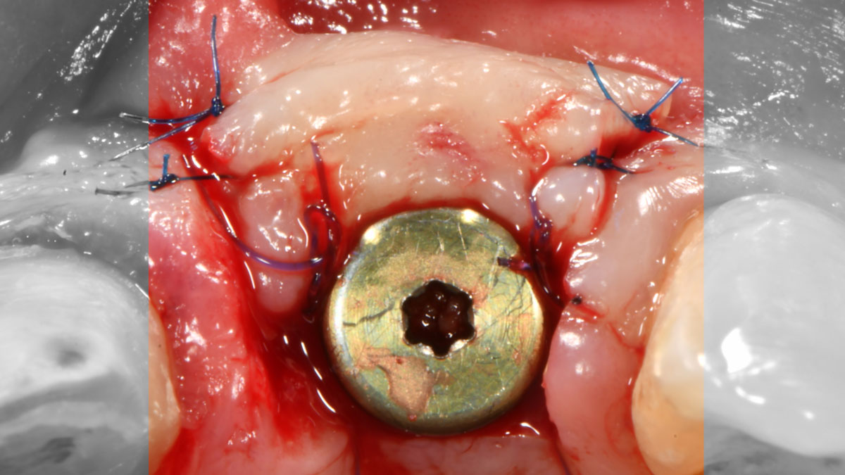
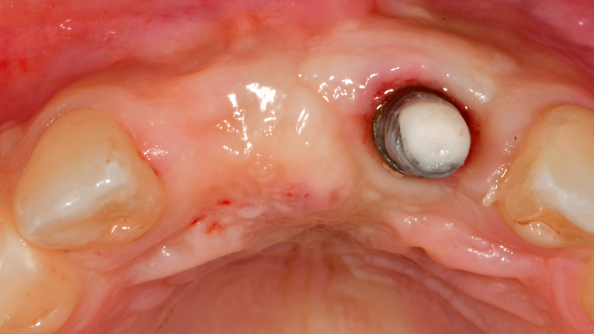
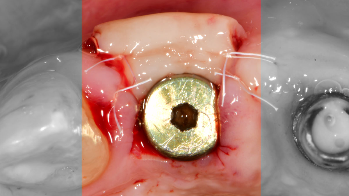
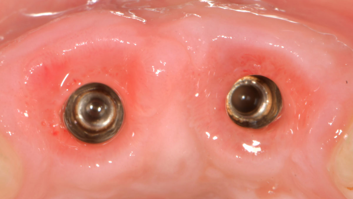
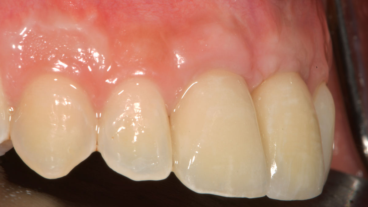
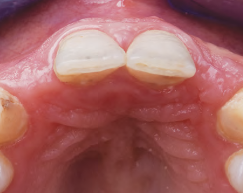
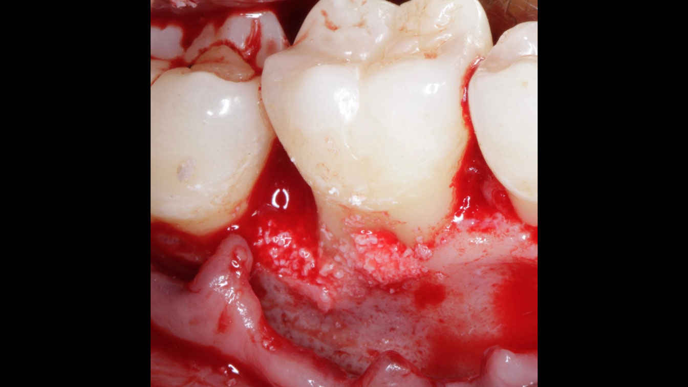
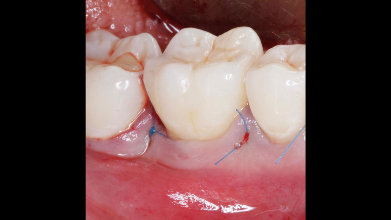
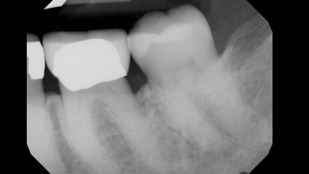
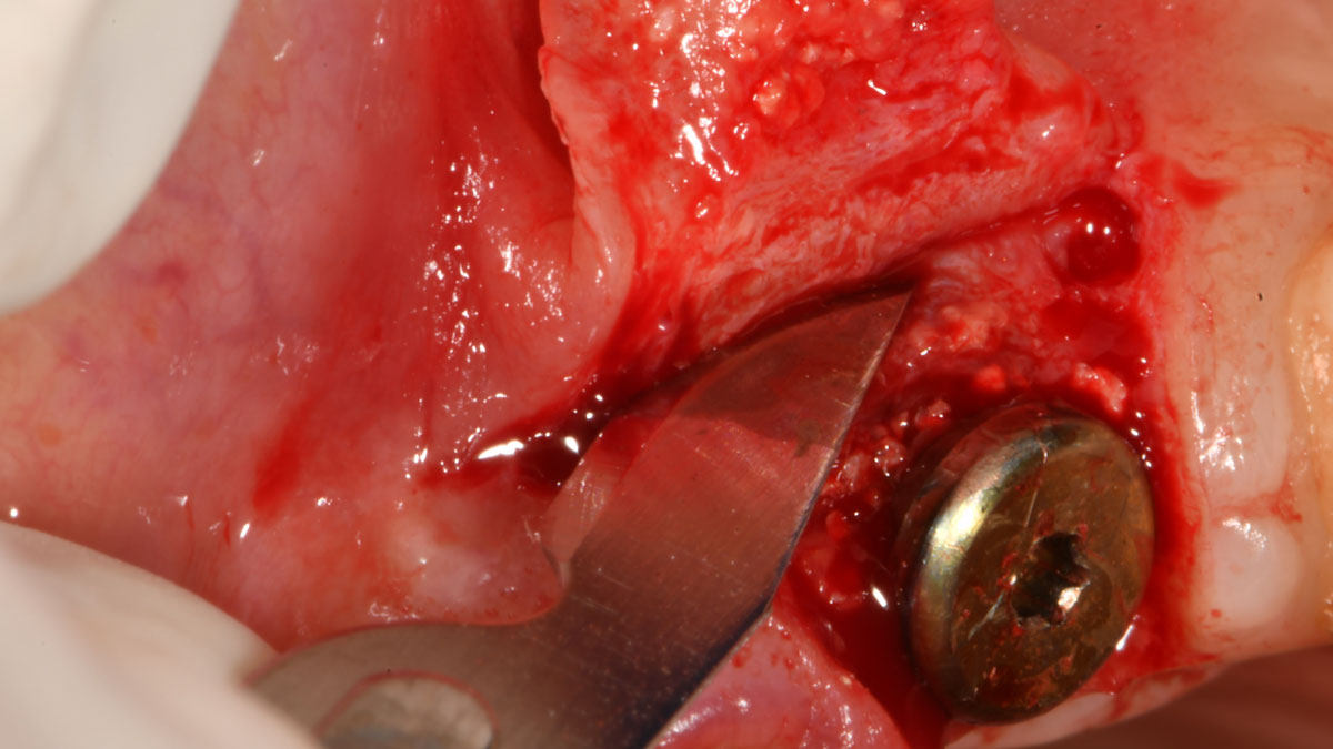
Teeth 30* and 31* had been previously extracted without alveolar ridge preservation. The site demonstrated that the site of tooth 7* had a history of implant removal with severe vertical bone deficiency & insufficient bone for implant placement. The treatment objective was bone regeneration using vallos® and Geistlich Bio-Gide® Forte.

*Universal Numbering System

Disclaimer: Case images courtesy of Karim Ghishan, BDS, MS (Periodontist, Ann Arbor, MI, USA) and Muhammad Saleh, BDS, MSD, PhD (Periodontist, Ann Arbor, MI, USA). Used with permission. Images are provided for illustrative and educational purposes only. Results are not guaranteed, and individual outcomes may vary depending on patient-specific circumstances and clinical factors. This information is for general educational purposes only and does not constitute medical advice, nor does it necessarily reflect the official position, opinion, or recommendations of Geistlich. Treatment decisions are made at the clinician’s discretion based on each patient’s unique needs.

CLINICAL CASE

THE SITUATION

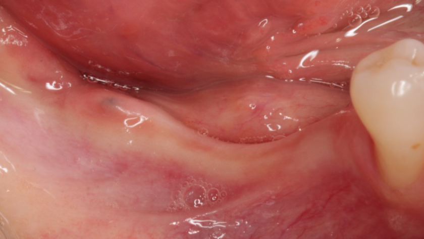
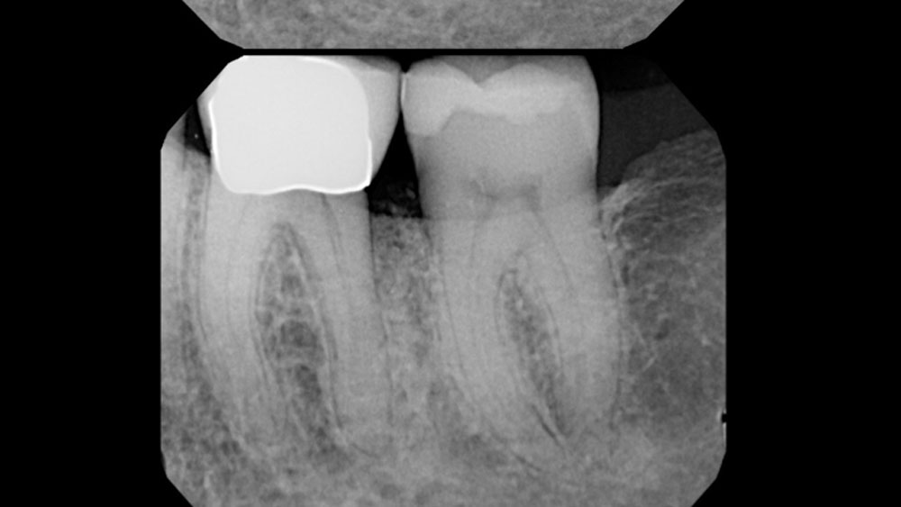
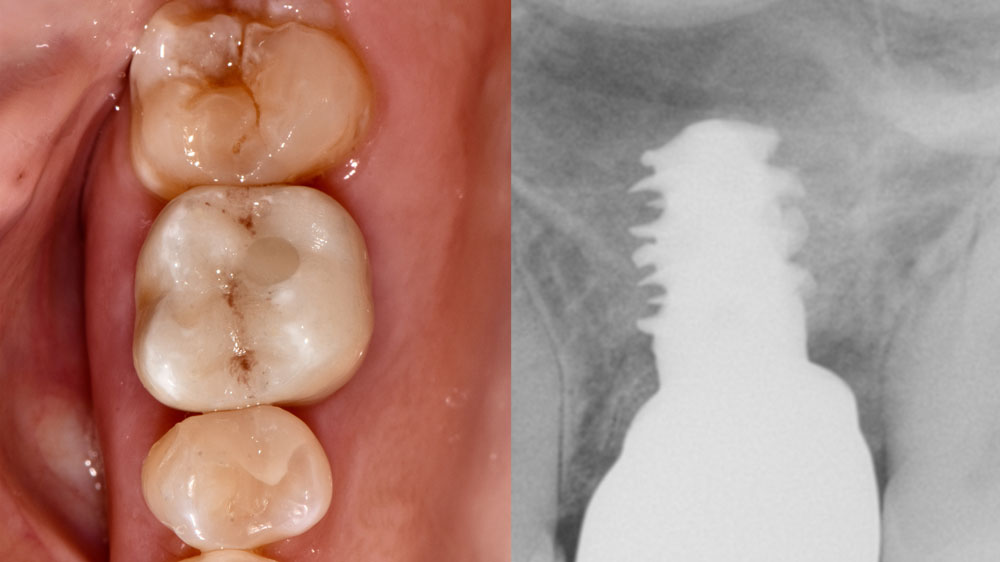
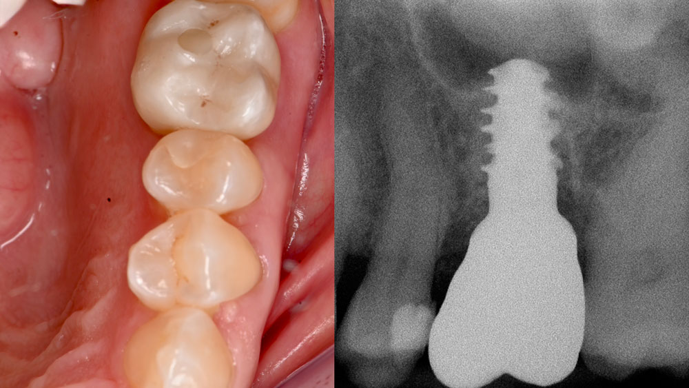
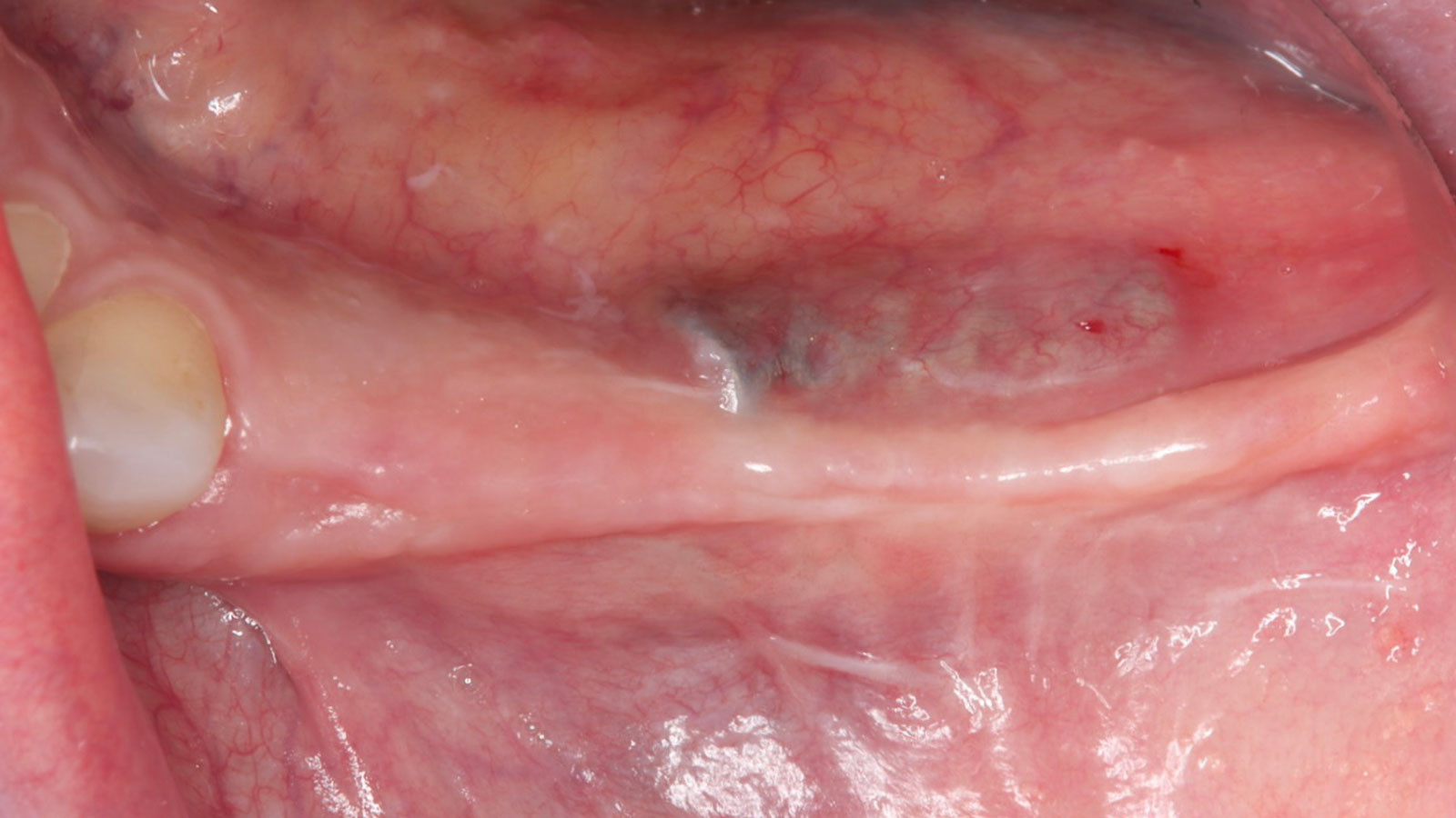
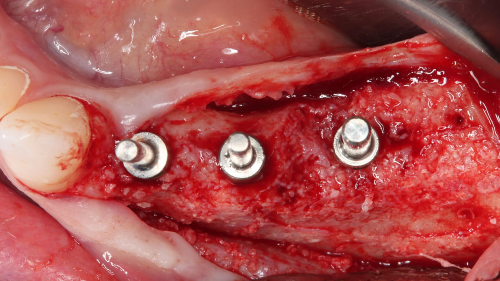
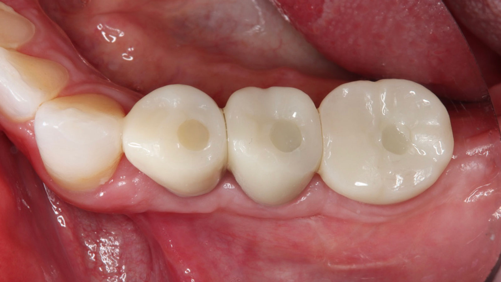
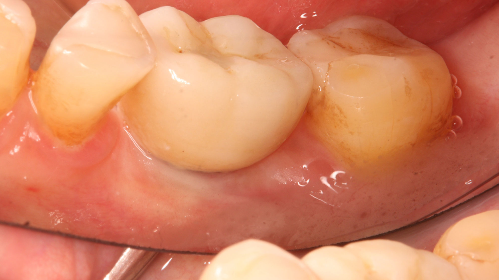
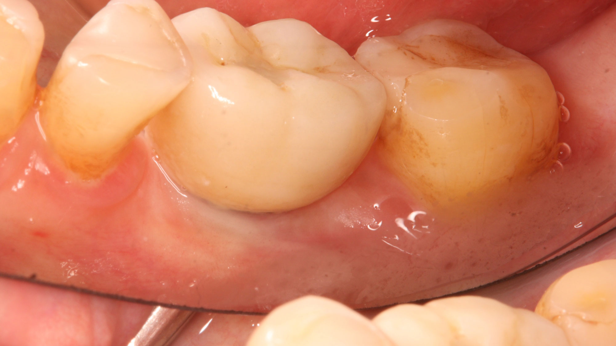
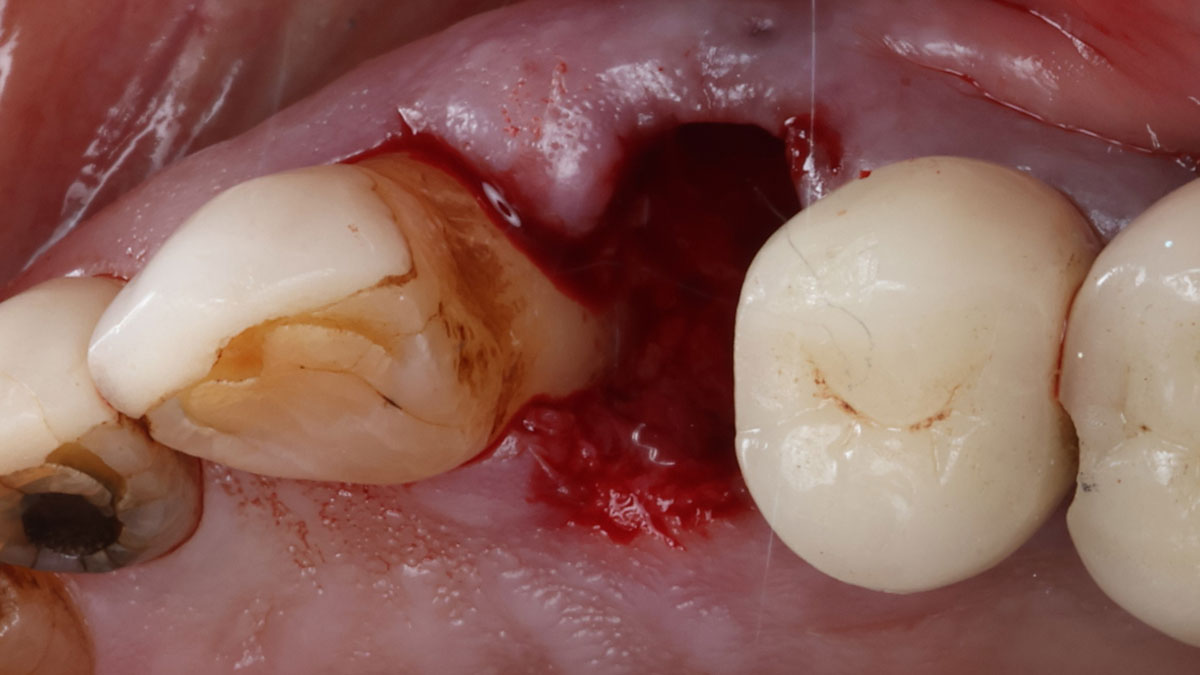
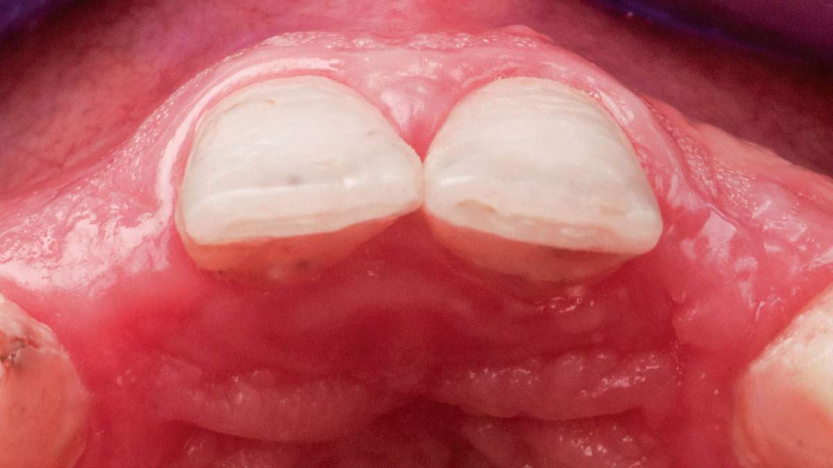
Teeth 30* and 31* had been previously extracted without alveolar ridge preservation. The site demonstrated approximately 1–2 mm of residual ridge width. The goal was to restore the site with implants.  

*Universal Numbering System

Disclaimer: These results are not guaranteed; individual outcomes may vary depending on patient circumstances. This information is for informational purposes only and may not reflect Geistlich’s official position, opinion, or recommendation. Treatment decisions are made at the physician’s discretion, based on the unique needs of each patient.

CLINICAL CASE

THE SITUATION

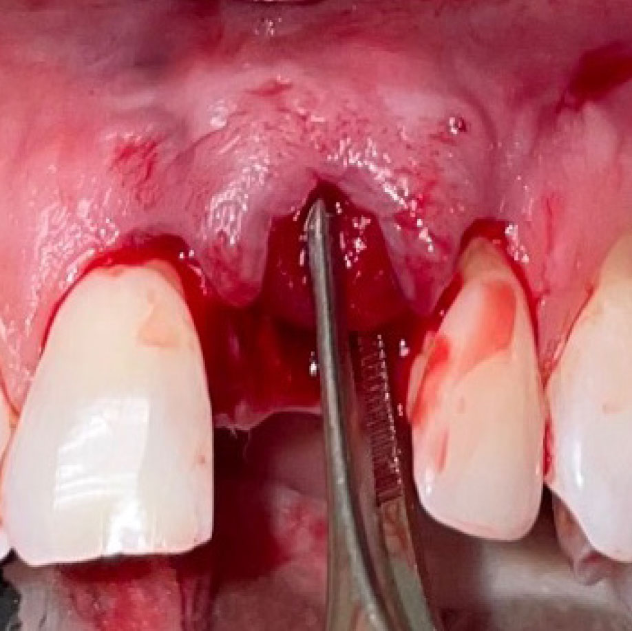
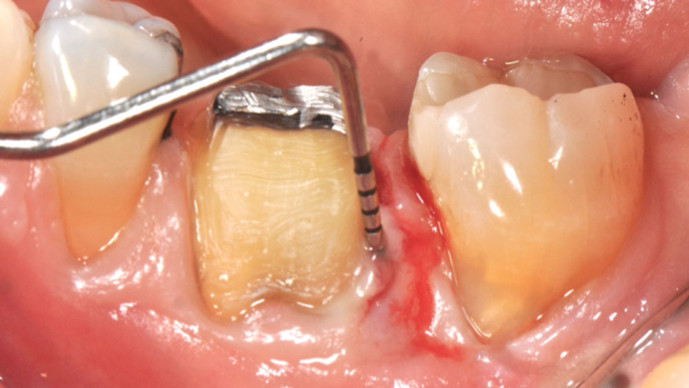
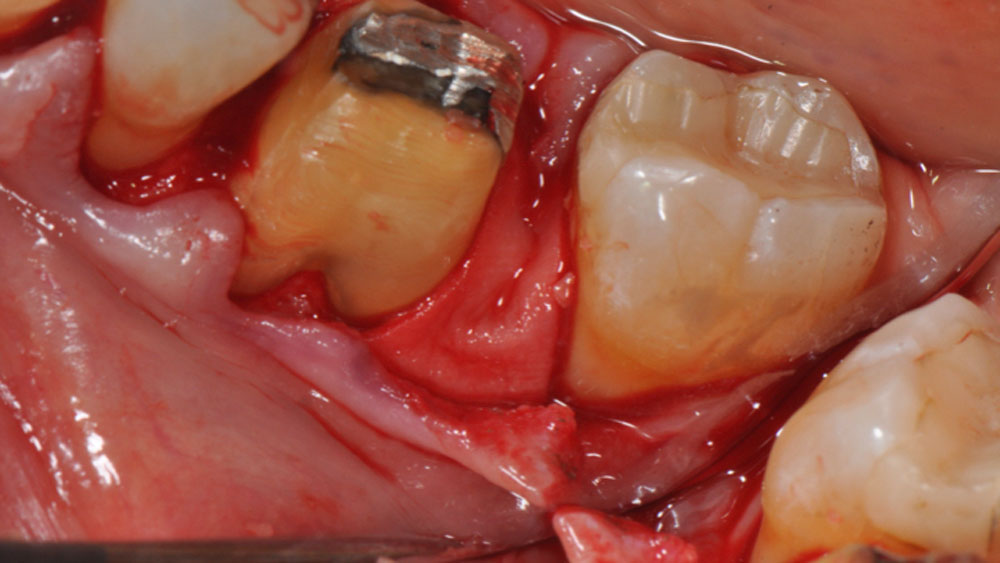
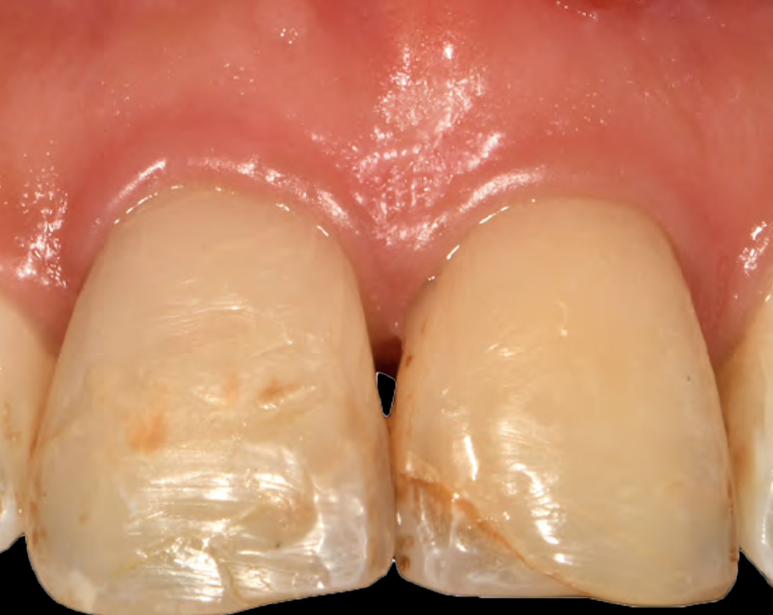
Tooth 22* required extraction in a 49-year-old female patient. The site demonstrated a Siebert Class III defect with a significant vertical discrepancy. The goal was to restore the site with an implant.  

Disclaimer: Case courtesy of Jasper Wu, DDS, MS (Periodontist) and Andrea Ravida, DDS, MS, PhD (Periodontist) | Pittsburgh, PA (USA). Used with permission. Images are provided for illustrative and educational purposes only. Results are not guaranteed, and individual outcomes may vary depending on patient-specific circumstances and clinical factors. This information is for general educational purposes only and does not constitute medical advice, nor does it necessarily reflect the official position, opinion, or recommendations of Geistlich. Treatment decisions are made at the clinician’s discretion based on the unique needs of each patient.

*Universal Numbering System

CLINICAL CASE

THE SITUATION

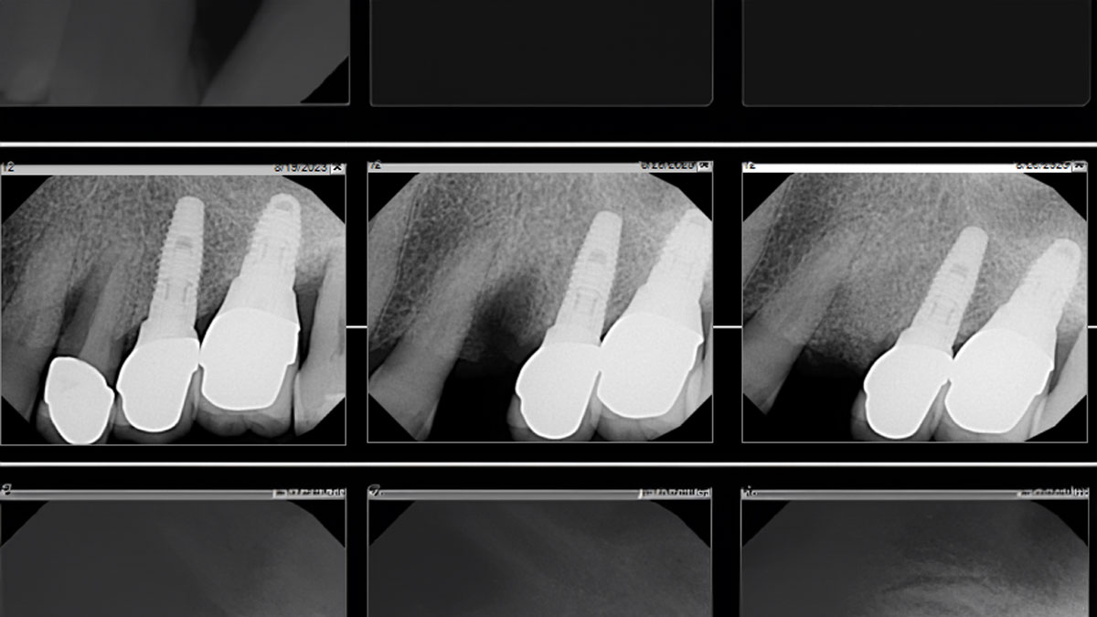
A 64-year-old female patient underwent extraction of Tooth 5*and Tooth 12* with GBR only, and implant placement at Tooth 7 and Tooth 8 with simultaneous buccal GBR.

*Universal Numbering System

Disclaimer: Case images courtesy of a board-certified oral and maxillofacial surgeon (Los Angeles, CA, USA). Used with permission. Images are provided for illustrative and educational purposes only. Results are not guaranteed, and individual outcomes may vary depending on patient-specific circumstances and clinical factors. This information is for general educational purposes only and does not constitute medical advice, nor does it necessarily reflect the official position, opinion, or recommendations of Geistlich. Treatment decisions are made at the clinician’s discretion based on the unique needs of each patient.

CLINICAL CASE

THE SITUATION

Tooth 13* required extraction in a 54-year-old male patient. The buccal bone was absent. Goal was to restore the site with an implant.

* Universal Numbering System

Disclaimer: Case images courtesy of a board-certified oral and maxillofacial surgeon (Los Angeles, CA, USA). Used with permission. Images are provided for illustrative and educational purposes only. Results are not guaranteed, and individual outcomes may vary depending on patient-specific circumstances and clinical factors. This information is for general educational purposes only and does not constitute medical advice, nor does it necessarily reflect the official position, opinion, or recommendations of Geistlich. Treatment decisions are made at the clinician’s discretion based on the unique needs of each patient.

BIOBRIEF

Vertical Bone Augmentation with Bone Plate Technique and Soft Tissue Phenotype Modification

Dr. Muhammad Saleh
Muhammad Saleh, BDS, MSD, PhD

THE SITUATION

A patient presented with a vertical bone defect in the maxillary anterior region and expressed high esthetic demands while explicitly wishing to avoid autogenous grafting. This required a regenerative strategy that could reliably achieve vertical augmentation, maintain space, and deliver a stable peri-implant soft tissue phenotype—all without harvesting autogenous blocks or connective tissue.

THE RISK PROFILE

Low RiskMedium RiskHigh Risk
Patient’s esthetic requirementsLowMediumHigh
Height of the smile lineLowMediumHigh
Gingival biotypeThick – “low scalloped”Medium – “medium scalloped”Thin – “high scalloped”
Infection at implant sightNoneChronicAcute
Bone height at adjacent tooth≤ 5 mm from contact point5.5 – 6.5 mm from contact point≥ 7 mm from contact point
Restorative status of adjacent toothIntactRestored (#8 has been restored)
Width of tooth gap1 tooth (≥ 7 mm)1 tooth (≤ 7 mm)2 teeth or more
Soft-tissue anatomyIntactCompromised
Bone anatomy of the alveolar ridgeNo defectHorizontal defectVertical defect

THE APPROACH

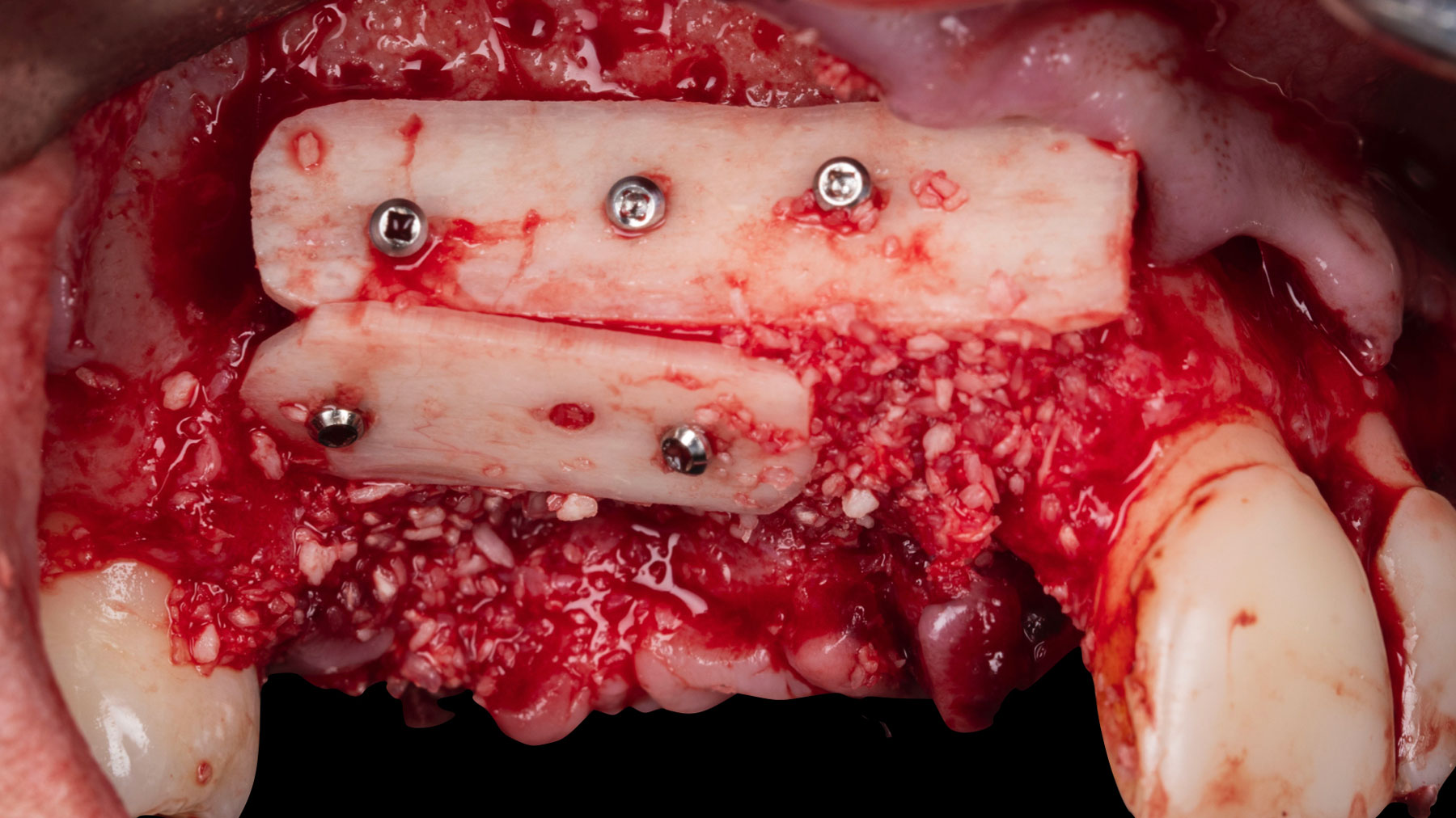
Vertical ridge augmentation was performed using allogenic cortical plates fixed with microscrews to create a stable, space-maintaining box, which was densely packed with vallos® mineralized cortico-cancellous granules hydrated with the rhPDGF-BB component of GEM 21S®. Five months later, the implants were placed, and the peri-implant soft tissue phenotype was enhanced using Geistlich Mucograft® and Geistlich Fibro-Gide® to improve soft-tissue height and thickness.

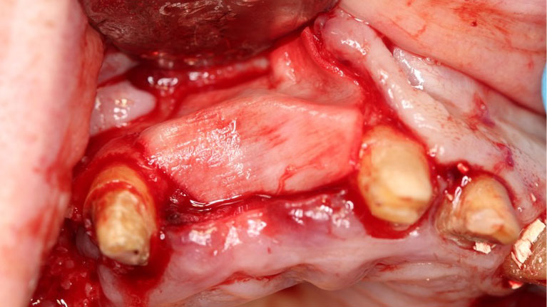
Preoperative view showing the anterior maxillary defect with loss of #6 and #7; #5 and #8 are planned to be extracted because of severe mobility and advanced bone loss.
Full-thickness mucoperiosteal flap elevation revealing a vertically and horizontally deficient ridge in the #5–8 region, prepared for augmentation.
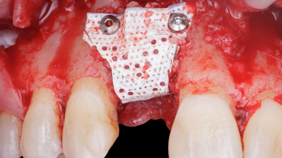
Adaptation and fixation of two allogenic cortical bone plates to the native ridge using micro screws to create a “box” configuration for vertical bone augmentation. The box between and around the plates is packed with vallos® mineralized cortico-cancellous granules mixed with rhPDGF-BB component of GEM 21S®, completing the three-dimensional reconstruction of the defect.
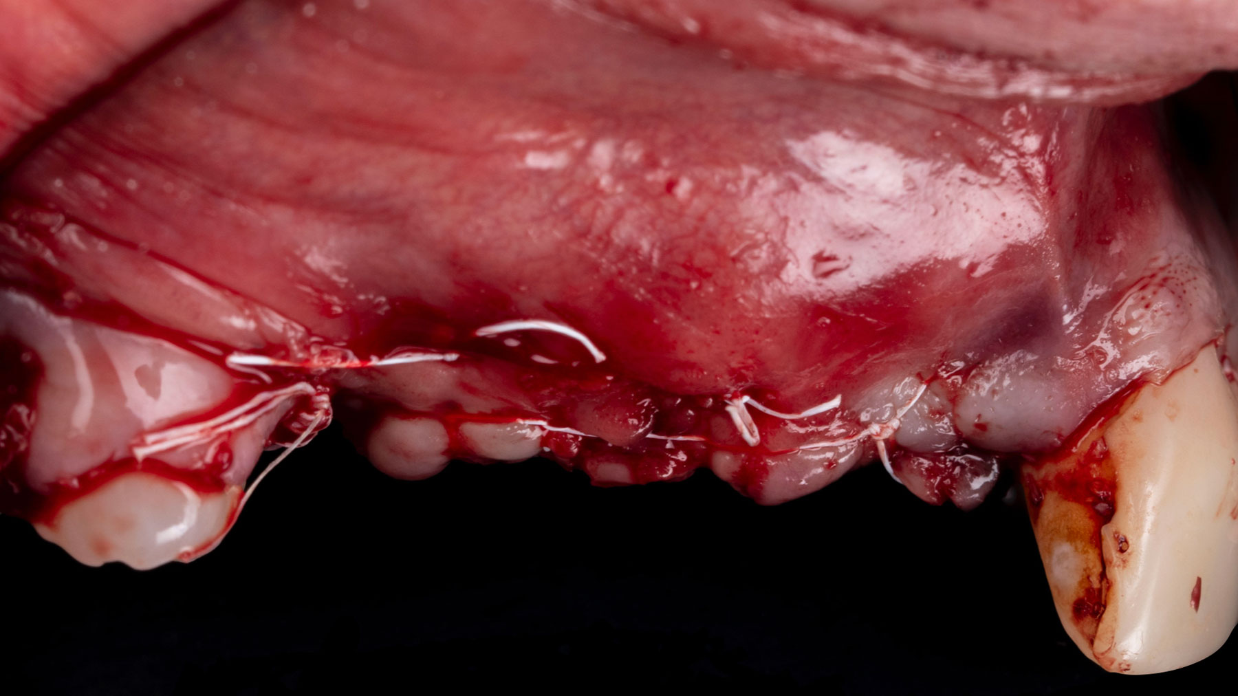
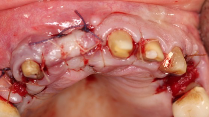
Immediate closure with PTFE sutures providing tension-free primary closure over the augmented site.
Site at five months post-op.
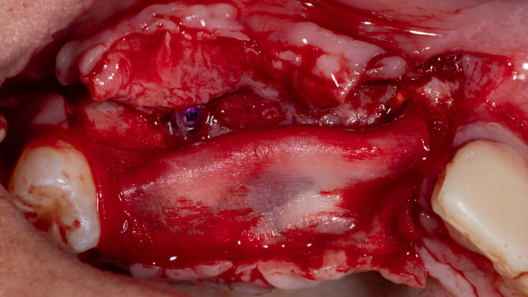
Re-entry after healing demonstrating well integrated cortical plates and a vertically augmented ridge suitable for implant placement.
Occlusal view showing well integrated particulate bone at relatively early stage.
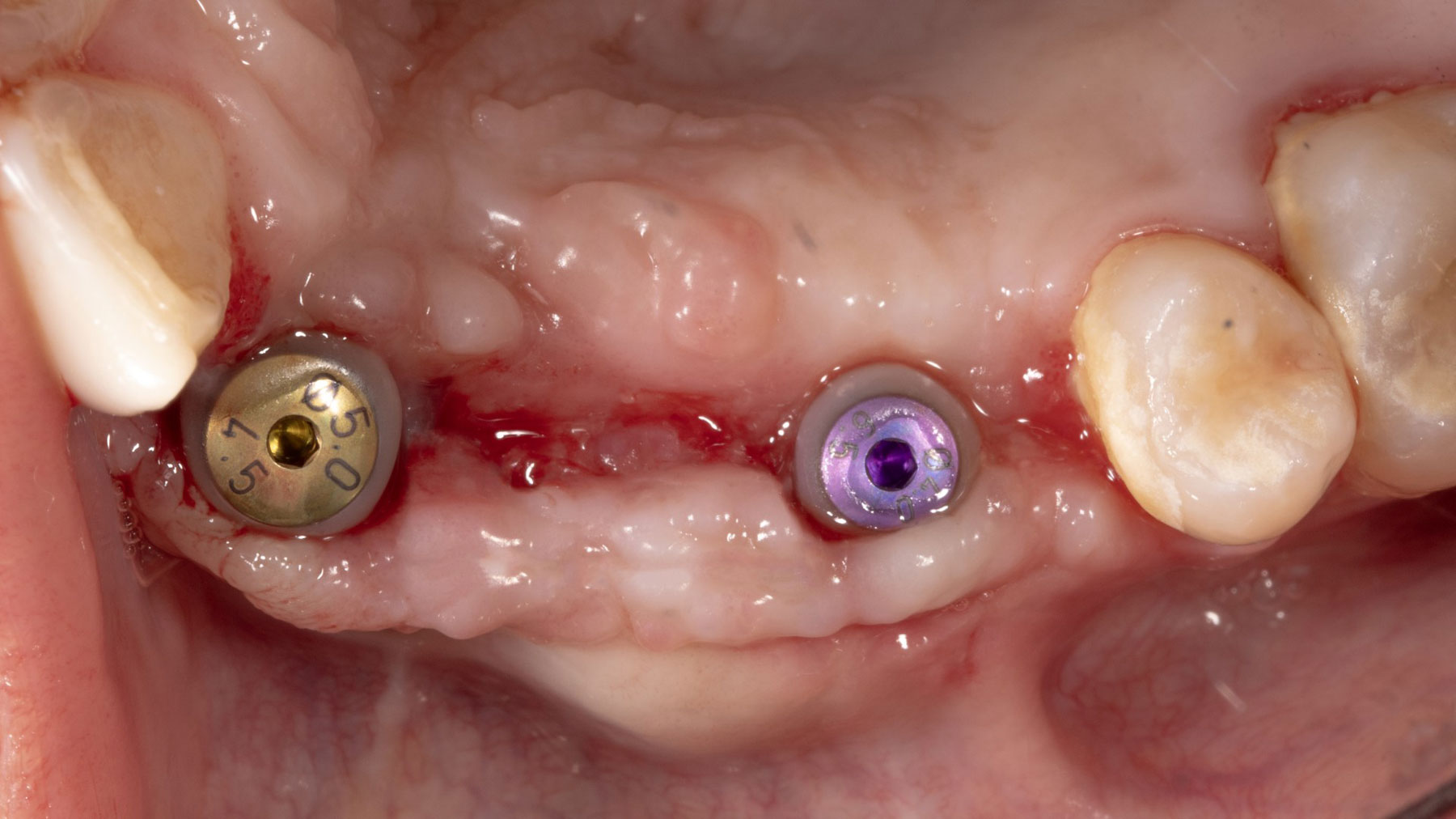
Placement of two implants into the regenerat ed ridge, with a regular diameter implant anteriorly and a wide diameter implant posteriorly.
Geistlich Mucograft® is layed on top of the implants before flap closure to improve the tissue phenotype.
Flap closure
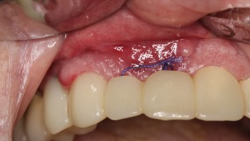
Occlusal view after the second stage for implant uncovery with Geistlich Fibro-Gide® left between the flap intentionally slightly exposed. Due to volume stability of Geistlich Fibro-Gide®, it aids in maintaining tissue verticality during healing.
Occlusal view after 2 weeks of the second stage showing excellent healing of Geistlich Fibro-Gide® with secondary intention with traces of keratinzed tissue forming between wound edges. Both healing abutments were replaced with bigger sizes to aid with tissue contouring before final restoration.

“The patient had high esthetic demands and specifically wanted to avoid autogenous grafting. Remarkably, this case was completed with 0% autogenous tissue, demonstrating that outcomes traditionally thought to require autogenous grafts can be achieved otherwise.”

— Dr. Muhammad Saleh

THE OUTCOME

Vertical ridge augmentation of the maxillary anterior was achieved using allogenic cortical bone plates fixed with microscrews to create a box configuration, filled with vallos® mineralized cortico-cancellous granules mixed with rhPDGF-BB component of GEM21S®. At implant placement,  soft tissue phenotype was enhanced using Geistlich Mucograft® and Geistlich Fibro-Gide®.

Rigid fixation of cortical plates creating a stable, space-maintaining box with tension-free soft tissue closure are the most critical factors for predictable vertical ridge augmentation.”

Muhammad Saleh, BDS, MSD, PhD
Dr. Muhammad Saleh

Muhammad Saleh, BDS, MSD, PhD

Dr. Muhammad H. Saleh, Diplomate of the American Board of Periodontology, is a full-time faculty member at the University of Michigan and an internationally recognized lecturer in periodontology and implant dentistry. He has published over 140 peer-reviewed articles and reviews for leading Q1 journals. His numerous honors include the 2021 AAPF Schoor Award, 2022 Ramfjord Symposium Award, 2022 AAP Institute Award, 2024 AAPF LEAD and Nevins Awards, and the 2025 Roy H. Roberts Award.

BIOBRIEF

Vertical and Horizontal Maxillary Ridge Reconstruction with Advanced Grafting

Nikolaos Soldatos, DDS, PhD, MSD

THE SITUATION

A 26-year-old edentulous female presented for implant-supported maxillary rehabilitation. She was systemically healthy and classified as ASA I. Clinical and radiographic evaluation revealed a Seibert Class III maxillary ridge defect with combined horizontal and vertical deficiencies. The patient exhibited a medium smile line and reported a highly active lifestyle with regular physical activity, indicating the need for a stable, durable, and esthetically driven implant rehabilitation capable of meeting long-term functional demands.

THE RISK PROFILE

Low RiskMedium RiskHigh Risk
Patient’s healthIntact immune system
Non-smoker
Light smokerImpaired immune system 
Patient’s esthetic requirementsLowMediumHigh
Height of the smile lineLowMediumHigh
Gingival biotypeThick – “low scalloped”Medium – “medium scalloped”Thin – “high scalloped”
Infection at implant sightNoneChronicAcute
Bone height at adjacent tooth≤ 5 mm from contact point5.5 – 6.5 mm from contact point≥ 7 mm from contact point
Soft-tissue anatomyIntactCompromised
Bone anatomy of the alveolar ridgeNo defectHorizontal defectVertical defect

THE APPROACH

Horizontal and vertical ridge augmentation was performed using a full-thickness maxillary flap with two distal vertical releasing incisions. Extensive buccal periosteal release and cortical perforations were carried out. A composite graft of large-particle vallos® allograft (demineralized cortical granules) and large-particle Geistlich Bio-Oss® xenograft, combined with rhPDGF-BB, a component of GEM 21S®, was placed and stabilized under a high-density PTFE membrane, which was secured with four horizontal mattress and multiple simple interrupted 4-0 PTFE sutures.
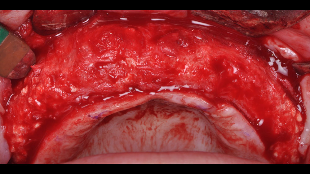
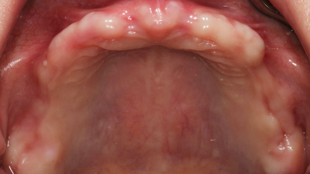
Pre‑operative occlusal photograph of the maxillary arch showing a pronounced Seibert Class III ridge deformity, with simultaneous horizontal and vertical soft and hard tissue loss in the edentulous area.
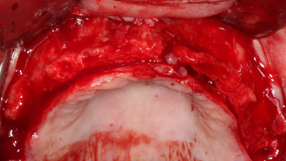
Intraoperative view following reflection of full‑thickness flaps, revealing a pronounced combined horizontal and vertical ridge deficiency.
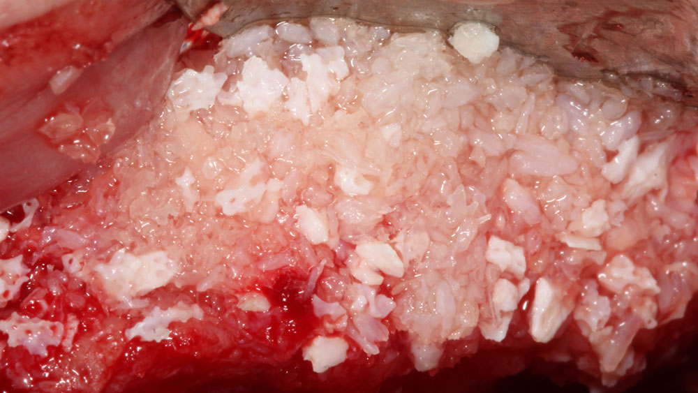
Upper left view showing the mixture of large-particle vallos® allograft, demineralized cortical granules, and large-particle Geistlich Bio-Oss® xenograft, combined with rhPDGF-BB, a component of GEM 21S®.
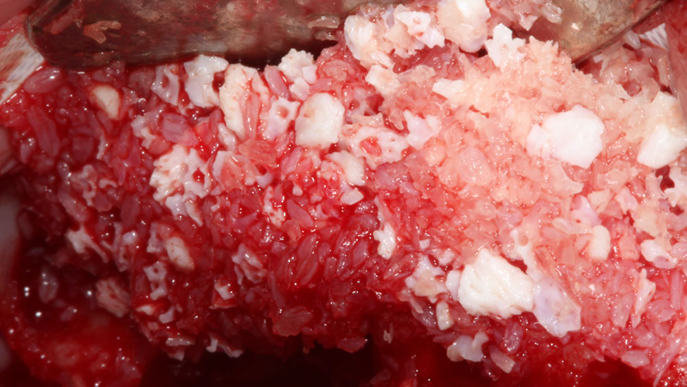
Upper right view showing the mixture of large-particle vallos® allograft, demineralized cortical granules, and large-particle Geistlich Bio-Oss® xenograft, combined with rhPDGF-BB, a component of GEM21S®.
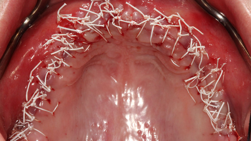
Closure of the maxilla was achieved using a PTFE barrier membrane and 4‑0 PTFE sutures. Horizontal mattress sutures were placed to provide initial tension relief and flap stabilization, while simple interrupted sutures were used for precise approximation of the wound margins.
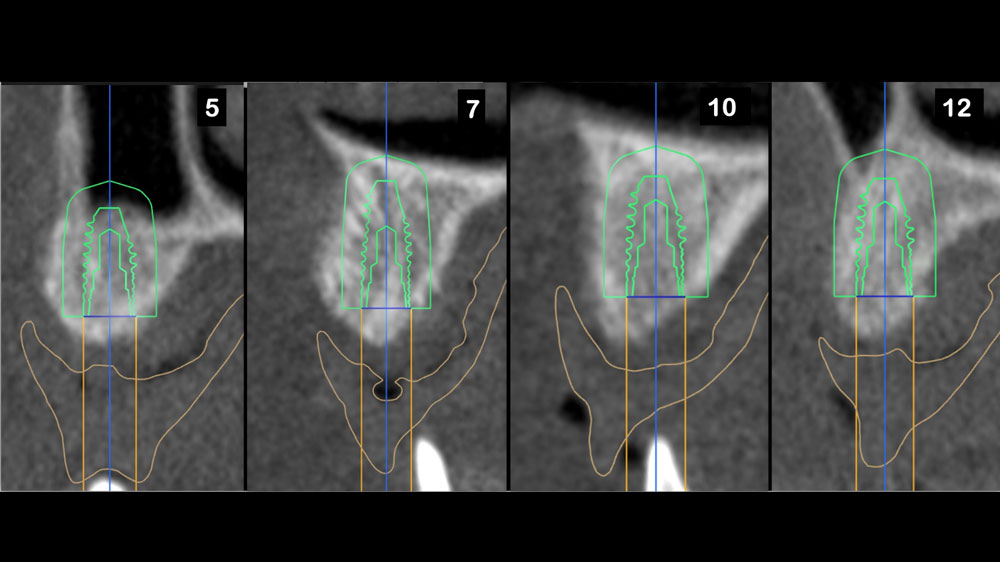
Six‑month postoperative CBCT images with virtual implant planning reveal sites #5, 7, 10, and 12, exhibiting 5–7 mm of horizontal ridge augmentation and 3–4 mm of vertical ridge augmentation.
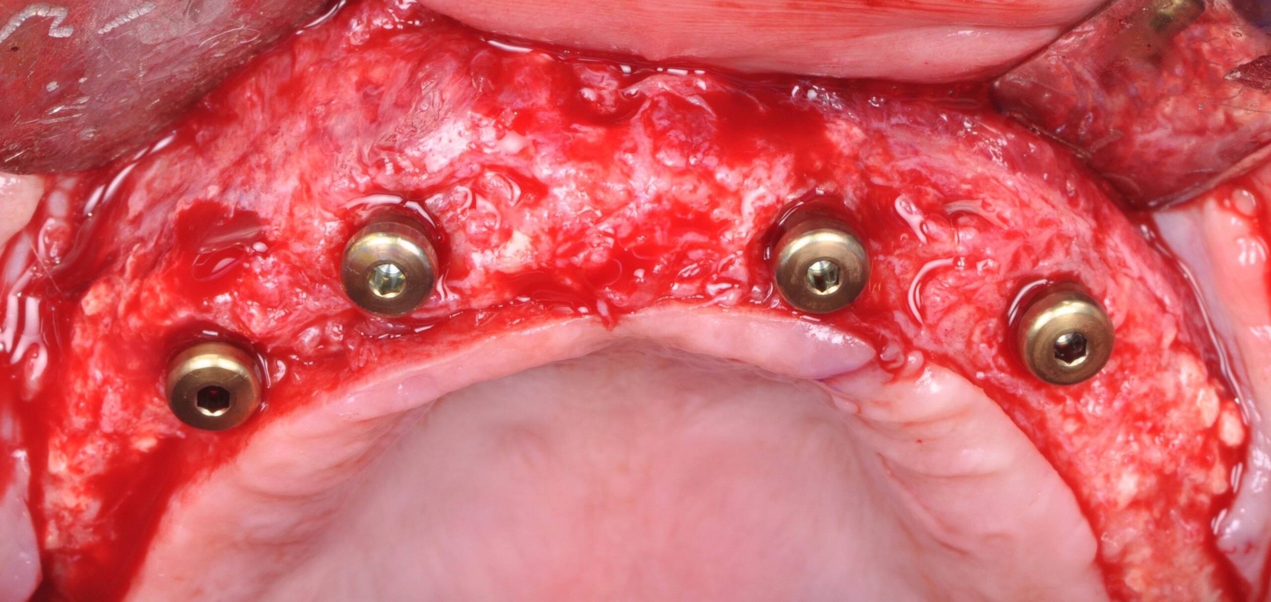
Intraoperative view demonstrating vertical and horizontal ridge augmentation at 7 months following the initial augmentation procedure.
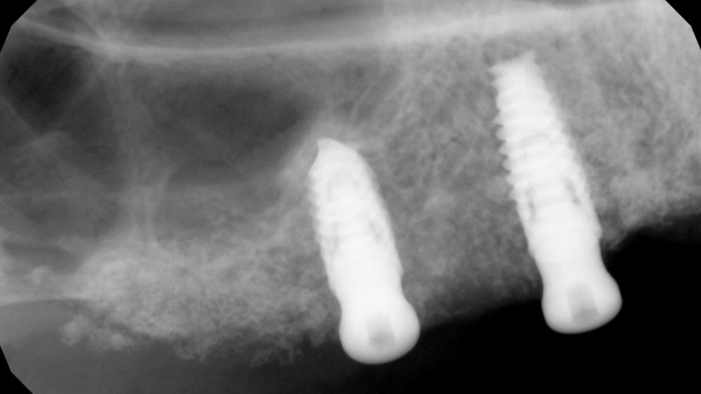
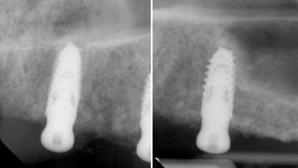
A 4.3 × 8 mm tapered implant with crestal sinus elevation was placed at site #5, and a 4.3 × 10 mm tapered implant was placed at site #7.
(Left) A 4.3 × 10 mm implant was inserted at the site. (Right) Placement of a 4.3 × 10 mm implant in conjunction with a crestal sinus elevation.

“The patient’s young age and
excellent systemic health are favorable prognostic factors, but achieving optimal outcomes requires meticulous surgical execution, careful soft tissue management, and strict adherence to post-operative instructions to minimize complications and ensure long-term success.”

Nikolaos Soldatos, DDS, PhD, MSD

THE OUTCOME

Horizontal (5–7 mm) and vertical (3–4 mm) ridge augmentation were successfully obtained, with stablewound management achieved through precise suturing techniques. The osteoinductive properties of the large vallos®, demineralized granules combined with rhPDGF-BB, a component of GEM21S® promoted high-quality bone regeneration, whereas the large Geistlich Bio‑Oss®, xenograft particles contributed to volume preservation by moderating resorption. As a result, four 4.3 mm implants were placed with high primary stability, each exceeding 35 N·cm of insertion torque, providing a strong foundation for a predictable esthetic and functional restoration.
The combination of rhPDGF-BB, a component of GEM 21S®, promotes enhanced bone regeneration by stimulating cellular proliferation and osteogenesis. When supplemented with Geistlich Bio-Oss®, which maintains graft volume through slow resorption kinetics, this approach provides predictable ridge augmentation. Together, these graft components support stable, high‑quality bone formation and foster optimal conditions for long‑term implant integration.”
Nikolaos Soldatos, DDS, PhD, MSD

Nikolaos Soldatos, DDS, PhD, MSD

Dr. Soldatos is a tenure-track Associate Professor and Clinical Director of the Postgraduate Advanced Program in Periodontics at Oregon Health & Science University. He is a Board-Certified Periodontist and Implant Surgeon, he holds a DDS, PhD, and MSD, and has completed advanced periodontal and implant training in both the U.S. and Europe. Dr. Soldatos’ research focuses on translational implant biology and bone regeneration. He is also a Fellow of the Academy of Osseointegration.

BIOBRIEF

Immediate Implant Placement and Provisionalization for Anterior Esthetics

David E. Urbanek, DMD, MS

THE SITUATION

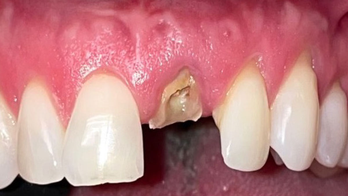
A healthy, 56-year-old female presented with fractured, endodontically treated tooth #9. The tooth was fractured at the gingival level and asymptomatic. Both the patient and the restorative dentist had high esthetic expectations, and preferred immediate implant placement with provisionalization if possible.

THE RISK PROFILE

Low RiskMedium RiskHigh Risk
Patient’s healthIntact immune system
Non-smoker
Light smokerImpaired immune system 
Patient’s esthetic requirementsLowMediumHigh
Height of the smile lineLowMediumHigh
Gingival phenotypeThick – “low scalloped”Medium – “medium scalloped”Thin – “high scalloped”
Shape of dental crownsRectangularTriangular
Infection at implant sightNoneChronicAcute
Bone height at adjacent tooth≤ 5 mm from contact point5.5 – 6.5 mm from contact point≥ 7 mm from contact point
Restorative status of adjacent toothIntactRestored
Soft-tissue anatomyIntactCompromised
Bone anatomy of the alveolar ridgeNo defectHorizontal defectVertical defect

THE APPROACH

The goals of this case were to: 1) maximize pink and white esthetic scores, 2) preserve the pre-operative soft tissue architecture, 3) minimize hard and soft tissue remodeling over time following tooth extraction, and 4) promote long-term implant health and stability. To achieve these objectives, immediate implant placement with immediate provisionalization was planned. The extraction was performed with minimal flap elevation, and the implant was placed in a guided manner with palatal bias to maximize the facial gap. This gap was then grafted with a slowly resorbing bovine xenograft (Geistlich Bio-Oss Collagen®) to minimize remodeling of the labial bone plate. To further enhance soft tissue volume and contour, the facial soft tissue was augmented after using a Geistlich Fibro-Gide® collagen matrix. Finally, an immediate provisional crown was placed to contain the bone graft and provide support for the soft tissue.

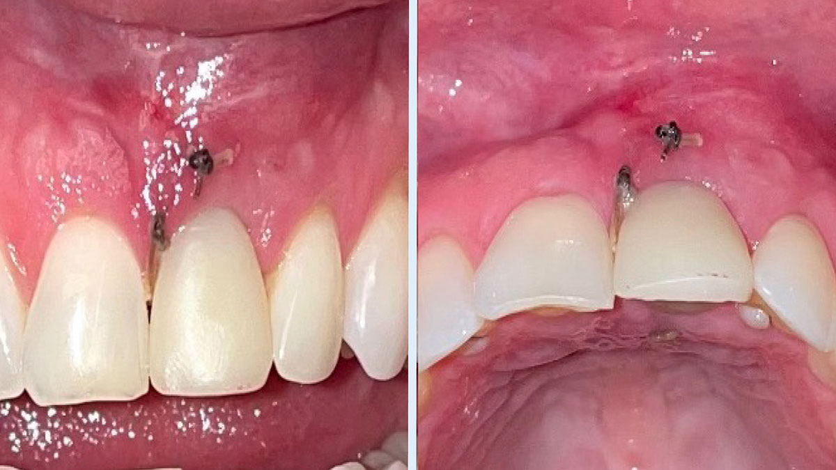
Pre-operative image showing a fractured, endodontically treated tooth #9.
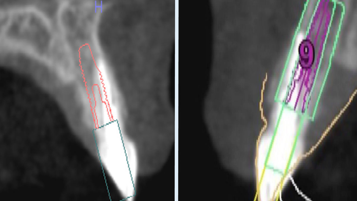
Left: Pre-operative sagittal CBCT image demonstrating a Kan Class 1 sagittal root position with initial digital implant planning conducted during the consultation appointment. Right: Definitive digital implant planning.
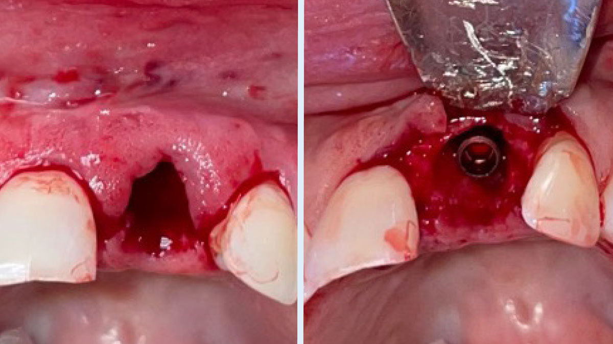
Left: Tooth #9 was extracted with minimal flap elevation, preserving the papillae. Right: A 3.6 x 15 mm implant was placed in a fully guided manner with a palatal bias to maximize the facial gap dimension, achieving a +2mm gap.
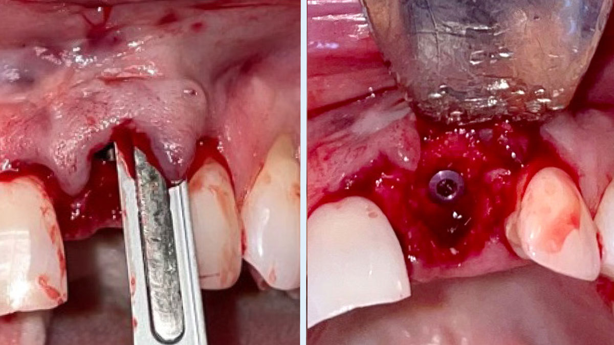
Left: The underside of the periosteum was incised just apical to the mucogingival junction, and a supra-periosteal pocket was created using sharp dissection with a #15 blade. Right: The facial gap was packed with 50 mg of Geistlich Bio-Oss Collagen®. A cover screw was temporarily placed to prevent bone graft granules from entering the implant chamber.
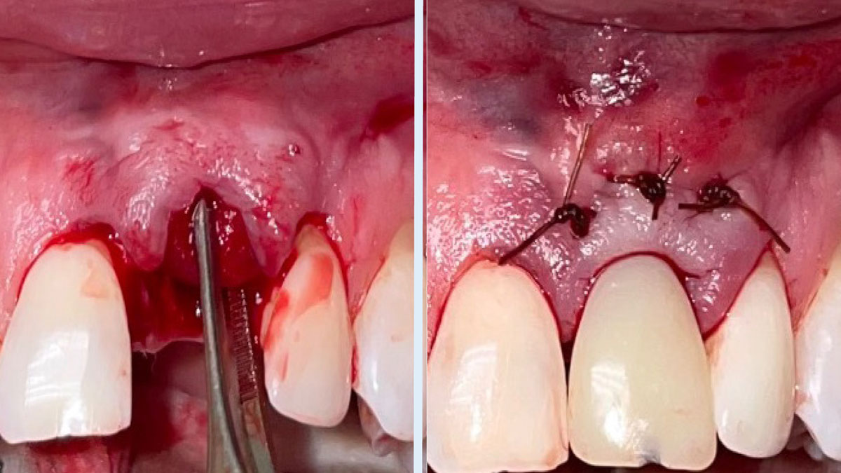
Left: A 15mm x 20mm x 3mm Geistlich Fibro-Gide® collagen matrix was inserted into the supra-periosteal pocket, with the coronal portion positioned to cover the facial bone crest. Right: The provisional crown was placed, and the facial mucosal flap was coronally advanced to cover the Geistlich Fibro-Gide® collagen matrix.
2-week post-operative follow-up
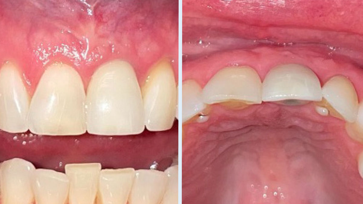
Final restoration at 16 months post-operative.
Final restoration with a screw-retained crown at 16 months post-operative.
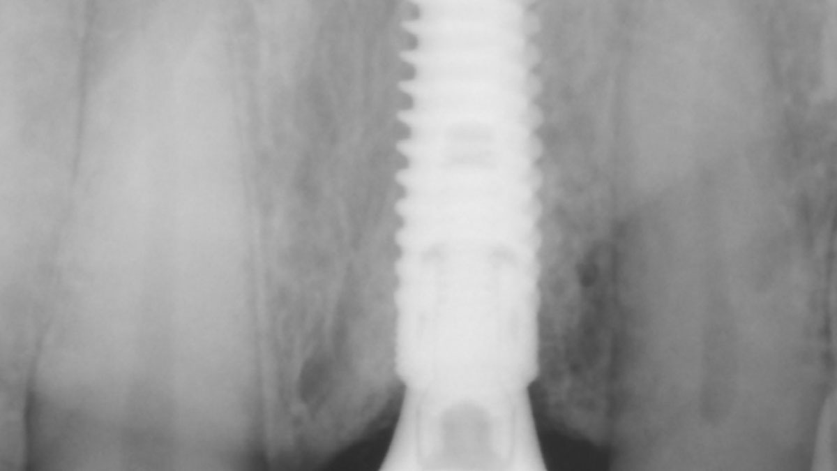
Periapical radiograph at 16 months post-operative demonstrating stable crestal bone levels.

“This was a challenging case in which the patient and her dentist had high esthetic expectations. The goal of this case was to preserve as much of the preoperative anatomy as possible and minimize the inevitable hard and soft tissue remodeling that occurs after a tooth is removed.”

David E. Urbanek, DMD, MS

THE OUTCOME

This case finished with excellent pink and white esthetic scores, and the patient and her dentist were very pleased with the results. Most importantly, the implant demonstrated excellent health and stability over one year since placement.

To obtain the best result with challenging cases, such as this one, I always approach them with thorough pre-surgical assessment, proper hard and soft tissue management, and the use of high-quality, evidence-based materials.”

David E. Urbanek, DMD, MS

David E. Urbanek, DMD, MS

Dr. Urbanek is a board-certified Oral & Maxillofacial Surgeon who practices in St. Louis, Missouri. He completed his OMS training at Carle Foundation Hospital in Champaign/Urbana, Illinois. He earned his Dental Degree from the Case Western Reserve University School of Dental Medicine, and a Master’s Degree with Honors in Applied Anatomy from CWRU. Dr. Urbanek serves as adjunct faculty at Carle Foundation Hospital and the A. T. Still University, Missouri School of Dentistry & Oral Health. In addition he avidly lectures to the dental and OMS community throughout the country.

BIOBRIEF

Horizontal Ridge Augmentation with a Layered Allograft-Xenograft Approach

Eswar Kandaswamy, BDS, MS
Amber Kreko, DDS

THE SITUATION

The patient presented to the clinic for a dental implant in the tooth #12 location. Clinical evaluation revealed a ridge deficiency. A Cone Beam Computed Tomography (CBCT) scan was taken, confirming insufficient ridge width for implant placement. As a result, the site was treatment planned for horizontal ridge augmentation.

THE RISK PROFILE

Low RiskMedium RiskHigh Risk
Patient’s healthIntact immune systemLight smokerImpaired immune system 
Patient’s esthetic requirementsLowMediumHigh
Gingival biotypeThick – “low scalloped”Medium – “medium scalloped”Thin – “high scalloped”
Infection at implant sightNoneChronicAcute
Bone height at adjacent tooth≤ 5 mm from contact point5.5 – 6.5 mm from contact point≥ 7 mm from contact point
Restorative status of adjacent toothIntactRestored
Soft-tissue anatomyIntactCompromised
Bone anatomy of the alveolar ridgeNo defectHorizontal defectVertical defect

watch video download pdf
THE APPROACH

Horizontal ridge augmentation was performed using a horizontal layering technique. An inner layer of demineralized freeze-dried bone allograft (DFDBA), featuring vallos® demineralized cortical particles (to promote osteoinduction), was followed by an outer layer of deproteinized bovine bone, Geistlich Bio-Oss® (to maintain space and volume). The graft was contained with a native bilayer collagen membrane, Geistlich Bio-Gide®, and secured with titanium pins (tacks).

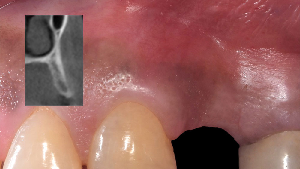
Image 1a depicts the pre-operative situation, while the inset shows a cross-sectional view of the deficient ridge in area #12.
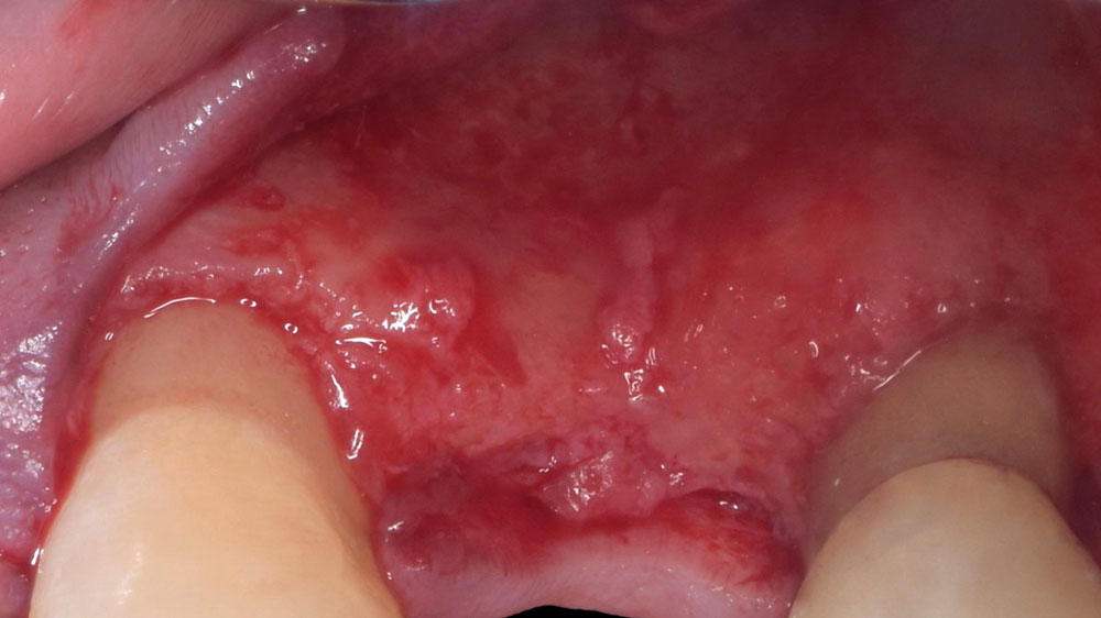
Pre-operative view, post-flap reflection.
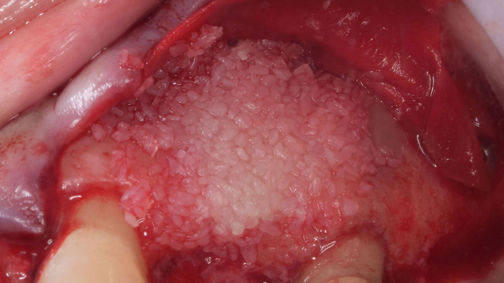
An inner layer of vallos® demineralized cortical particles (allograft) was applied to the defect.
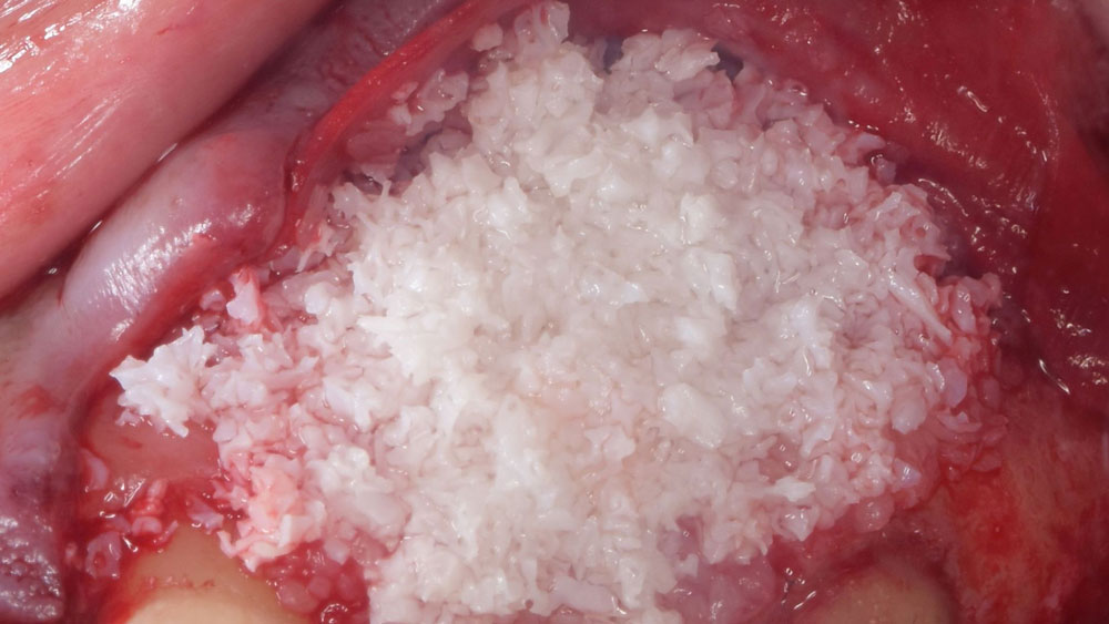
An outer layer of Geistlich Bio-Oss® (xenograft) was applied on top of vallos® (allograft).
Bone grafts covered with Geistlich Bio-Gide® and secured via tacks
Final sutured closure over the bone grafts covered with Geistlich Bio-Gide® and secured via tacks
Four-week post-operative image demonstrating good soft tissue healing.
Sequential CBCT imaging comparisons, from pre-operative to 6 and 9 months post-operative, confirm the re-establishment of horizontal ridge width achieved through bone augmentation.
Comparative CBCT cross-sectional analysis of pre-operative (left) and 6-month post-operative (right) images reveals an increase in horizontal ridge width.

“By horizontally layering two distinct bone graft materials—Geistlich Bio-Oss® and vallos®—this approach was designed to tailor the regenerative environment, harnessing the unique osteoinductive potential of the allograft and the long-term space-maintaining properties of the xenograft to optimize both early bone formation and dimensional stability.”

Eswar Kandaswamy, BDS, MS

THE OUTCOME

The 6-month post-operative CBCT  evaluation demonstrated sufficient ridge width for restoratively driven implant placement, a result achieved through the utilization of vallos® and Geistlich Bio-Oss® bone graft materials.

Four-week post-operative image demonstrating good soft tissue healing.

The incorporation of vallos® demineralized cortical particles, leveraging its reliable osteoinductive properties, was paramount for achieving predictable and successful bone regeneration in this case.”

Eswar Kandaswamy, BDS, MS

The patient required horizontal ridge augmentation for successful placement of a dental implant.”

Eswar Kandaswamy, BDS, MS

Eswar Kandaswamy, BDS, MS

Dr. Eswar Kandaswamy, BDS MS, is an Assistant Professor at Louisiana State University Health Sciences Center, School of Dentistry. He earned his Dental Degree from Sri Ramachandra University, India, and practiced general dentistry for two years. He then completed his specialty training in Periodontics and a Master of Science at The Ohio State University.

Amber Kreko, DDS

Dr. Amber Kreko, DDS is a third-year Periodontics resident at Louisiana State University School of Dentistry, soon to earn her Master of Science. With a foundation in dental hygiene and six years of clinical practice in Southeast Louisiana,she returned to LSU for her DDS. Her comprehensive background enriches her approach to periodontal care. Upon graduation, she will transition to private practice.

BIOBRIEF

Lateral and Vertical Bone Regeneration with Simultaneous Soft Tissue Augmentation

Arnd Lohmann, MSc

THE SITUATION

After extraction of the periodontally damaged tooth #20 the preoperative Cone-Beam Computed Tomography (CBCT) imaging shows reduced vertical bone volume in the area of tooth #s 18 – 20. A lateral and vertical bone  regeneration was necessary.

The goal of treatment was a late implant placement after bone regeneration and creation of stable periimplant soft tissue for long-term implant preservation.

THE RISK PROFILE

Low RiskMedium RiskHigh Risk
Patient’s healthIntact immune systemLight smokerImpaired immune system 
Patient’s esthetic requirementsLowMediumHigh
Gingival biotypeThick – “low scalloped”Medium – “medium scalloped”Thin – “high scalloped”
Infection at implant sightNoneChronicAcute
Bone height at adjacent tooth≤ 5 mm from contact point5.5 – 6.5 mm from contact point≥ 7 mm from contact point
Width of tooth gap1 tooth (≥ 7 mm)1 tooth (≤ 7 mm)2 teeth or more
Soft-tissue anatomyIntactCompromised
Bone anatomy of the alveolar ridgeNo defectHorizontal defectVertical defect

Additional Risk Factors: Roots were divergent, and intra-radicular bone (septal bone) was excellent, with more than 5 mm of remaining apical bone to achieve optimal primary stability.

THE APPROACH

A customized bone regeneration procedure utilizing Yxoss CBR®. Followed by coverage of the graft with Geistlich Bio-Gide® for the purpose of Guided Bone Regeneration (GBR). Soft tissue thickening using Geistlich Fibro-Gide®. Delayed implantation into the augmented tissue. A vestibuloplasty with Geistlich Mucograft® for the regeneration of keratinized mucosa.

Baseline situation and planning: vertical bone deficit of 5.29mm in section 18-20.
Central incision in the keratinized mucosa and perforated cortical bone
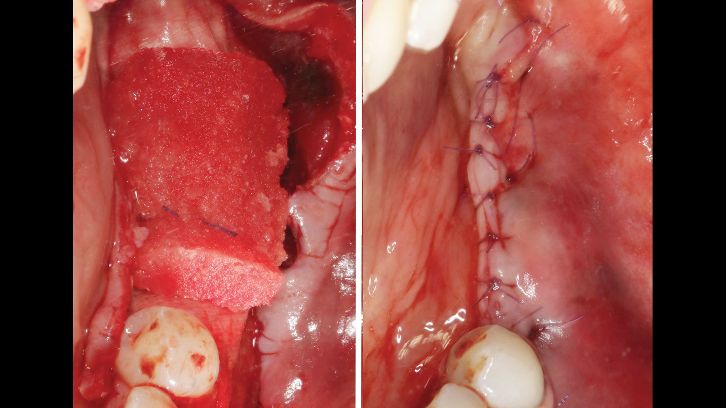
Yxoss CBR® filled with a 50:50 mix of autologous bone and Geistlich Bio-Oss®. Coverage of the titanium scaffold with Geistlich Bio-Gide®
Geistlich Fibro-Gide® is tailored to the defect and a tension-free suture is done.
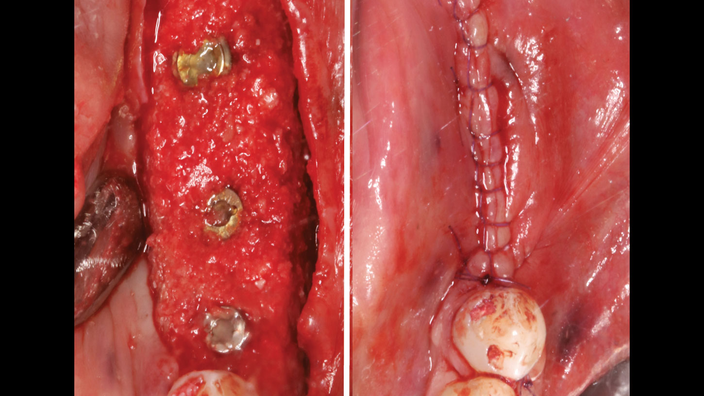
4 months after augmentation shows the bone situation after removal of the titanium scaffold and insertion of implants. Autologous bone chips are applied on top of the implants. Occlusal view of the mucosa level after implantation and wound closure.
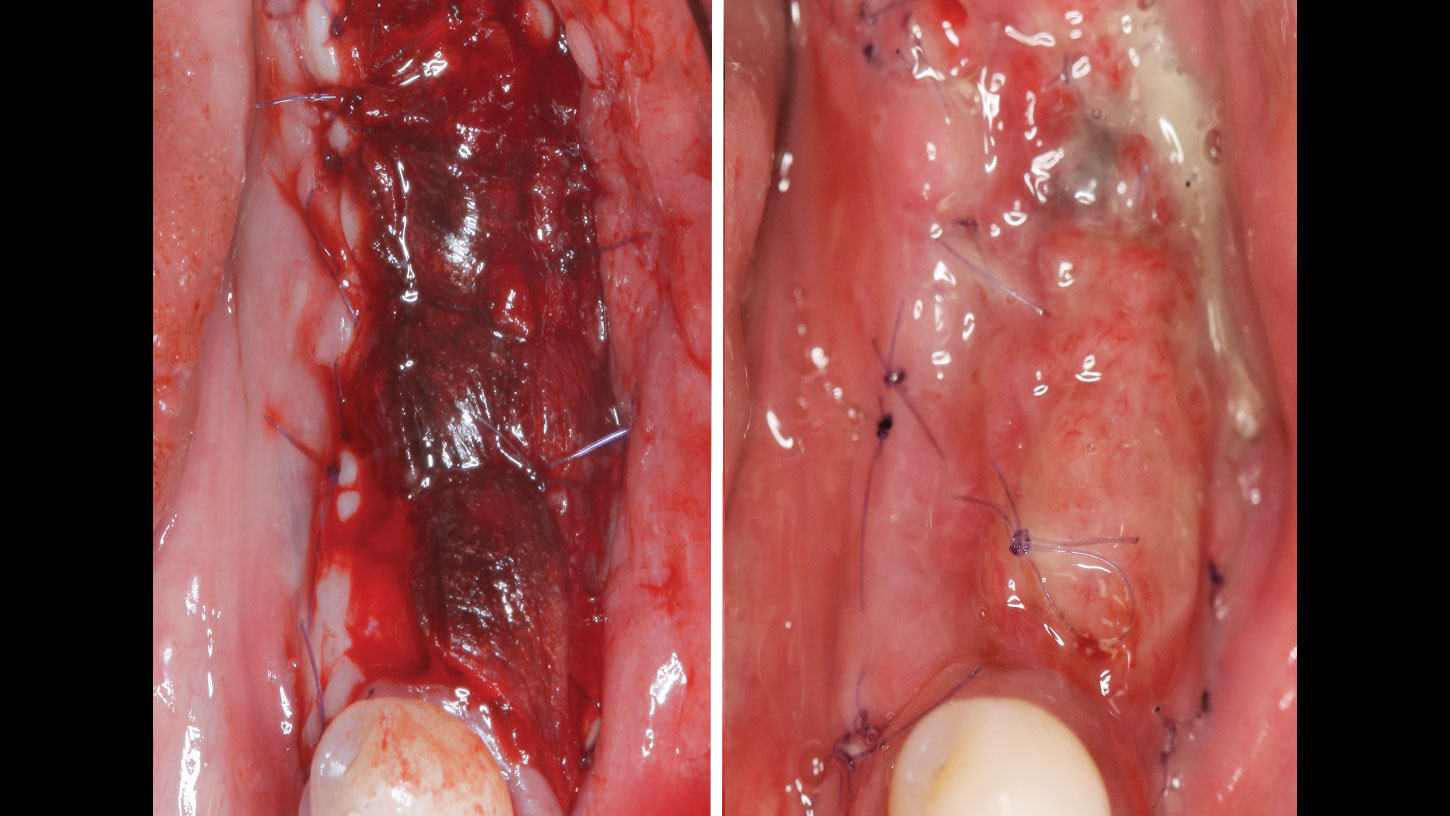
6 weeks after implantation and 5 1/2 months after augmentation. The mucosa is prepared apically; the preparation reaches the periosteum apically. The recipient site is fully surrounded by keratinized mucosa. Geistlich Mucograft® is sutured into the area.
6 months after regeneration shows about 5mm of vertical bone regeneration. Panoram ic X-Ray after implant placement shows clearly recognizable mucosa shadows.
One year after augmentation shows the dental prosthesis inserted occlusally.

“Using the Geistlich Fibro-Gide® matrix enabled concurrent augmentation of hard
and soft tissues without any postoperative complications. At the same time, the soft
tissue thickening facilitated floor of the mouth surgery and vestibuloplasty.”

— Arnd Lohmann, MSc

THE OUTCOME

Treatment resulted in approximately 5 mm of vertical bone regeneration. The potential occurrence of a dehiscence associated with a wound opening and exposure of Yxoss CBR® was able to be prevented with Geistlich Fibro-Gide®.

On one hand, the quality of the peri-implant soft tissue was improved by the
soft tissue thickening with Geistlich Fibro‑Gide® and, on the other, by increasing the width of keratinized mucosa with Geistlich Mucograft®. The treatment method chosen resulted in a reduced invasiveness and morbidity by avoiding a donor site for sourcing a transplant.

One year after augmentation shows the dental prosthesis inserted occlusally.

Implant therapy should restore the natural anatomical structures as closely as possible in order to avoid subsequent peri-implant problems.”

Arnd Lohmann, MSc

Arnd Lohmann, MSc

Dr. Arnd Lohmann is a recognized specialist in implantology and periodontology. He earned his dental license in Hamburg in 2002, completed his doctorate in 2003, and has been a partner at a private practice in Bremen since then.

With a Master of Science in Implantology (2007), he specializes in dental implantology and bone augmentation. He is an active speaker at national and international congresses, leads the Bremen study group of the German Society of Oral Implantology (DGOI), and is a member of DGOI, DGZI, and DGI. His practice is equipped with state-of-the-art technology, ensuring high-quality patient care.

CLINICAL CASE

THE OUTCOME

At 6 months, healing was complete with a bone fill of 10x10x6.5 mm. Tooth #7, affected by the combined defect, was fully regenerated. The adjacent ridge defect was also restored, enabling the placement of a regular-sized implant without the need for additional grafting.

Disclaimer: These results are not guaranteed; individual outcomes may vary depending on patient circumstances. This information is for informational purposes only and may not reflect Geistlich’s official position, opinion, or recommendation. Treatment decisions are made at the physician’s discretion, based on the unique needs of each patient.

GEM 21S® has not been approved by FDA for use in ridge augmentation in the US and the safety and effectiveness of GEM 21S® for this use has not been established by FDA.

CLINICAL CASE

THE APPROACH

Considering the patient’s age, condition, and good compliance, a regenerative approach was chosen to preserve all teeth. Due to the extent and severity of the defect, a bone graft, biologics, and a membrane were utilized. The goal was to restore lost attachment and stabilize the teeth through splinted therapy.

THE OUTCOME

At the 7-month follow-up, probing depths decreased from 7 mm (MB, DB) to 3 mm, with radiographic bone fill indicating a 90% success. Attachment gain of 3 mm was achieved, improving from 7 mm to 4 mm. The patient reported satisfactory outcomes, and splinting effectively maintained tooth stability with no mobility. 

Disclaimer: These results are not guaranteed; individual outcomes may vary depending on patient circumstances. This information is for informational purposes only and may not reflect Geistlich’s official position, opinion, or recommendation. Treatment decisions are made at the physician’s discretion, based on the unique needs of each patient.

CLINICAL CASE

THE APPROACH

Following flap elevation and thorough defect debridement, a xenograft bone substitute mixed with PDGF (GEM 21S®) was used to fill the defect. The graft was left uncovered without a membrane.

THE OUTCOME

The 1-year follow-up radiograph demonstrated near-complete bone fill in a previously deep defect. Clinically, shallow probing depths and healthy tissue were also observed at the 1-year follow-up.

Disclaimer: These results are not guaranteed; individual outcomes may vary depending on patient circumstances. This information is for informational purposes only and may not reflect Geistlich’s official position, opinion, or recommendation. Treatment decisions are made at the physician’s discretion, based on the unique needs of each patient.

CLINICAL CASE

THE APPROACH

The crown was biologically shaped, and the root surface was detoxified using Ellman burs. Following flap elevation and thorough defect debridement, an allograft mixed with GEM 21S® was used to fill the defect. The graft was then covered with Geistlich Bio-Gide®.

THE OUTCOME

At the 10-month follow-up, radiographs revealed evidence of bone fill, and clinically, the interdental tissue showed signs of maturation. At the 9-year follow-up, clinical photos demonstrated long-term stability, with maintained bone levels, minimal interproximal recession, and lack of facial recession. Radiographic analysis further confirmed the sustained stability of the bone.

Disclaimer: These results are not guaranteed; individual outcomes may vary depending on patient circumstances. This information is for informational purposes only and may not reflect Geistlich’s official position, opinion, or recommendation. Treatment decisions are made at the physician’s discretion, based on the unique needs of each patient.

BIOBRIEF

Bone Grafting and Immediate Implant Placement in the Maxillary First Molar Region

Waldemar D. Polido, DDS, MMS, PhD
Wel-Shao Lin, DDS, FACP, PhD, MBA

THE SITUATION

Patient presented with unrestorable left maxillary first molar. After data collection with Cone Beam Computed Tomography (CBCT) and intra-oral scanning, and clinical examination, the situation was considered favorable for minimally traumatic extraction and immediate implant placement.

THE RISK PROFILE

Low RiskMedium RiskHigh Risk
Patient’s healthIntact immune systemLight smokerImpaired immune system 
Patient’s esthetic requirementsLowMediumHigh
Height of smile lineLowMediumHigh
Gingival biotypeThick – “low scalloped”Medium – “medium scalloped”Thin – “high scalloped”
Shape of dental crownsRectangularTriangular
Infection at implant sightNoneChronicAcute
Bone height at adjacent tooth≤ 5 mm from contact point5.5 – 6.5 mm from contact point≥ 7 mm from contact point
Restorative status of adjacent toothIntactRestored
Width of tooth gap1 tooth (≥ 7 mm)1 tooth (≤ 7 mm)2 teeth or more
Soft-tissue anatomyIntactCompromised
Bone anatomy of the alveolar ridgeNo defectHorizontal defectVertical defect

Additional Risk Factors: Roots were divergent, and intra-radicular bone (septal bone) was excellent, with more than 5 mm of remaining apical bone to achieve optimal primary stability.

THE APPROACH

A fully guided approach was utilized, with an immediate provisional Computer-Aided Design (CAD) and Computer Aided Manufacturing (CAM) crown. Alveolar socket gaps were grafted with Geistlich Bio-Oss Collagen®, after implant placement. The provisional crown was used also as a socket seal, optimizing healing. After 3 months, a final ceramic crown was delivered. A one-year and a three-year follow-up show excellent clinical contour of the alveolar bone, and integration of the implant.

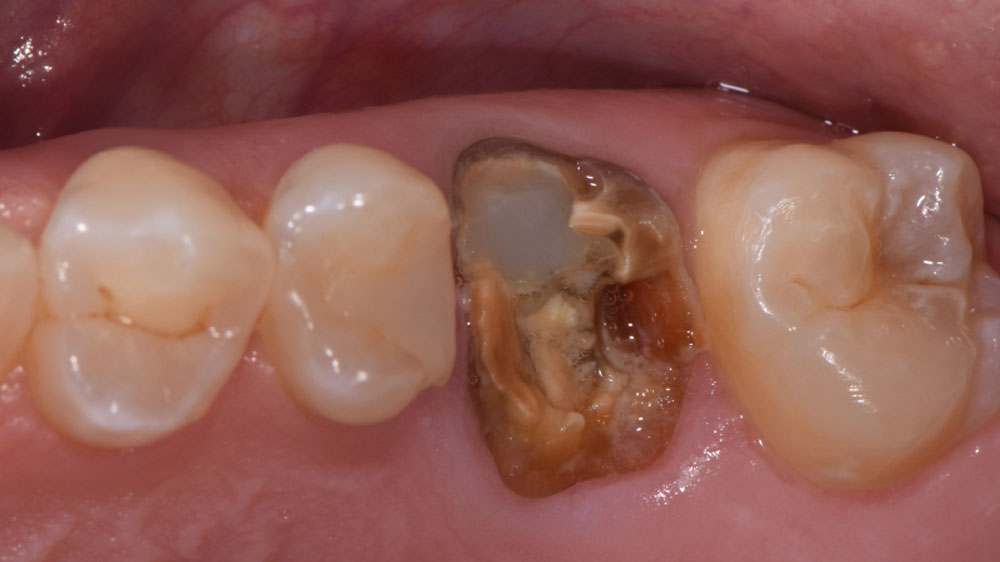
Pre-operative occlusal view showing the involved tooth’s condition.
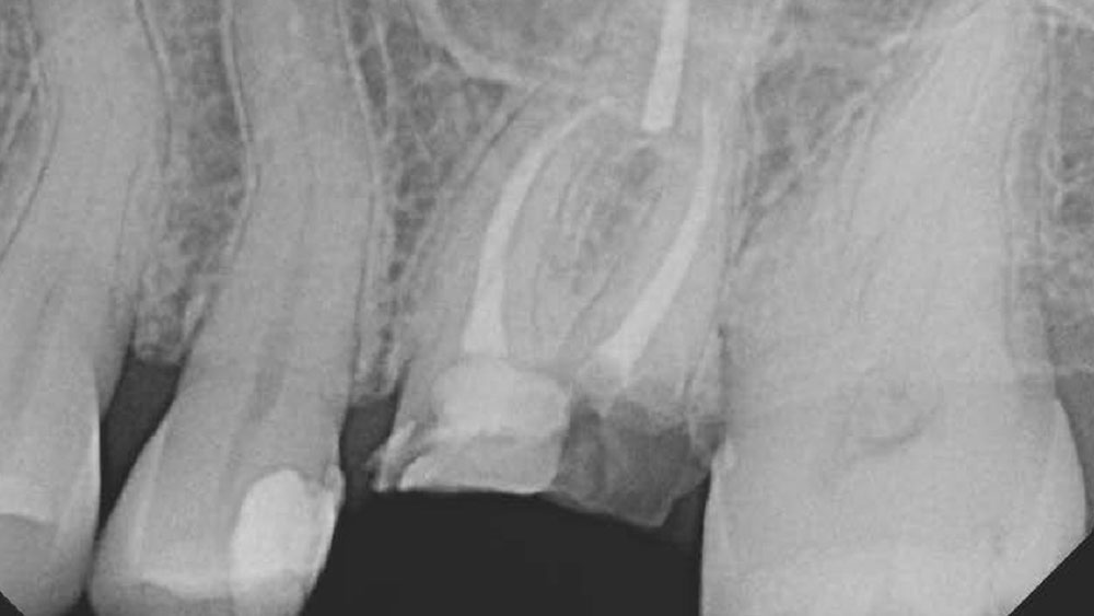
Pre-operative periapical radiograph capturing the cross-section of the involved tooth.
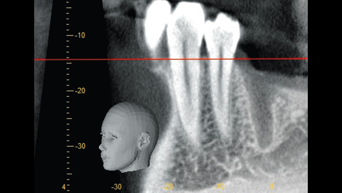
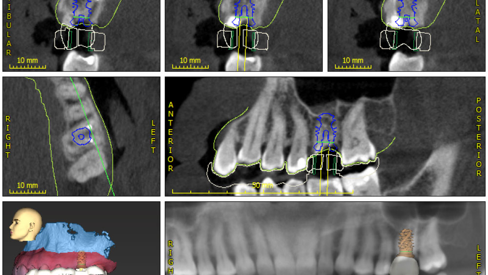
Comprehensive Digital Planning for Implant Placement – A multi-view CBCT and 3D reconstruction showcasing precise anatomical assessment and guided surgical approach.
Geistlich Bio-Oss Collagen® placement, demonstrating the material packed around the implant within the socket.
Immediate provisional crown, occlusal view showcasing restoration alignment technique.
Immediate crown periapical view, reflecting the initial stability of the implant and prosthesis.
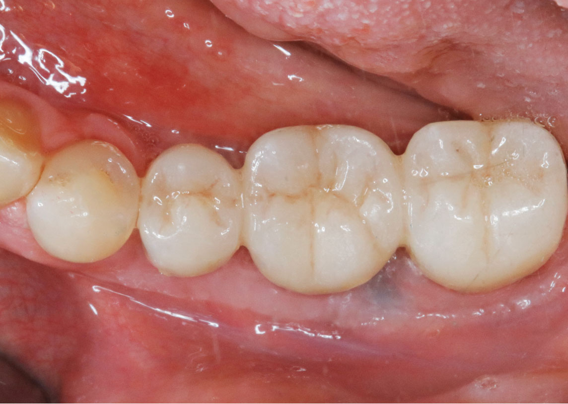
Final crown occlusal view, illustrating the restoration’s integration and final crown periapical view, emphasizing implant stability post-restoration.
Occlusal view at the 3-year post-implant placement, highlighting tissue health and crown durability. Periapical CBCT view at the 3-year post-implant placement, providing insight into bone stability over time.

“Immediate implant placement and loading in molars is a feasible technique, with excellent long-term outcomes, if case selection is adequate, treatment planning is optimized by digital technology, and proper surgical and restorative techniques are applied.”

— Waldemar D. Polido, DDS, MS, PhD

THE OUTCOME

This case shows a three-year follow-up of an immediate implant placement, using Geistlich Bio-Oss Collagen® as a graft material on the gap. Careful tissue management, minimally traumatic extraction, and proper planning, including guided implant surgery can optimize treatment outcomes.

Immediate implant placement usually requires a bone graft to fill the gap between the implant and the socket walls. The use of bovine granules with the addition of porcine collagen (Geistlich Bio-Oss Collagen®) has demonstrated long-term stability to maintain alveolar contour and optimal bone level and soft tissue support around implants.”

Waldemar D. Polido, DDS, MS, PhD

Waldemar D. Polido, DDS, MMS, PhD

Dr. Polido is an Oral and Maxillofacial Surgeon with MS and PhD degrees from the PUCRS School of Dentistry in Porto Alegre, RS, Brazil. He completed his residency in Oral and Maxillofacial Surgery at The University of Texas, Southwestern Medical Center in Dallas, Texas. Currently, Dr. Polido is a Clinical Professor of Oral and Maxillofacial Surgery at the Indiana University School of Dentistry. He is also the Co-Director of the Center for Implant, Esthetic, and Innovative Dentistry at Indiana University School of Dentistry in Indianapolis.

Wel-Shao Lin, DDS, FACP, PhD, MBA

Dr. Lin is a tenured Professor and Chair of Prosthodontics at Indiana University School of Dentistry. He earned his DDS from Chung-Shan and Surgical Implant Fellowship at the University of Rochester (2010). He holds a PhD in Educational Leadership (2020) and an MBA in Healthcare Administration (2022) and is currently pursuing a Master’s Intelligence. Dr. Lin specializes in dental implants, digital dentistry, and AI applications, with over 120 peer-reviewed publications. A Diplomate of the American Board of Prosthodontics and Fellow of ITI and ACP, he also serves as an associate editor for the Journal of Prosthodontics and maintains a clinical practice at Indiana University.

BIOBRIEF

Prosthetic-Surgical Approach to Regenerative Treatment for Peri-Implantitis

Andrea Ravidà, DDS, MS, PhD
Anu Viswanathan DDS, MDS

THE SITUATION

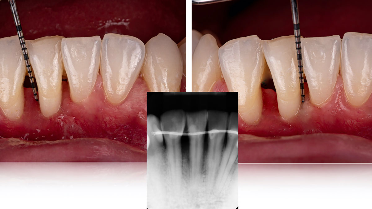
A 68-year-old male patient, who received an implant in tooth position #31 about 8 years prior, presented for an examination. He reports bleeding during brushing around the implant and some discomfort. Clinically, there was severe vertical bone loss, profuse bleeding on probing, and deep probing depths, but no pain. The condition was diagnosed as peri-implantitis according to the 2018 classification.

THE RISK PROFILE

Low RiskMedium RiskHigh Risk
Patient’s healthIntact immune systemLight smokerImpaired immune system 
Patient’s esthetic requirementsLowMediumHigh
Height of smile lineLowMediumHigh
Gingival biotypeThick – “low scalloped”Medium – “medium scalloped”Thin – “high scalloped”
Infection at implant sightNoneChronicAcute
Restorative status of adjacent toothIntactRestored
Soft-tissue anatomyIntactCompromised
Bone anatomy of the alveolar ridgeNo defectHorizontal defectVertical defect

Additional Risk Factors: The patient exhibited bleeding on probing and deep pocket depths. He also reported occasional marijuana use and was inconsistent with periodontal maintenance and oral hygiene visits.

watch video download pdf
THE APPROACH

The treatment goals were to eliminate peri-implant infection, regenerate lost hard and soft tissues, and ensure long-term implant stability. A closed regenerative approach was utilized, including crown removal, thorough implant decontamination with Perioflow®, an airpolishing technology, application of the correct bone grafting materials (Geistlich Bio-Oss®, vallos® and GEM 21S®), enclosed healing, and fabrication of a new crown to facilitate hygiene.

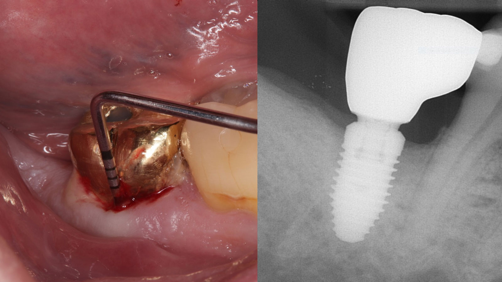
Clinical presentation of tooth #31 showing radiographic evidence of bone loss, profuse bleeding on probing (BOP), deep probing depths, and suppuration, indicative of peri-implantitis with a Class I-infraosseous (c) circumferential-type defect, as described in the study by Monje et al. (2019) Clin Implant Relat Res, 21(4)635-643.
Crown removal by the prosthodontist, followed by placement of a healing abutment for non-surgical therapy using PerioFlow®. After therapy, a cover screw was placed, and the tissue was allowed to heal over the implant for eight weeks.
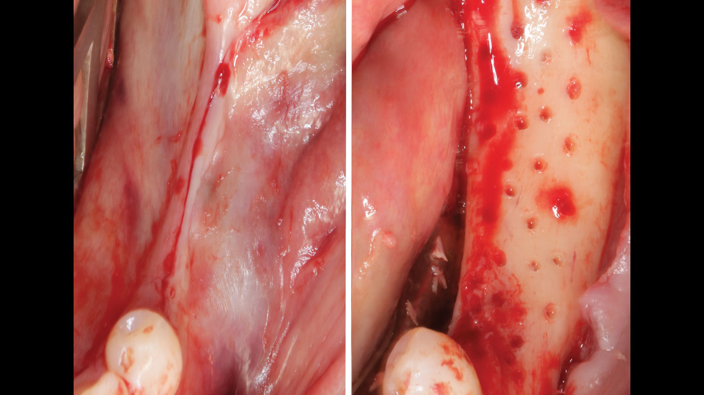
Surgical treatment initiated with a midcrestal incision and full-thickness flap elevation. Granulation tissue was removed using a surgical curette, revealing a deep infrabony defect.
Implant thoroughly decontaminated using Perioflow® with erythritol powder to ensure a clean surface for regeneration.
rhPDGF-BB was used to hydrate bone grafting materials (vallos® Demineralized Cortical Granules and Geistlich Bio-Oss®), which were first hydrated with sterile water before rhPDGF-BB was added. The materials were mixed in a 1:1 ratio and allowed to sit for 10 minutes before being applied to the deep infrabony defect to promote regeneration.
Flap closed primarily with 5-0 PTFE horizontal mattress and single interrupted suture for secure closure.
Collagen membrane stabilized with 5-0 chromic gut sutures using the lasso technique.
After 5 months of healing, significant bone gain is evident. Geistlich Bio-Oss® was placed on the buccal site to enhance thickness, covered with an amnion-chorion membrane. A healing abutment was placed at this stage.
Two-year follow-up shows disease resolution with shallow probing depths, no bleeding or suppuration, and complete bone gain. A new crown was fabricated with an increased final abutment height (>2mm), contributing to optimal maintenance and long-term stability based on evidence supporting its role in promoting long-term success. A second surgery may be necessary to gain additional tissue thickness or cover residual thread exposure to achieve the desired long-term results.

“The implant presented with significant bone loss, deep probing depths, and bleeding on probing, placing it at risk of failure and requiring intervention to preserve function and longevity.”

— Andrea Ravidà, DDS, MS, PhD

THE OUTCOME

At the two-year follow-up, clinical and radiographic assessments showed disease resolution, complete bone gain, and stable peri-implant tissues. Probing depths were within healthy ranges, and no bleeding on probing was observed, confirming the long-term success of the treatment.

Enclosed healing, meticulous implant decontamination, appropriate selection of bone grafting materials, and customized crown design, combined with patient compliance and regular maintenance, contributed to disease resolution and complete bone regeneration.”

Andrea Ravidà, DDS, MS, PhD

The air polishing device with erythritol powder ensured thorough implant decontamination, while the bone grafting materials combined with rhPDGF-BB provided essential biologic support for regeneration and improved peri-implantitis treatment outcomes.”

Andrea Ravidà, DDS, MS, PhD

Andrea Ravidà, DDS, MS, PhD

Dr. Andrea Ravidà is the Director of the Graduate Periodontics Program in the department of Periodontics at the University of Pittsburgh. He conducts clinical research focusing on peri-implantitis, periodontitis and short implants. He has published more than 70 peer-reviewed articles and conference abstracts/presentations related to periodontics and implant therapy. He is section editor of the International Journal of Oral Implantology and the Journal of Translational Medicine.

Anu Viswanathan DDS, MDS

Dr. Anu Viswanathan is a Diplomate of the American Board of Periodontology and Implant Dentistry. She earned her Doctor of Dental Surgery degree from the University of Colorado School of Dental Medicine in 2019. Dr. Viswanathan completed a Certificate in Periodontics and earned a Master of Dental Science at the University of Pittsburgh School of Dental Medicine. She also obtained a Certificate in IV Sedation. Dr. Viswanathan is currently in private practice in Shoreline, Connecticut.

BIOBRIEF

Mandibular Ridge Augmentation Using Customized Titanium Mesh

Shaun R. Young, DMD

THE SITUATION

A 60-year-old healthy male presented with a failing lower left bridge. Due to a long history of missing teeth, he had a significantly atrophic mandibular ridge. We decided to use a customized titanium mesh to achieve the necessary vertical and horizontal bone augmentation for dental implant rehabilitation.

THE RISK PROFILE

Low RiskMedium RiskHigh Risk
Patient’s healthIntact immune systemLight smokerImpaired immune system 
Patient’s esthetic requirementsLowMediumHigh
Restorative status of adjacent toothIntactRestored
Width of tooth gap1 tooth (≥ 7 mm)1 tooth (≤ 7 mm)2 teeth or more
Bone anatomy of the alveolar ridgeNo defectHorizontal defectVertical defect

THE APPROACH

The goal of this procedure was to regenerate sufficient bone to place restoratively driven dental implants. Due to the horizontal and vertical ridge deficiency, we used a customized titanium mesh to predictably achieve this outcome.


Autogenous bone collected with SafeScraper Twist and Geistlich Bio-Oss® filled the Yxoss CBR® Protect and a Geistlich Bio-Gide® collagen membrane covered the mesh.

.

Pre-op mandibular ridge after the extraction of teeth #17 and #20. Note the significant horizontal ridge deficiency.
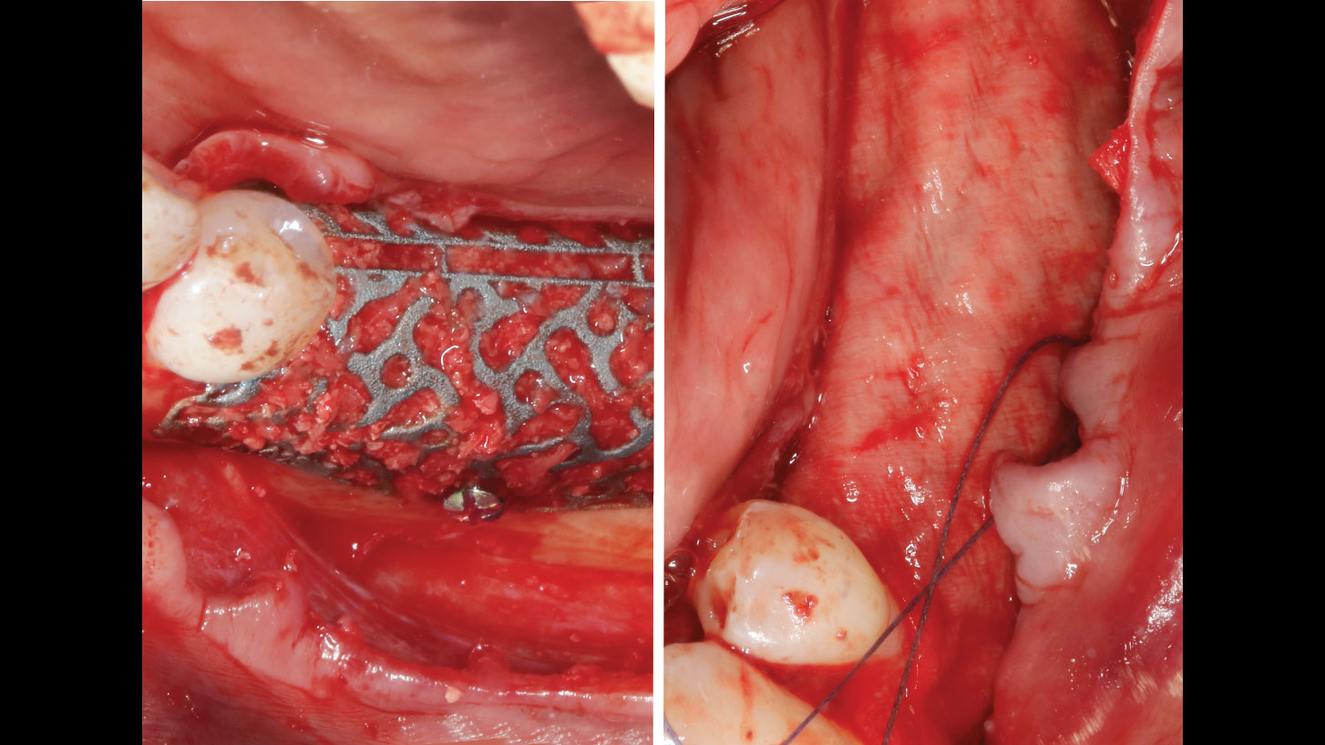
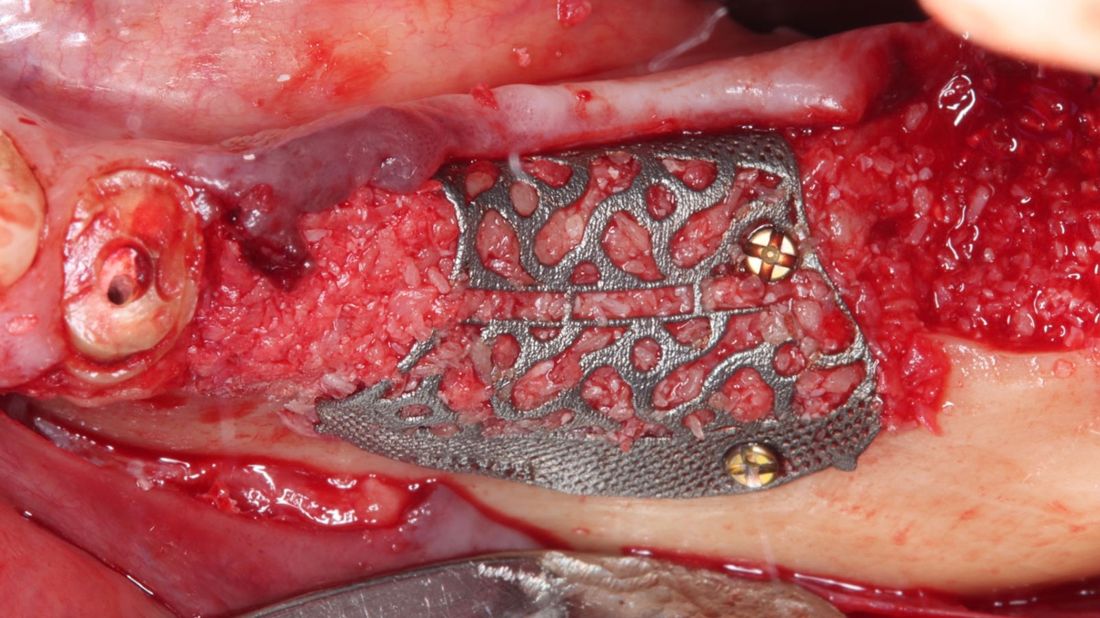
Yxoss CBR® Protect Customized Bone Regeneration Titanium Mesh in place, secured with two Stryker screws, 1.2 mm in diameter and 6 mm in length.
Note the excellent adaptation of the Yxoss CBR® Protect and that the edges are apical to the adjacent bony sockets.
Geistlich Bio-Gide® (resorbable collagen membrane) draped over the Yxoss CBR® Protect to separate osteoblasts from fibroblasts.
Implants restored with single-unit crowns.
Six months post-ridge augmentation.
Guide pins in place for implants #19, #20, and #21, with adequate ridge height and width.
Screw-retained final implant crowns with healthy keratinized tissue on the facial side.

“Success in these cases primarily depends on proper mesh design and careful handling of soft tissue to ensure zero-tension primary closure.”

— Shaun R. Young, DMD

THE OUTCOME

A left mandibular ridge deficiency was corrected using a Yxoss CBR® Protect Customized Bone Regeneration Titanium Mesh, designed from the patient’s CBCT scan. 

Screw-retained final implant crowns with healthy keratinized tissue on the facial side

Guided bone regeneration using Yxoss CBR® Protect to correct vertical and horizontal mandibular ridge deficiencies is a predictable procedure.”

Shaun R. Young, DMD

Shaun R. Young, DMD

Dr. Shaun Young, an Oral and Maxillofacial Surgeon based in Tampa, Florida, specializes in complex ridge augmentation, immediate implants, and All-on-X full arch rehabilitation. He earned his Doctor of Dental Medicine degree from the University of Florida and completed his OMFS residency at Emory University in Atlanta, Georgia, where he served as Administrative Chief Resident. Dr. Young brings his expertise to a full-scope group practice, serving Tampa, Clearwater, and New Port Richey, Florida.

WEBINAR

BIOBRIEF

Alveolar Ridge Preservation with vallos® Mineralized Cortico-Cancellous Allograft

Hanae Saito, DDS, MS, CCRC
Andrew Tong, DDS

THE SITUATION

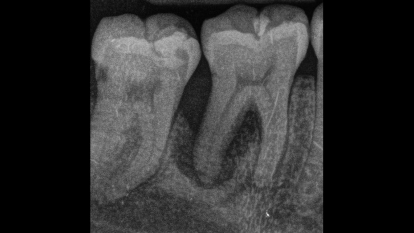
A 68 year old female patient was referred from her general dentist for persistent minor discomfort on #31, suspected endo-perio lesion. Upon the examination, deep probing depth and grade 1 mobility were noted. Radiographic interpretation indicating a large J shaped lesion and possible root fracture. Patient had missing #30 and #32 has been mesially drifted and left a restorative space more than > 13 mm mesio-distally.

THE RISK PROFILE

Low RiskMedium RiskHigh Risk
Patient’s healthIntact immune system/Non-smoker Light smokerImpaired immune system 
Patient’s esthetic requirementsLowMediumHigh
Height of smile lineLowMediumHigh
Gingival biotypeThick – “low scalloped”Medium – “medium scalloped”Thin – “high scalloped”
Shape of dental crownsRectangularTriangular
Infection at implant sightNoneChronicAcute
Bone height at adjacent tooth site≤ 5 mm from contact point5.5 – 6.5 mm from contact point≥ 7 mm from contact point
Restorative status of adjacent toothIntactRestored
Width of tooth gap1 tooth (≥ 7 mm)1 tooth (≤ 7 mm)2 teeth or more
Soft-tissue anatomyIntactCompromised
Bone anatomy of the alveolar ridgeNo defectHorizontal defectVertical defect
* The esthetic related risk factors are not needed for this case. Restorative space of more than 13 mm may be an additional risk factor.
watch video download pdf
THE APPROACH

A successful treatment outcome comes with proper selection of the technique and materials. In order to facilitate an implant supported restoration in the site with > 13mm mesio-distal space, a staged approach was selected, with alveolar ridge preservation (ARP) performed using an atraumatic extraction technique and vallos® mineralized cortico-cancellous bone allograft chosen as the material.

Preoperative radiograph of #31 with guarded prognosis due to potential root fracture and infection.
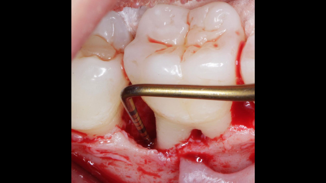
Atraumatic extraction was performed and intact furcation bone is noted.
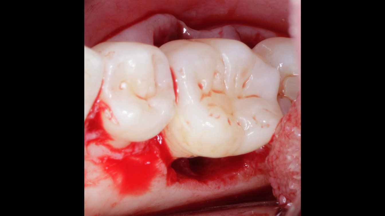
vallos® mineralized cortico-cancellous bone allograft was placed.
A collagen plug was used to cover the bone allograft
Immediate post alveolar ridge preservation radiograph.
Healing at 2 weeks prior to implant placement.
Healing at 4 months prior to implant placement.
Implant was placed with 32 Ncm. Post implant placement radiograph.
6 months after the implant placement, the final restoration was delivered.

“Ensuring atraumatic extraction techniques, regardless of whether it’s in the anterior or posterior regions, is crucial for preserving the integrity of both hard and soft tissues. Equally important is the selection of biomaterials that not only offer structural support but also possess bone regeneration properties.”

— Dr. Hanae Saito

THE OUTCOME

The planned treatment of replacing a tooth with a dental implant in the regenerated alveolar ridge was achieved. By employing secondary intention healing following ARP and utilizing a lingual paracrestal incision, adequate keratinized tissue was preserved on the buccal side of the implant-supported restoration. 

6 months after the implant placement, the final restoration was delivered.

Removal of the infection and maintaining the ridge dimension for the implant supported restoration in the site with > 13 mm mesio-distal space were required.”

Dr. Hanae Saito

Hanae Saito, DDS, MS, CCRC

Hanae Saito, DDS, MS, CCRC serves as a clinical associate professor and oversees the Dual Perio-Pros program and predoctoral periodontal education within the Division of Periodontics, at the University of Maryland School of Dentistry. She is a Diplomate of the American Board of Periodontology. Dr. Saito obtained a Master of Science in Clinical Research and a certificate in Periodontics from New York University College of Dentistry. Additionally, she operates a faculty practice focused on periodontology and implant dentistry.

Andrew Tong, DDS

Andrew Tong, DDS earned his Bachelor of Science degree from the University of Maryland at College Park in 2015 before completing his Doctor of Dental Surgery (D.D.S) degree at the University of Maryland School of Dentistry in 2019. Following this, he undertook a General Practice Residency at the Newark Beth Israel Medical Center in New Jersey from 2019 to 2020. Dr Tong now practices general dentistry at Tong Dental Care in Gaithersburg, MD. Concurrently, he is pursuing a Master’s degree in Periodontics at the University of Maryland School of Dentistry.

Sorry, you do not have permission to view this content.

Sorry, you do not have permission to view this content.

BIOBRIEF

Selecting Biomaterials for Combined Complex Defects

Irina F. Dragan, DDS, DMD, MS, eMBA

THE SITUATION

The patient called the office complaining of sensitivity and swelling in the maxillary left quadrant. He was seen and prescribed an antibiotic. Tooth #12 was deemed hopeless, and the peri-apical and radicular lesion presented on the radiograph extended significantly on the mesial aspect, impacting the interproximal bone level for tooth #11. Patient presents with implant supported restorations distal to the affected area and was concerned about the infection spreading to that area as well. The area was treated successfully, and the patient was pleased with the outcome, allowing him to preserve the tooth, on the mesial aspect of the lesion and the implant distally.

THE RISK PROFILE

Low RiskMedium RiskHigh Risk
Patient’s healthIntact immune system/Non-smoker Light smokerImpaired immune system 
Patient’s esthetic requirementsLowMediumHigh
Height of smile lineLowMediumHigh
Gingival biotypeThick – “low scalloped”Medium – “medium scalloped”Thin – “high scalloped”
Shape of dental crownsRectangularTriangular
Infection at implant sightNoneChronicAcute
Bone height at adjacent tooth site≤ 5 mm from contact point5.5 – 6.5 mm from contact point≥ 7 mm from contact point
Restorative status of adjacent toothIntactRestored
Width of tooth gap1 tooth (≥ 7 mm)1 tooth (≤ 7 mm)2 teeth or more
Soft-tissue anatomyIntactCompromised
Bone anatomy of the alveolar ridgeNo defectHorizontal defectVertical defect

THE APPROACH

The goals of the procedure were to eliminate infection, the source of pain, and reduce periodontal problems to the adjacent tooth and implant. Full thickness flap was reflected, #12 was removed and the socket was debrided and irrigated. A peri-radicular lesion was removed and submitted for histopathological exam.

Initial presentation – buccal view.
Full thickness flap elevation exposing the complex clinical situation.
Alveolar socket after the tooth removal exposing the loss of bone on the distal of tooth #11, prior to the debridement of the granulation tissue and root preparation.
Adaptation on the buccal defect prior to placement of bone grafting with vallos® mineralized cortical cancellous mix granules (bottom) followed Geistlich Bio-Oss® (top).
Post adaptation with Geistlich Bio-Gide® for alveolar ridge preservation and guided tissue regeneration, followed by final suturing of the site using ePTFE material.
Radiographic overview of the clinical procedure: initial presentation with the bony defect impacting distal of #11 and #12 – mesial and inter-radicular, site after the tooth #12 was extracted, radiographic bone fill of the defect post-operative.
Post-operative healing of the site, 4 weeks after the procedure was completed.

“A localized infection can easily spread and impact adjacent teeth and implants. It is critical for clinicians to intervene as soon as possible to prevent further complications. Patient education and motivation is key to successfully treat these types of clinical situations encountered in a daily practice.”

— Dr. Irina Dragan

THE OUTCOME

The combined defect: #11 distal guided tissue regeneration and #12 alveolar ridge preservation for #12. This area was treated with vallos®, Geistlich Bio-Oss Collagen®, and Geistlich Bio-Gide®. The xenograft was placed in the apical portion of the socket and the allograft towards the coronal surface.

Healing of the site at 4 weeks post-operative.

Considering today’s advancements in regeneration we are able to successfully treat complex clinical scenarios that involve combined therapeutic applications, such as guided tissue regeneration and alveolar ridge preservation.”

Dr. Irina Dragan

Periotomes were able to support with an atraumatic extraction of tooth #12 and maintaining as much as possible the soft and hard tissue present in this compromised area.”

Dr. Irina Dragan

Irina F. Dragan, DDS, DMD, MS, eMBA

Periodontology and Implant Dentistry
Dr. Irina Dragan is board certified and an examiner for the American Board of Periodontology and Implant Dentistry. She is part-time faculty in postgraduate periodontics at Harvard School of Dental Medicine and an adjunct associate professor of periodontology at Tufts University School of Dental Medicine. She is a periodontist and clinical researcher at The Perio Studio, a practice limited to periodontology and implant dentistry in Boston, MA.

Sorry, you do not have permission to view this content.

Sorry, you do not have permission to view this content.

Sorry, you do not have permission to view this content.

Sorry, you do not have permission to view this content.

Sorry, you do not have permission to view this content.

Sorry, you do not have permission to view this content.

Sorry, you do not have permission to view this content.

Sorry, you do not have permission to view this content.

Sorry, you do not have permission to view this content.

Sorry, you do not have permission to view this content.

Sorry, you do not have permission to view this content.

Sorry, you do not have permission to view this content.

Sorry, you do not have permission to view this content.

Sorry, you do not have permission to view this content.

Sorry, you do not have permission to view this content.

Sorry, you do not have permission to view this content.

Sorry, you do not have permission to view this content.

Sorry, you do not have permission to view this content.

Sorry, you do not have permission to view this content.

Sorry, you do not have permission to view this content.

Sorry, you do not have permission to view this content.

Sorry, you do not have permission to view this content.

Sorry, you do not have permission to view this content.

Sorry, you do not have permission to view this content.

BIOBRIEF

The Buccal Pedicle Flap for Peri-Implant Soft Tissue Volume

Dr. Giorgio Tabanella

THE SITUATION

Patient presented with a fistula buccal on tooth #9 associated with a chronic peri-apical lesion and external root resorption. Also tooth #8 showed a chronic peri-apical lesion. Her chief complaint was the misalignment of her teeth. The clinical situation revealed the presence of bleeding upon probing and generalized moderate periodontal disease (Stage II, Grade I) as well as multiple endodontic failures.

THE RISK PROFILE

Low RiskMedium RiskHigh Risk
Patient’s healthIntact immune system Light smokerImpaired immune system 
Patient’s esthetic requirementsLowMediumHigh
Height of smile lineLowMediumHigh
Gingival biotypeThick – “low scalloped”Medium – “medium scalloped”Thin – “high scalloped”
Shape of dental crownsRectangularTriangular
Infection at implant sightNoneChronicAcute
Bone height at adjacent tooth site≤ 5 mm from contact point5.5 – 6.5 mm from contact point≥ 7 mm from contact point
Restorative status of adjacent toothIntactRestored
Width of tooth gap1 tooth (≥ 7 mm)1 tooth (≤ 7 mm)2 teeth or more
Soft-tissue anatomyIntactCompromised
Bone anatomy of the alveolar ridgeNo defectHorizontal defectVertical defect
Note: request for reducing the healing time, long-term maintenance
watch video download pdf
THE APPROACH

The aim of the treatment is to eradicate periodontal disease and restore esthetics and function. Treatment planning: non-surgical and surgical periodontal treatment, orthodontic alignment, extraction of both central incisors, immediate implant placement and Guided Bone Regeneration with Geistlich Bio-Oss®, peri-implant soft tissue boosting with a buccal pedicle flap and full ceramic CAD-CAM restorations.

The clinical picture is showing a fistula buccal to #21 as well as leakage on old composite restorations. A thin biotype is evident.
The sagittal cuts are reporting chronic peri-apical lesions on both central incisors and a thin buccal plate with minor vertical bone loss but fenestration apical to #21.
The intrasurgical picture is showing the bony defect, the buccal fenestration and the thin buccal plate.
After allowing the tissue to heal for 4 months a first “Buccal Pedicle Flap” was performed during the uncovery of the dental implant. Simultaneously, Geistlich Fibro-Gide® was inserted into the envelope created by the flap design.
Geistlich Fibro-Gide® is reduced to a thickness of 4 mm at its borders so that it is easier to get adapted to the recipient site.
The Fibro-Gide® is trimmed so that its borders don’t approach the vertical incisions of the Buccal Pedicle Flap.
Polypropylene 6.0 sutures are used to compressed the Fibro-Gide® underneath the flap thus creating the “wrinkles” on the mucosa.
The wrinkles are visible also on the occlusal view. The mucogingival line is repositioned at its original level.
Four months after immediate implant placement and GBR in area #11 a second Buccal Pedicle Flap is performed to reduce the buccal concavity, boost the peri-implant mucosa and increase the thickness as well as the band of the keratinezed mucosa.
As in the previous surgery the Fibro-Gide® is inserted underneath the Buccal Pedicle Flap and stabilized with e-PTFE 6.0 sutures.
8 weeks post surgery, the occlusal view is showing a biomimetic countouring of the peri-implant mucosa.
The final esthetic result is emphasizing an excellent blending of “white” and “pink” esthetics.

“Orthodontic treatment must be postponed because of the presence of periodontal disease. A thin biotype and a high smile line needs to be taken into consideration.”

THE OUTCOME

The final outcome at 8 weeks is showing pink esthetics as well as biomimetics and function. The use of the buccal pedicle flap allowed the increased volume of the peri-implant mucosa with a minimally invasive approach. The combination of Geistlich Fibro-Gide® and a buccal pedicle flap had the main advantage of reducing the morbidity generally associated with CT harvesting.

The final esthetic result is emphasizing an excellent blending of “white” and “pink” esthetics.

Dr. Giorgio Tabanella

Dr. Tabanella is a Diplomate of the American Board of Periodontology, an Active Member of the Italian Academy of Esthetic Dentistry and author of the book “Retreatment of Failures in Dental Medicine”. He graduated from the University of Southern California, Los Angeles, USA where he obtained his Certificate in Periodontics as well as a Master of Science in Craniofacial Biology. He is Director of O.R.E.C. – Oral Reconstruction and Education Center (www.tabanellaorec.com), reviewer and author of original articles.

BIOBRIEF

Use of Geistlich Fibro-Gide® for Correction of Maxillary Anterior Soft Tissue Peri-implant Ridge Deficiencies

Dr. Israel Puterman

THE SITUATION

A 27-year-old female with congenitally missing maxillary lateral incisors was referred for implant placement. Following completion of orthodontics, a plan was developed to place dental implants at the #7 and #10 positions. Based on CBCT evaluation, alveolar ridge height and width was deemed sufficient for implant placement. Despite sufficient bone volume, facial ridge volume deficiencies were noted at both edentulous sites, requiring augmentation to allow for optimal esthetics.

THE RISK PROFILE

Low RiskMedium RiskHigh Risk
Patient’s healthIntact immune system Light smokerImpaired immune system 
Patient’s esthetic requirementsLowMediumHigh
Height of smile lineLowMediumHigh
Gingival biotypeThick – “low scalloped”Medium – “medium scalloped”Thin – “high scalloped”
Shape of dental crownsRectangularTriangular
Infection at implant sightNoneChronicAcute
Bone height at adjacent tooth site≤ 5 mm from contact point5.5 – 6.5 mm from contact point≥ 7 mm from contact point
Restorative status of adjacent toothIntactRestored
Width of tooth gap1 tooth (≥ 7 mm)1 tooth (≤ 7 mm)2 teeth or more
Soft-tissue anatomyIntactCompromised
Bone anatomy of the alveolar ridgeNo defectHorizontal defectVertical defect
watch video download pdf
THE APPROACH

The goal of treatment was to replace missing maxillary lateral incisors with dental implants, while providing an esthetic result with predictable and minimally invasive techniques. Employing a surgical guide for implant placement, implants were placed in precise 3-dimentional positions. The use of xenograft biomaterials (Geistlich Fibro-Gide®) allowed for the augmentation of gingival biotype and elimination of the buccal ridge deficiencies while avoiding the harvesting of autogenous tissue.

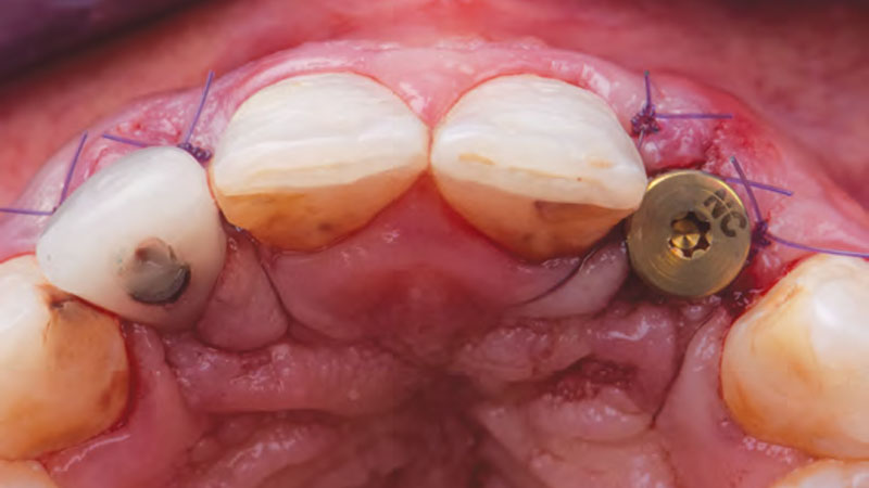
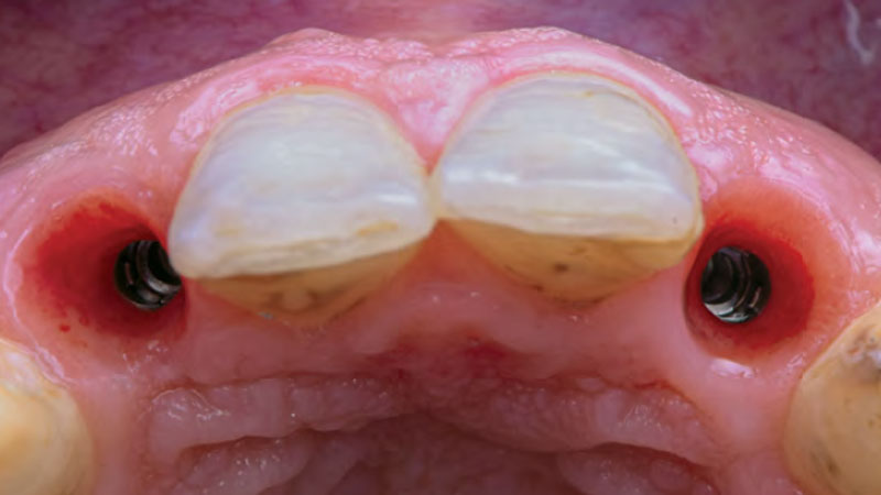
Pre-surgical, occlusal view, demonstrating buccal ridge concavities at edentulous sites, #7 and #10.
Geistlich Fibro-Gide® collagen matrix is cut and shaped prior to placement into surgical sites.
Immediate post-surgical occlusal view following placement of implants and Geistlich Fibro-Gide® on facial aspects. Implant #7 was provisionalized immediately, however implant #10 did not achieve sufficient stability and a healing abutment was placed.
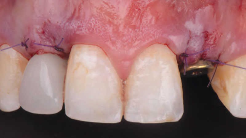
Immediate post-surgical facial view. One can note the buccal prominences being developed by the presence of Geistlich Fibro-Gide®.
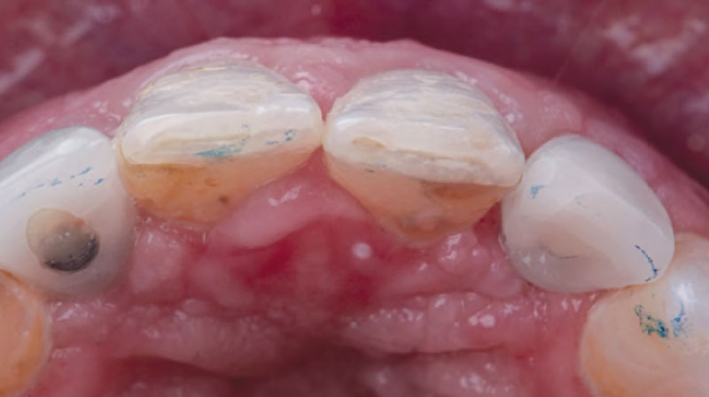
2-month post-surgical view, following provisionalization of implant #10. Buccal convexities at the implant sites are still evident.
8 month view of implant sites at time of torque test, following maturation of gingival emergence. Of note is thick, convex buccal tissue free of any sign of inflammation.
Occlusal view following final restoration, (Dr. Paul Krainson). Natural-appearing buccal gingival convexities remain 1.5 years post-surgery.
Frontal view of final restoration of implants. The tissue health with stippling and root-like gingival prominences are noted.

“A buccal ridge deficiency with congenitally missing lateral incisors in a high-scallop, high-smile young female patient.”

THE OUTCOME

The presented case involves a female patient with congenitally missing maxillary lateral incisors and soft tissue ridge deficiencies. Implants were placed and a volume-stable collagen matrix Geistlich Fibro-Gide® was placed to provide labial soft tissue volume. The tissue emergence was then developed with the use of provisional restorations, one placed at the time of surgery, the other following implant integration. The implants were restored with gingival tissue transformed to mimic convex root emergence.

Correction of labial soft tissue ridge deficiencies at implant sites through use of a Geistlich Fibro-Gide® volume stable, collagen matrix.”

Dr. Israel Puterman

A volume-stable collagen matrix can be used to correct a labial soft tissue deficiency, eliminating the potential negative sequelae of an autogenous connective tissue graft.”

Dr. Israel Puterman

Various materials can be used to restore a soft tissue deficiency; use of a volume-stable collagen matrix provides numerous advantages when used in the proper indication.”

Dr. Israel Puterman

Dr. Israel Puterman

Dr. Puterman, originally from Montreal Canada, received his DMD from Boston University in 2002 and dual graduate certificates in Implant Dentistry and in Periodontics from Loma Linda University in 2008. He is a published author in various journals including the Journal of Prosthetic Dentistry and the Journal of Prosthodontics. He practices in the Washington, DC area.

BIOBRIEF

Successful Implant Placement and Horizontal Augmentation for Bilateral Congenitally Missing Maxillary Incisors

Dr. Avinash Bidra

THE SITUATION

A 30-year-old male patient was referred to me with bilateral congenitally missing lateral incisors in the maxilla. The referring general dentist had previously made a resin-bonded bridge which was successful for a few years but had frequent debondings. Clinical examination revealed lack of ridge contour but the CBCT revealed existence of adequate width for placement of narrow-diameter implants with additional bone grafting and contour augmentation. The existing bone anatomy precluded placement of implants for screw-retained restorations without a pre-surgical lateral ridge augmentation procedure. The patient accepted a treatment plan for placement of two narrow-diameter implants and simultaneous bone grafting and contour augmentation followed by restoration with zirconia cement-retained crowns.

THE RISK PROFILE

Low RiskMedium RiskHigh Risk
Patient’s healthIntact immune system Light smokerImpaired immune system 
Patient’s esthetic requirementsLowMediumHigh
Height of smile lineLowMediumHigh
Gingival biotypeThick – “low scalloped”Medium – “medium scalloped”Thin – “high scalloped”
Shape of dental crownsRectangularTriangular
Infection at implant sightNoneChronicAcute
Bone height at adjacent tooth site≤ 5 mm from contact point5.5 – 6.5 mm from contact point≥ 7 mm from contact point
Restorative status of adjacent toothIntactCompromised
Width of tooth gap1 tooth (≥ 7 mm)1 tooth (≤ 7 mm)2 teeth or more
Soft-tissue anatomyIntactCompromised
Bone anatomy of the alveolar ridgeNo defectHorizontal defectVertical defect
watch video download pdf
THE APPROACH

My treatment goals were to preserve the existing soft-tissue architecture, especially the interdental papilla, mesial and distal to the lateral incisors, improve the facial contour at the lateral incisor sites by bone grafting with a low substitution biomaterial, and harmonize esthetics and function with optimal implant-supported restorations.
Frontal view revealed adequate contours of soft-tissue especially in gingival height and presence of interdental papilla which needed to be preserved.
Bilateral papilla-sparing incisions were used to preserve the existing papilla and the osteotomies were prepared to allow implant trajectories for cement retained restorations.
An apical fenestration was noted in the osteotomies. After placement of a narrow diameter implant at patient’s right lateral incisor, the site was grafted with a mixture of autologous bone chips and Geistlich Bio-Oss®.
Geistlich Bio-Gide® is trimmed to match the trapezoidal flap design and placed over the graft material.
Healing Abutments 3.5 mm were placed at time of surgery for single-stage healing
After a 3-month healing period, the implants were osseointegrated and then screw-retained provisional crowns were fabricated over both implants. The soft-tissues showed an excellent response to the bone graft materials.
The soft tissues around the implants show excellent maturation and support especially in the interdental papilla region which was preserved during surgery.
Occlusal view shows adequate restoration of the facial contour around the implant restorations indicating excellent outcome from the contour augmentation procedure.
Frontal close-up view of the implant restorations shows pleasing dental and gingival esthetics.

“The patient had failed resin-bonded bridges with deficient contours for bilateral congenitally missing lateral incisors.”

THE OUTCOME

Single-stage implant placement with bilateral papilla-sparing incision design and simultaneous contour augmentation using a mixture of Geistlich Bio-Oss® autologous bone chips and Geistlich Bio-Gide®.

The low substitution bone graft, combined with a rapidly vascularizing membrane, helped to achieve the biological integration of the biomaterial.”

Dr. Avinash Bidra

The use of Geistlich Bio-Gide® and Geistlich Bio-Oss® mixed with autologous bone can lead to a successful outcome in single-stage implant placement with simultaneous contour augmentation.”

Dr. Avinash Bidra

Dr. Avinash Bidra

Dr. Bidra is a Board Certified Maxillofacial Prosthodontist and Director of the Prosthodontics Residency Program at UCONN School of Dental Medicine. He has extensive surgical experience and maintains a part-time private practice restricted to Implant Surgery and Prosthodontics in Meriden, CT. He has lectured at national and international meetings, as well as published extensively in international scientific journals. He has invented prosthetic components and is a co-inventor of a new implant design.