Buy products 5, get 1 free. Use code b5g1. Check it out!
Clinical Resources
Use the search and filter form below to find informative clinical content by format, therapeutic areas, or product.
FEATURED RESOURCE
BIOBRIEF
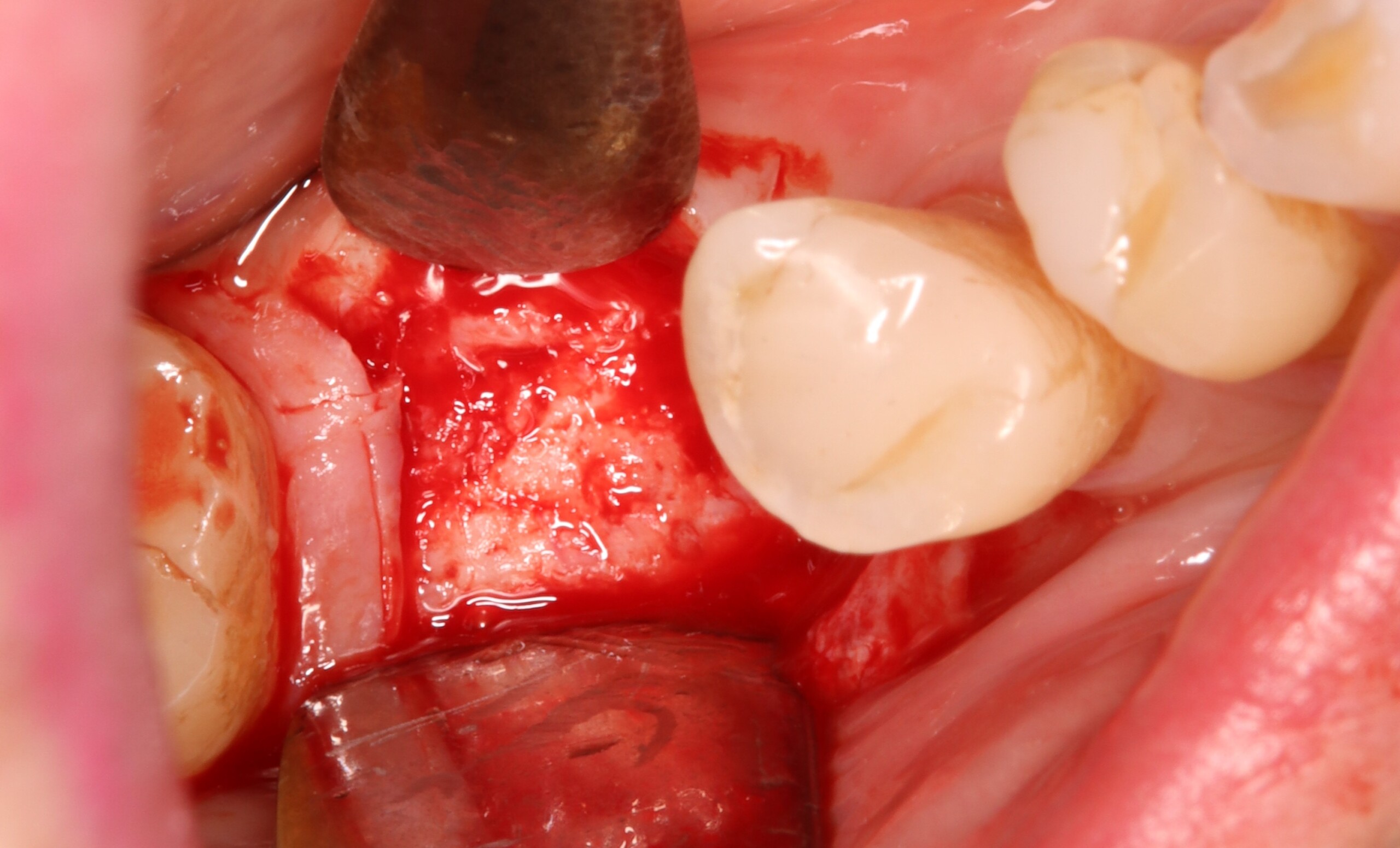
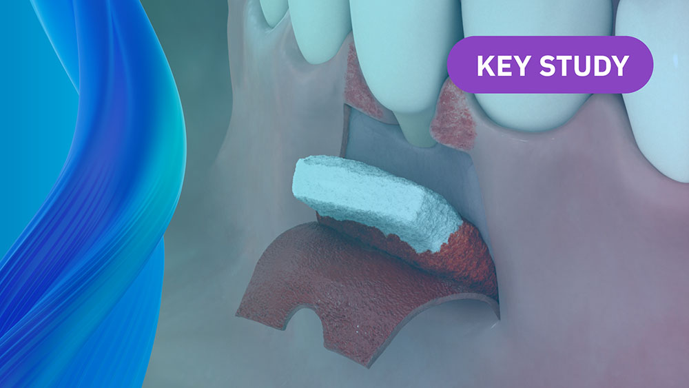
Alveolar Ridge Preservation with vallos® Mineralized Cortico-Cancellous Allograft
Hanae Saito, DDS, MS, CCRC
, Andrew Tong, DDS
On-Demand Webinars
Learn on your own Time and Earn CE
Geistlich is committed to educating our clinician partners in regenerative dentistry through in-person and virtual sessions. We look forward to growing with you on this journey.
Coronally Advanced Flap with a Xenogeneic Collagen Matrix or an rhPDGF-BB Regenerative Approach for Bilateral Multiple Gingival Recessions: A Split-Mouth Clinical Feasibility Study
Efficacy of biologics for the treatment of periodontal infrabony defects: An American Academy of Periodontology best evidence systematic review and network meta-analysis
Efficacy of biologics in root coverage and gingival augmentation therapy: An American Academy of Periodontology best evidence systematic review and network meta-analysis
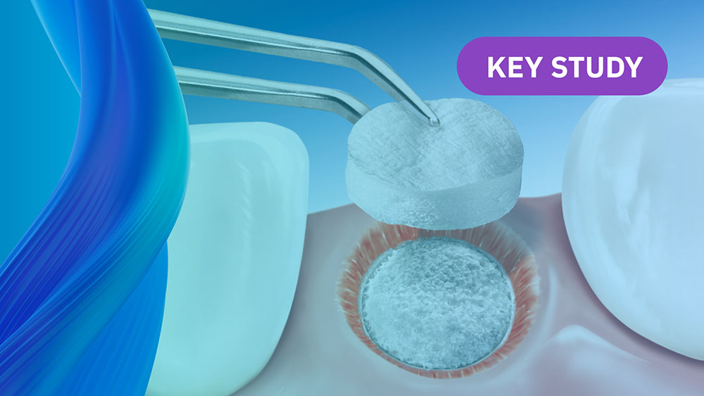
Efficacy of Xenogeneic Collagen Matrix to Augment Peri-Implant Soft Tissue Thickness Compared to Autogenous Connective Tissue Graft: A Systematic Review and Meta-Analysis
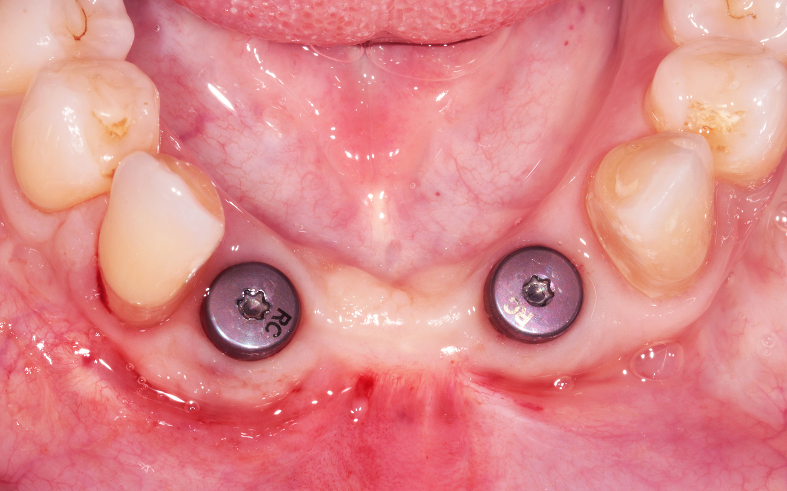
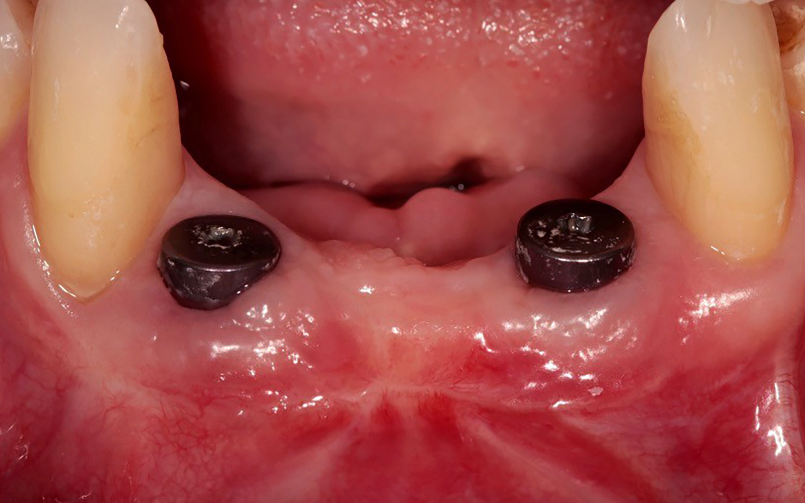
A mid-facial incision was made, with the intent to preserve the minimal keratinized tissue that was available,
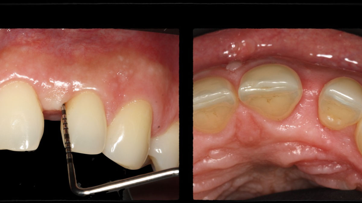
as well as vertical incisions along the line angles of the canines to reflect a partial thickness flap.Platelet-rich fibrin clots were formed by centrifugation. The leukocyte-PRF (L-PRF) was extracted, and the L-PRF was used to hydrate Geistlich Mucograft®Geistlich Mucograft® and PRF stabilized via glycolon sutures.Clinical situation at three-week follow-up.Occlusal view at twelve-month follow-up.Frontal view at twelve-month follow-up.
Dr. Alan Fetner, DMD
Jacksonville, Florida, USA
Esthetic Soft-Tissue Augmentation of Two Implant Sites in the Same Patient
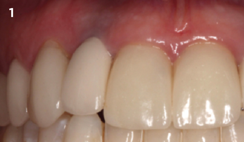
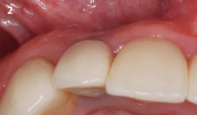
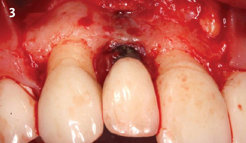
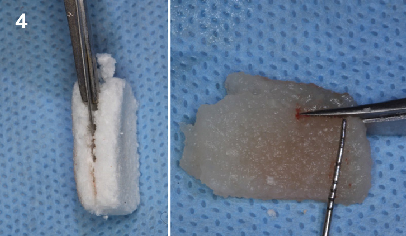
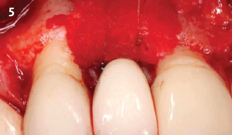
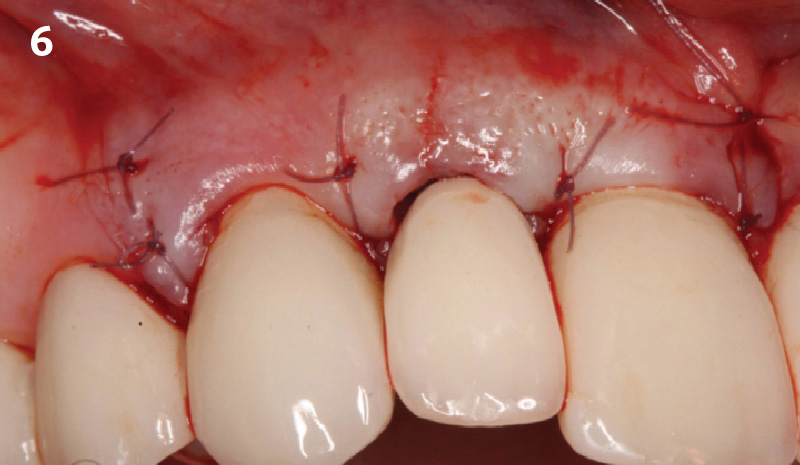
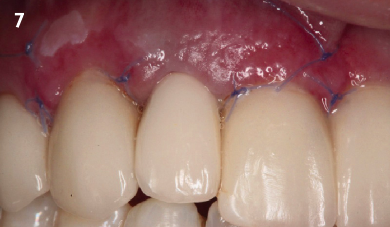
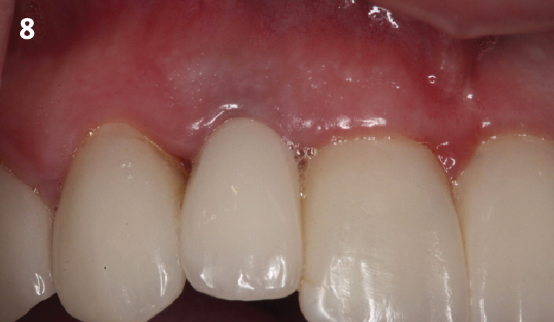
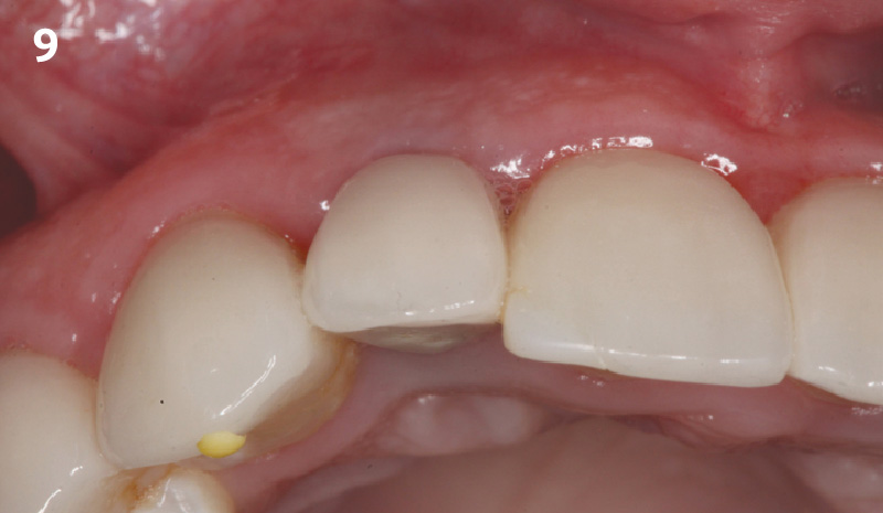
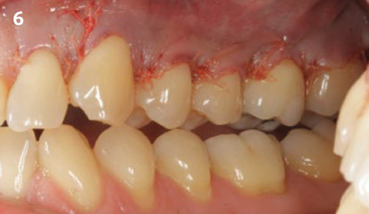
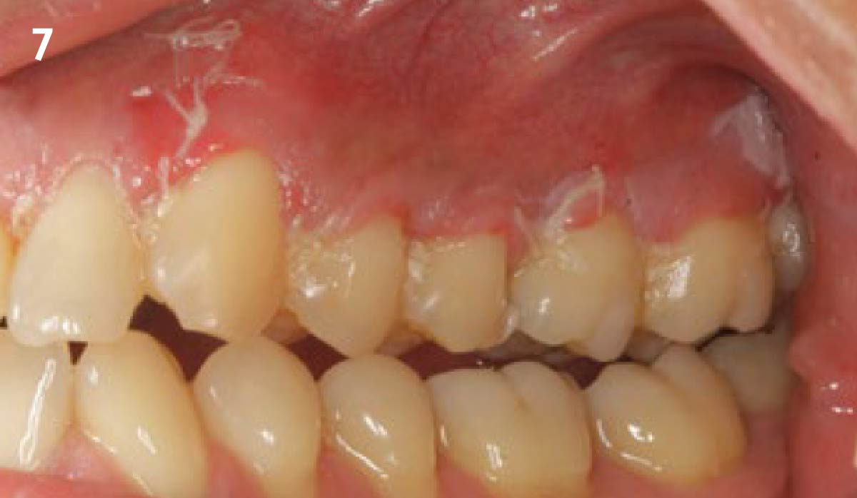
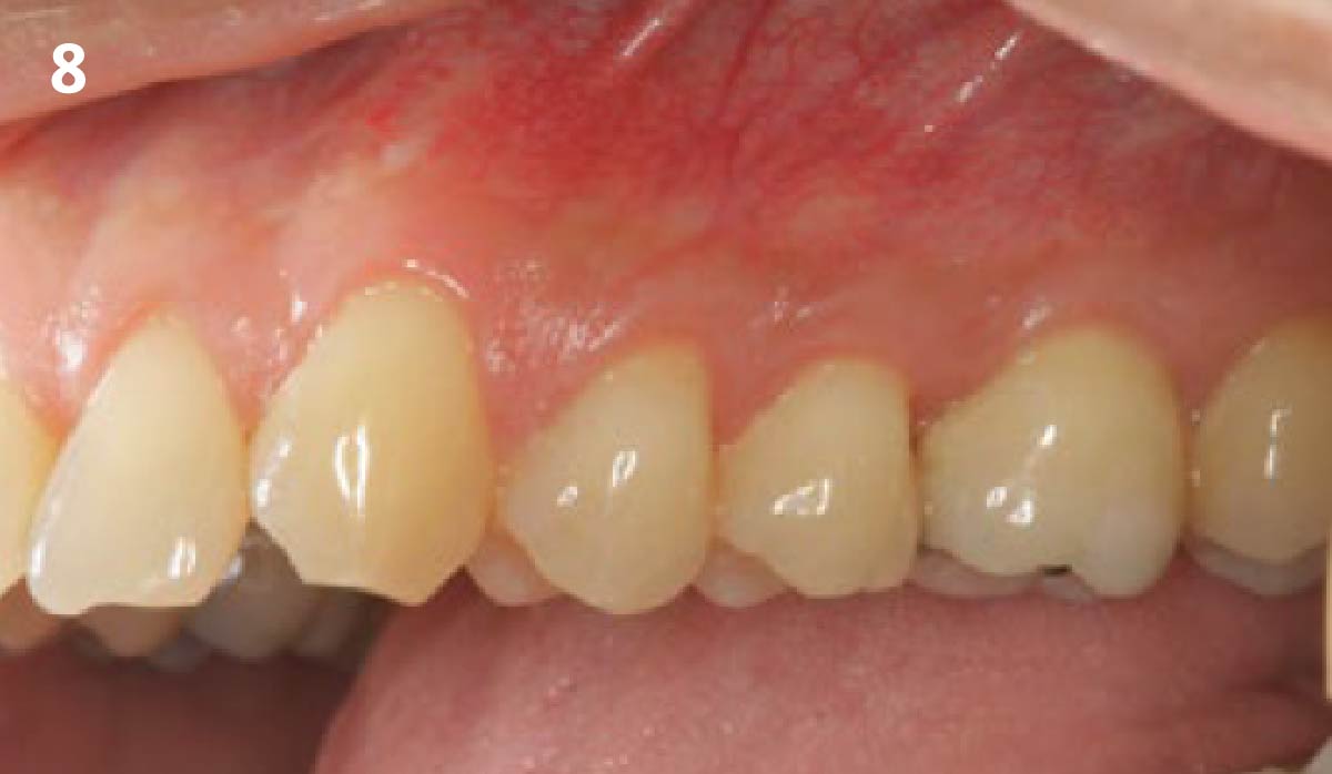
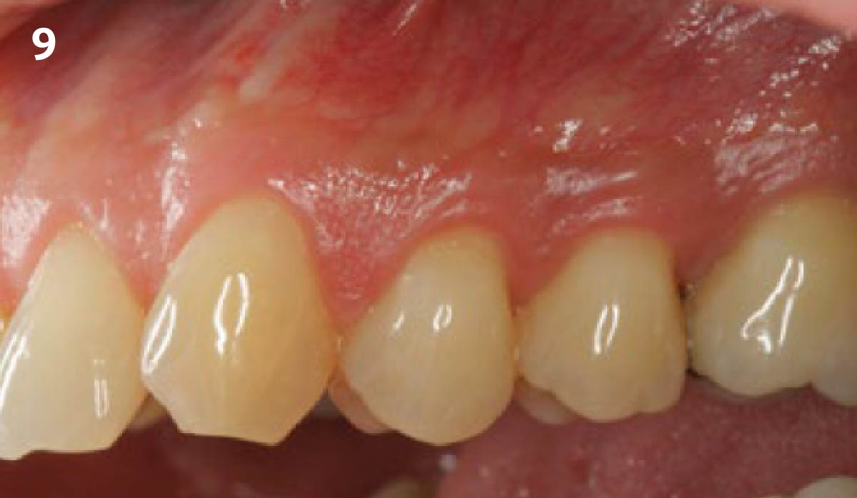
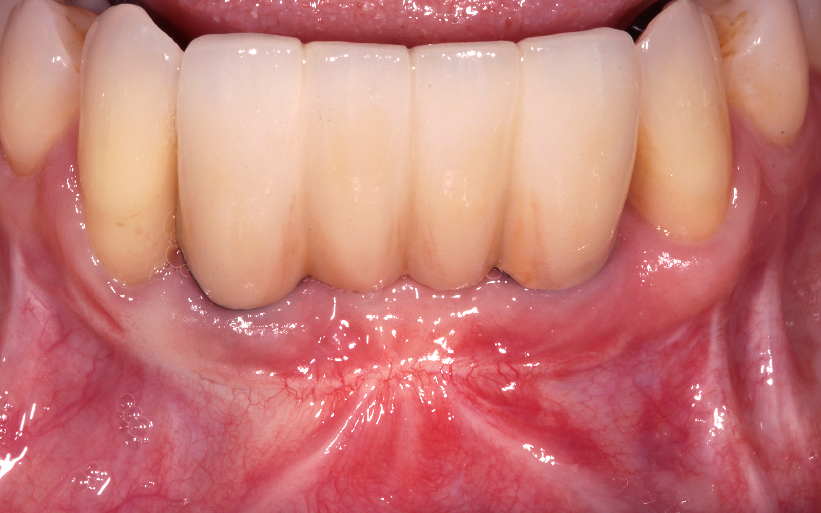
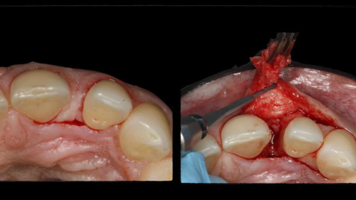
1 - Thin mucosal tissue over implant #7 creating an unesthetic appearance.2 - Occlusal view of #7 reveals buccal soft-tissue defects.3 - Augmentation of the soft-tissue on two implant sites, #7 and #10, using a conventional full thickness flap with vertical releasing incisions on site #7 and an envelope flap on site #10.4 - Geistlich Fibro-Gide® is split into two 3mm thick matrices to be used in both sites, hydrated with sterile saline and trimmed to the defect dimensions.5 - Geistlich Fibro-Gide® is secured to the periosteum apically and the interproximal papillae with 5-0 chromic gut sutures.6 - Passive closure of the flap achieved with 6-0 vicryl sutures.7 - 1 week post-surgery reveals uneventful healing with maintenance of tissue volume.8 - Buccal view at 8 weeks reveals uneventful healing with maintenance of tissue volume.9 - Occlusal view at 8 weeks post-surgery.
Evaluation of the tissue reaction to a new bilayered collagen matrix in vivo and its translation to the clinic
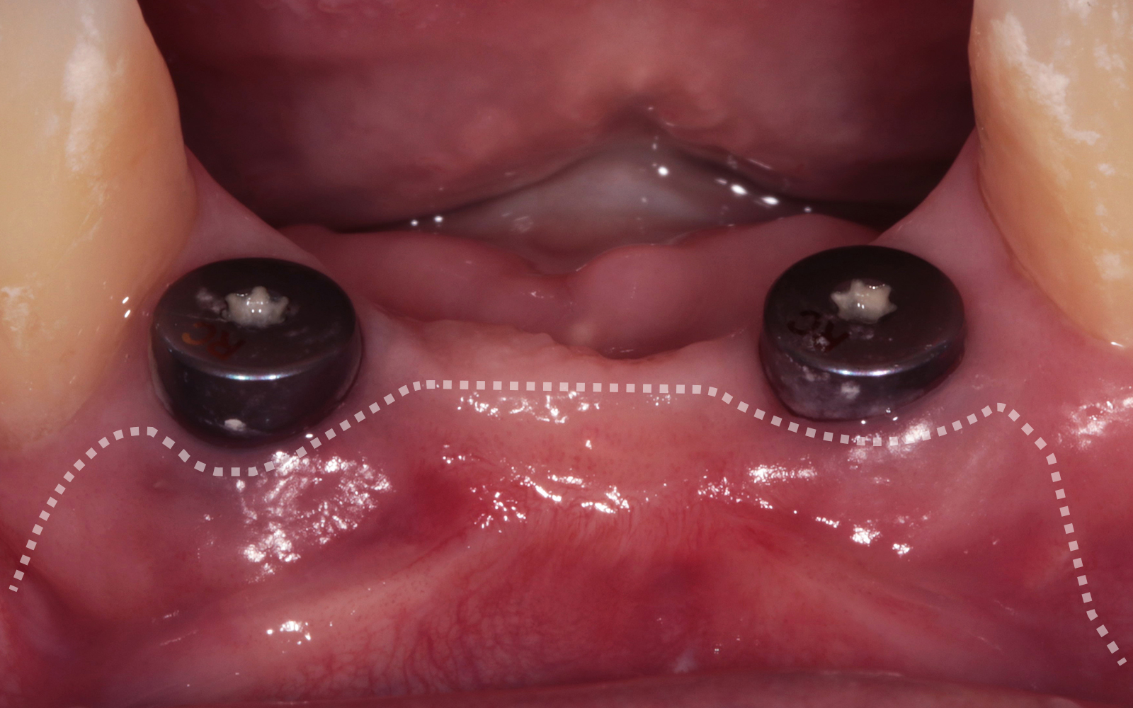
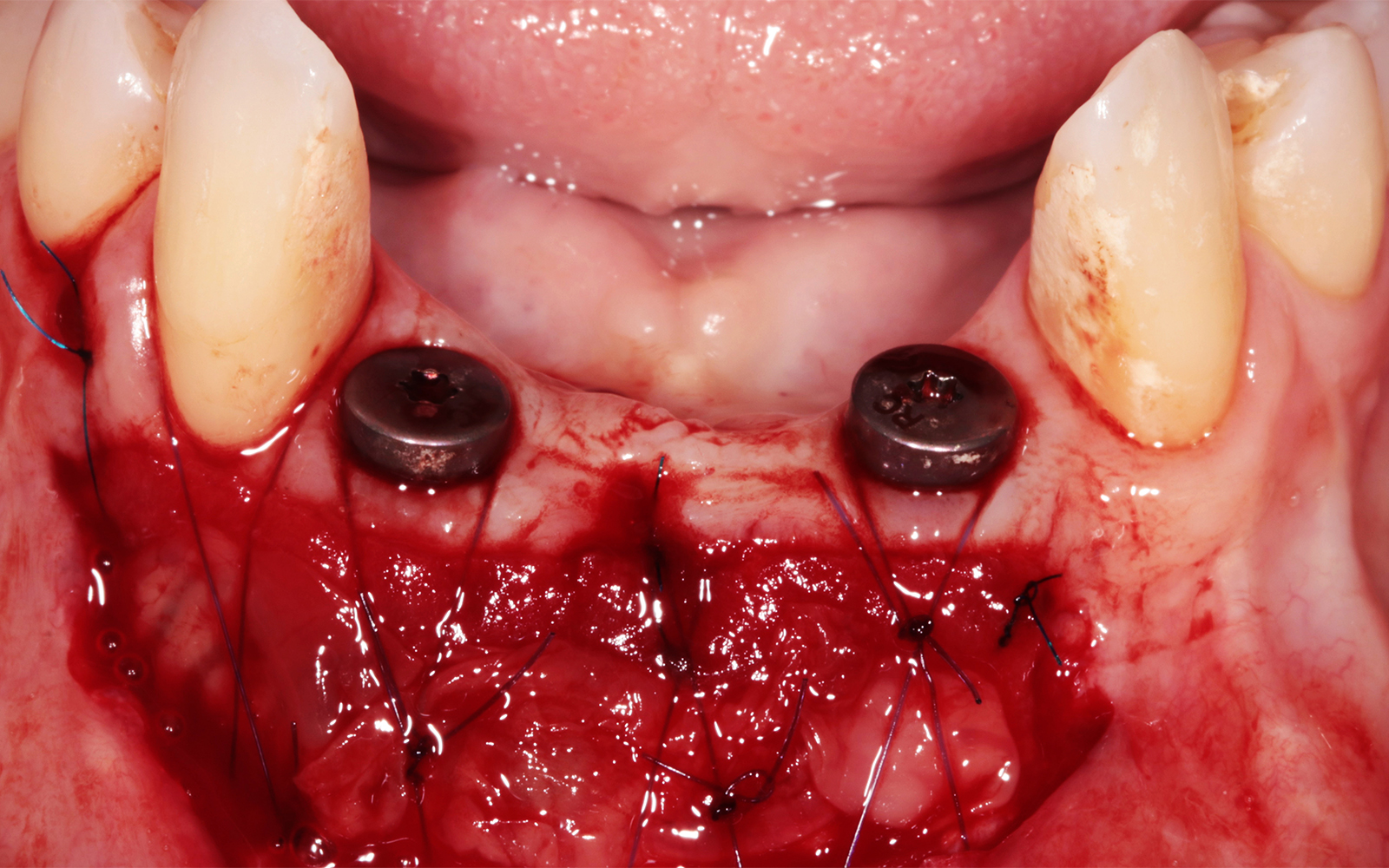
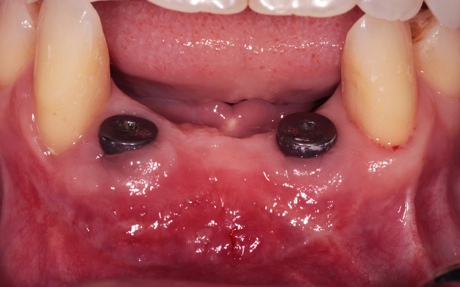
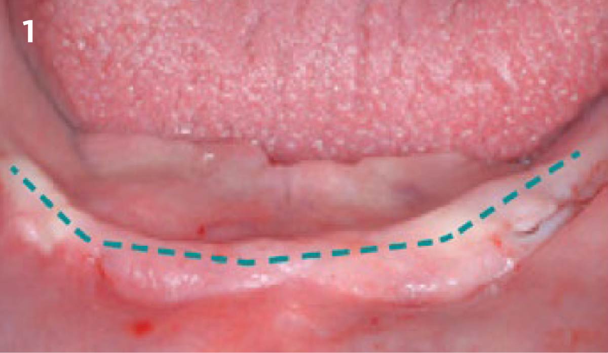
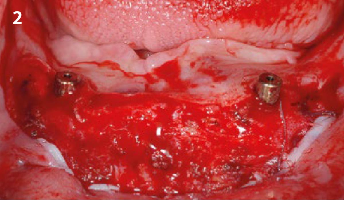
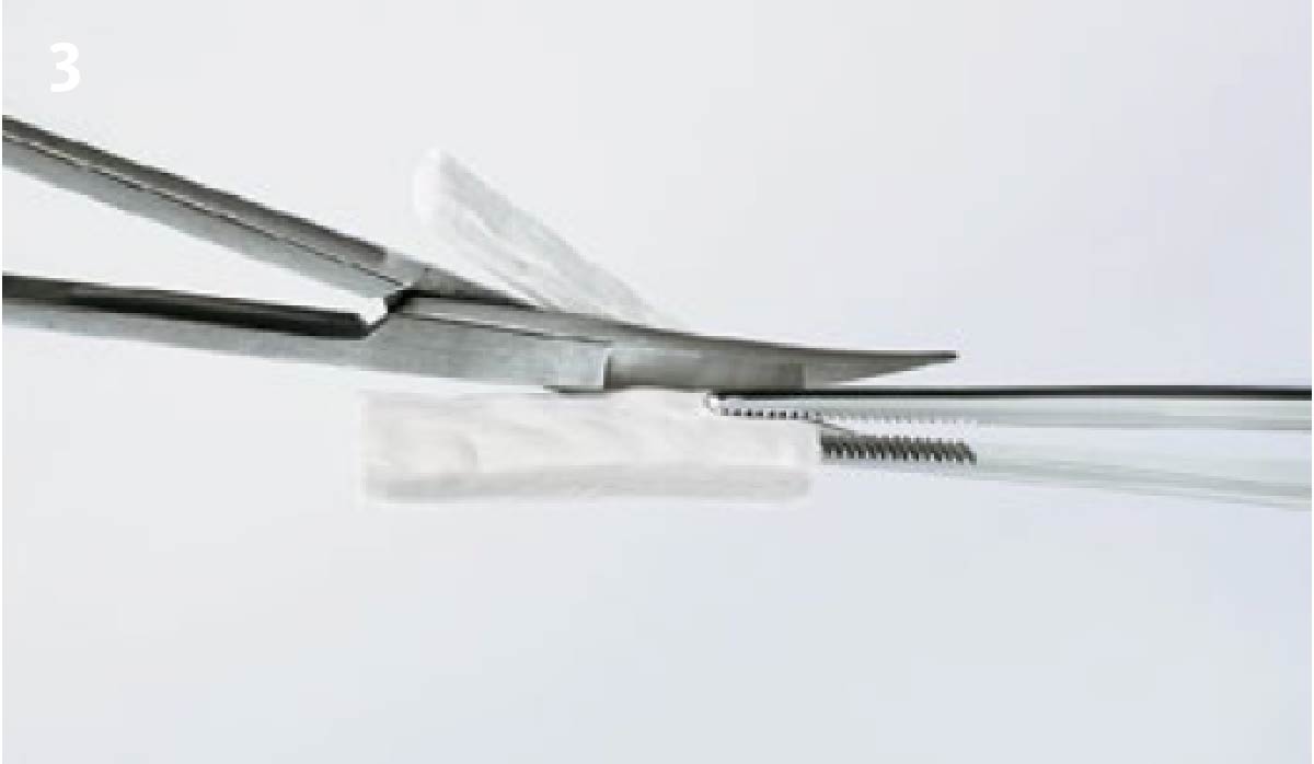
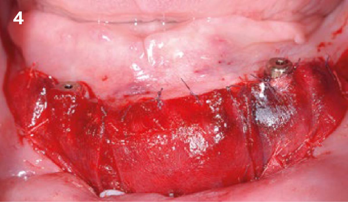
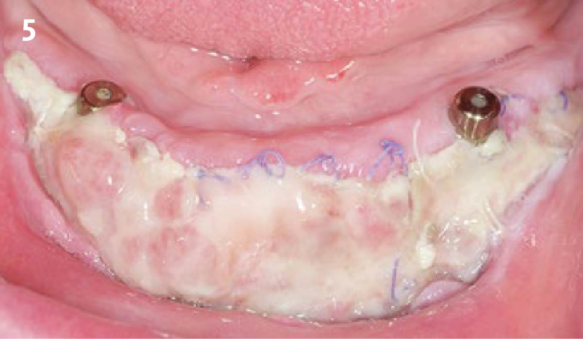
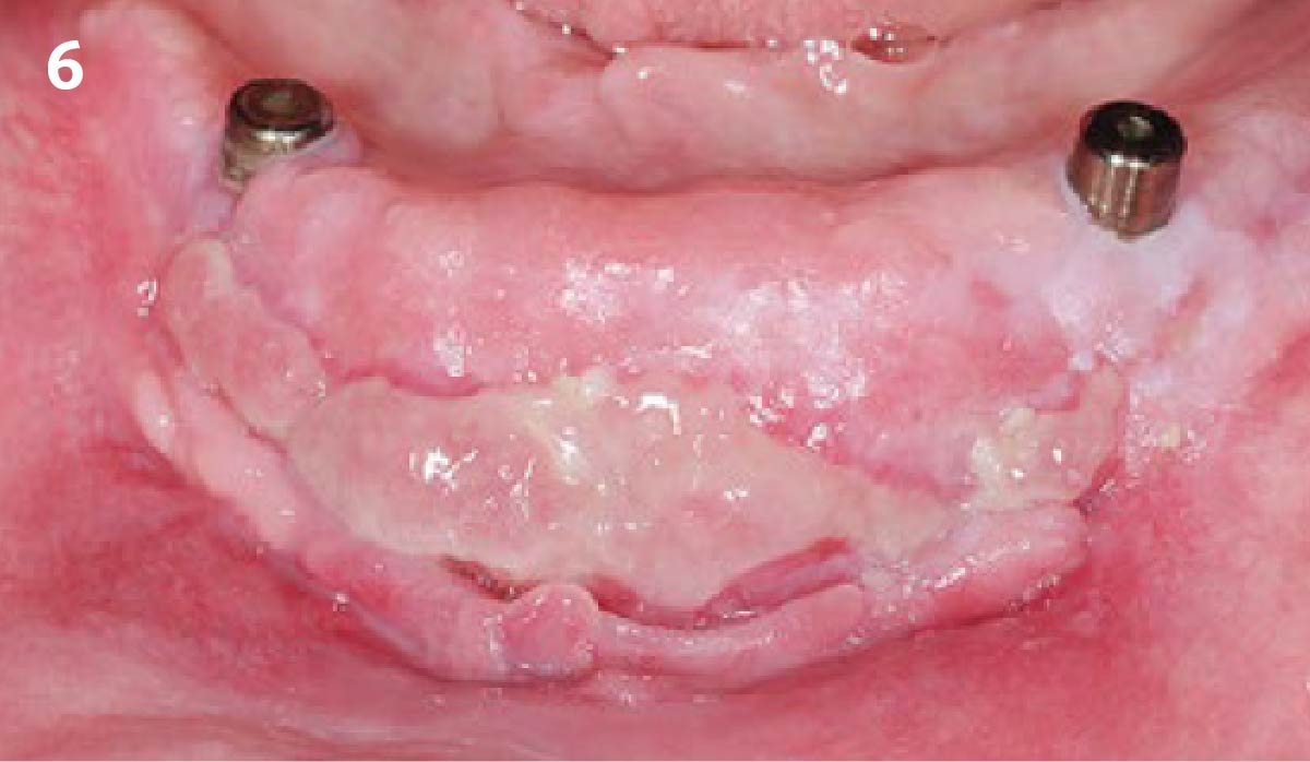
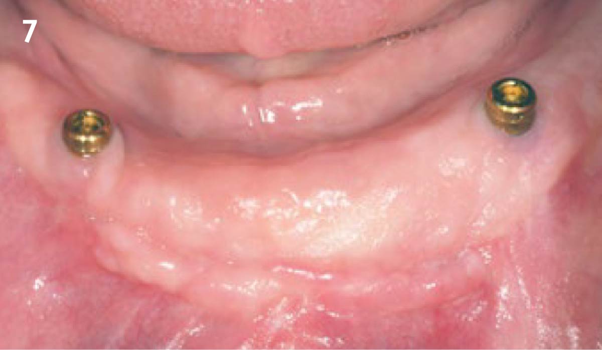
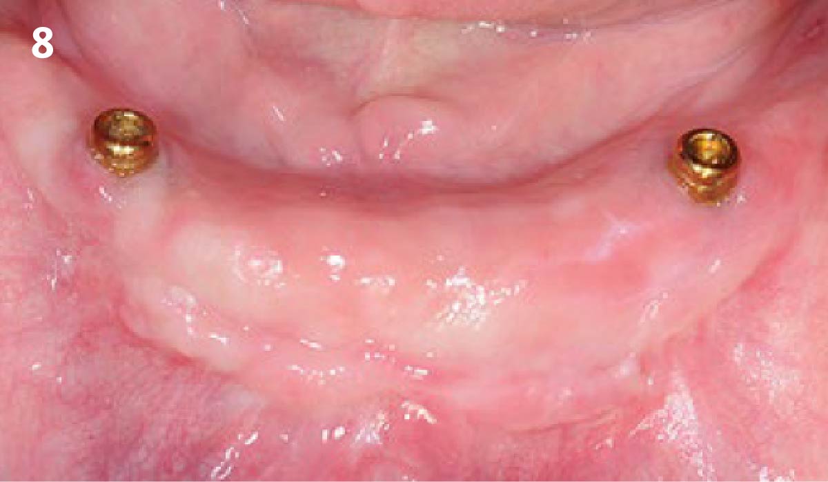
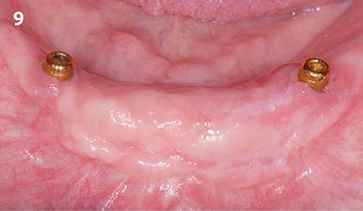
1 - This case shows a shallow vestibule and almost no keratinized tissue on the lower crestal part. The horizontal incision has to be made within the keratinized tissue (see depicted line). 2 - Ensure the incision is within the keratinized tissue: an apically fixed split flap is used here. Remove the muscles, scar fibers and ligaments before application of Geistlich Mucograft®.3 - Measure the defect carefully with a periodontal probe and cut Geistlich Mucograft® in a dry state. In the case of a large vestibuloplasty, several pieces of Geistlich Mucograft® can be sutured side by side.4 - Geistlich Mucograft® is sutured directly onto the periosteum with 5.0 resorbable sutures and left exposed for open healing (healed by secondary intention). 5 - 10 days post-operative: favorable healing where granulation tissue and new soft tissues have formed. 6 - 1 month post-operative: excellent wound healing with complete integration and epithelialization of Geistlich Mucograft® as well as keratinization of the crestal aspect could be observed. 7 - 3 months post-operative: excellent esthetic outcome with full integration and epithelialization of Geistlich Mucograft® to the surrounding tissue and keratinization of the crestal aspect. 8 - 1 year post-operative: the formed keratinized tissue fulfills its functionality and shows an esthetically pleasing result matching the structure and color of the surrounding tissues. The mean width of the peri-implant keratinized mucosa was 4.0 mm, and a low shrinkage was assessed at 38%. 9 - 5 years post-operative: the mucogingival appearance remains stable. The resulting keratinized tissue fulfills its functionality and shows an esthetically pleasing result.
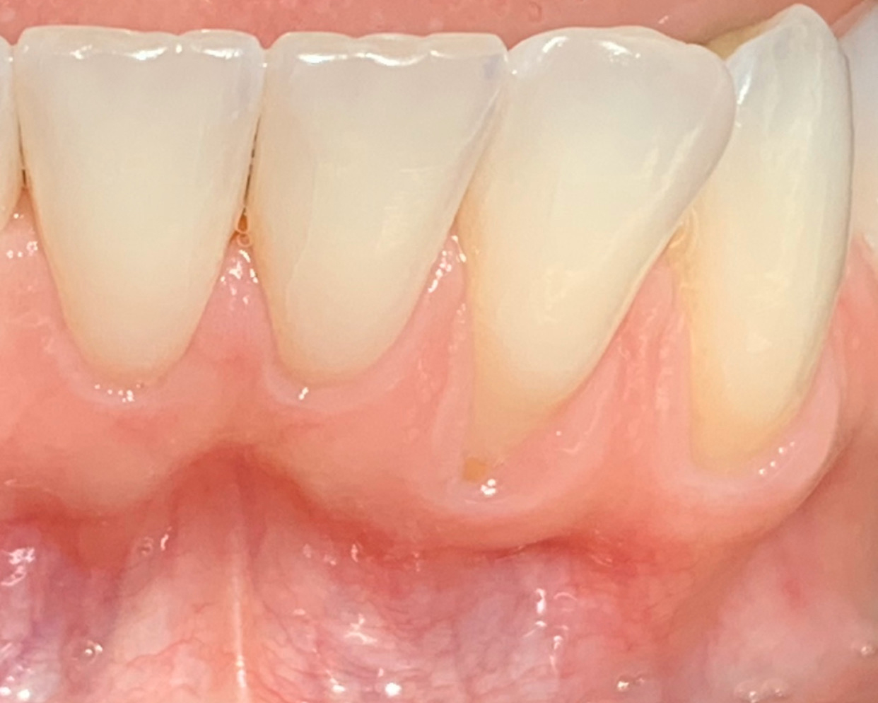
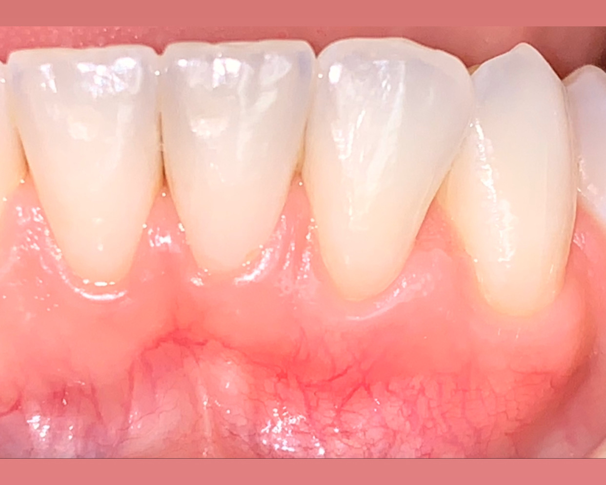
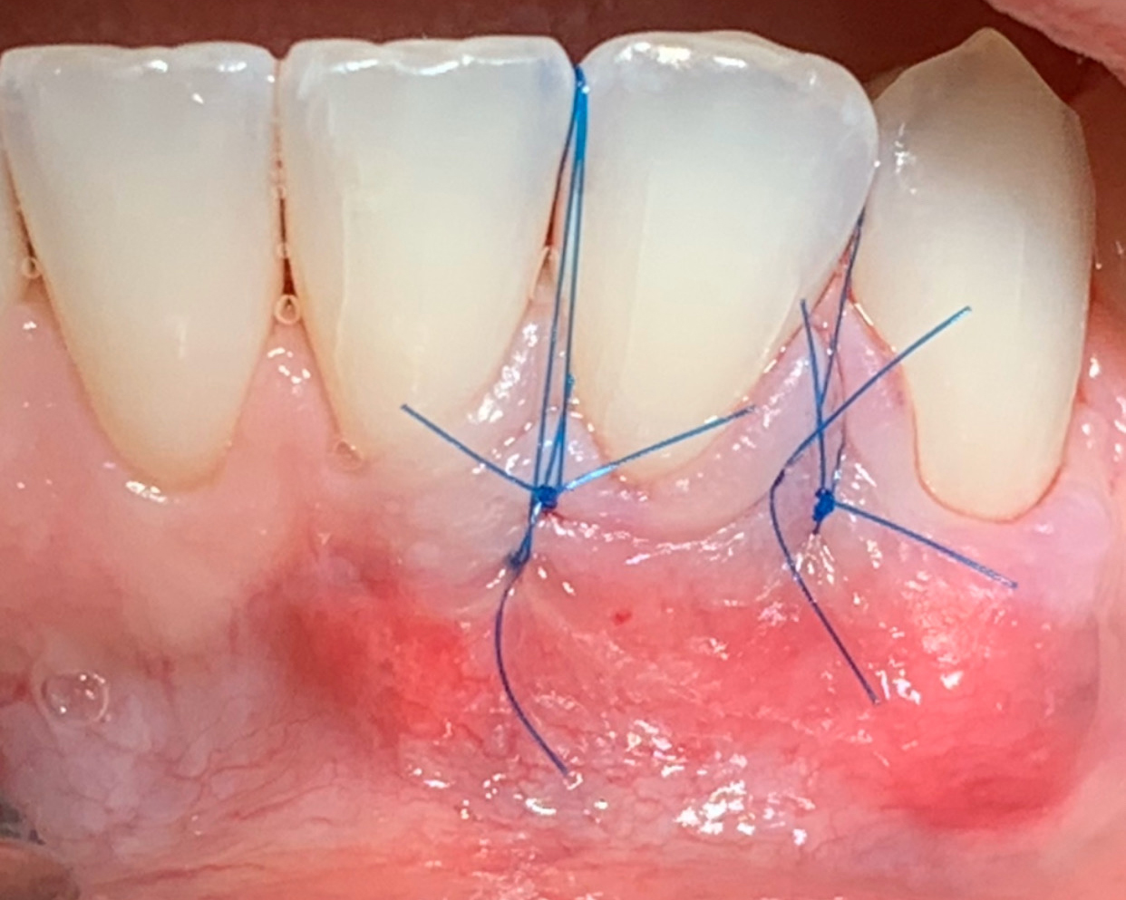
1 - Gingival recessions #23 and #223 - 10 week post op healing. Root coverage achieved. Patient reports no problems and is pleased with the results. More follow ups to come.2 - Sites at the day of the procedure after sutures placed. Tunnel between #23,24 and partial thickness flap raised between #23,22.
Dr. Daniel Gober
Cedarhurst, New York, USA
Geistlich Mucograft® for the Treatment of Multiple Adjacent Recession Defects
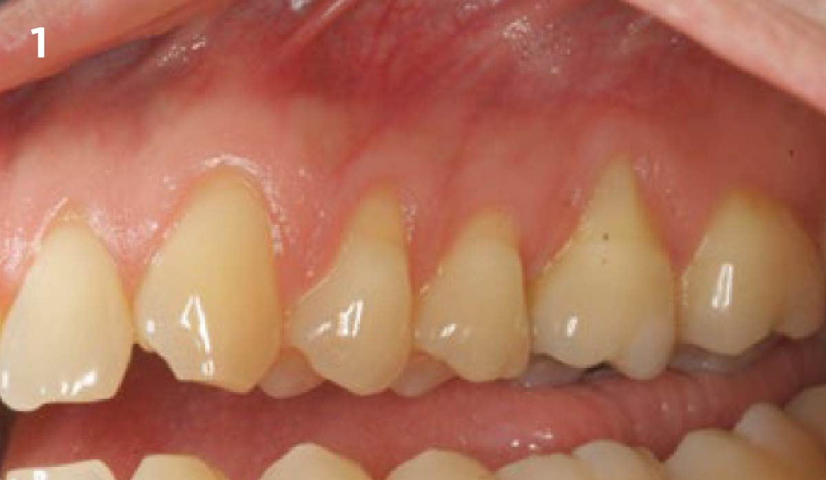
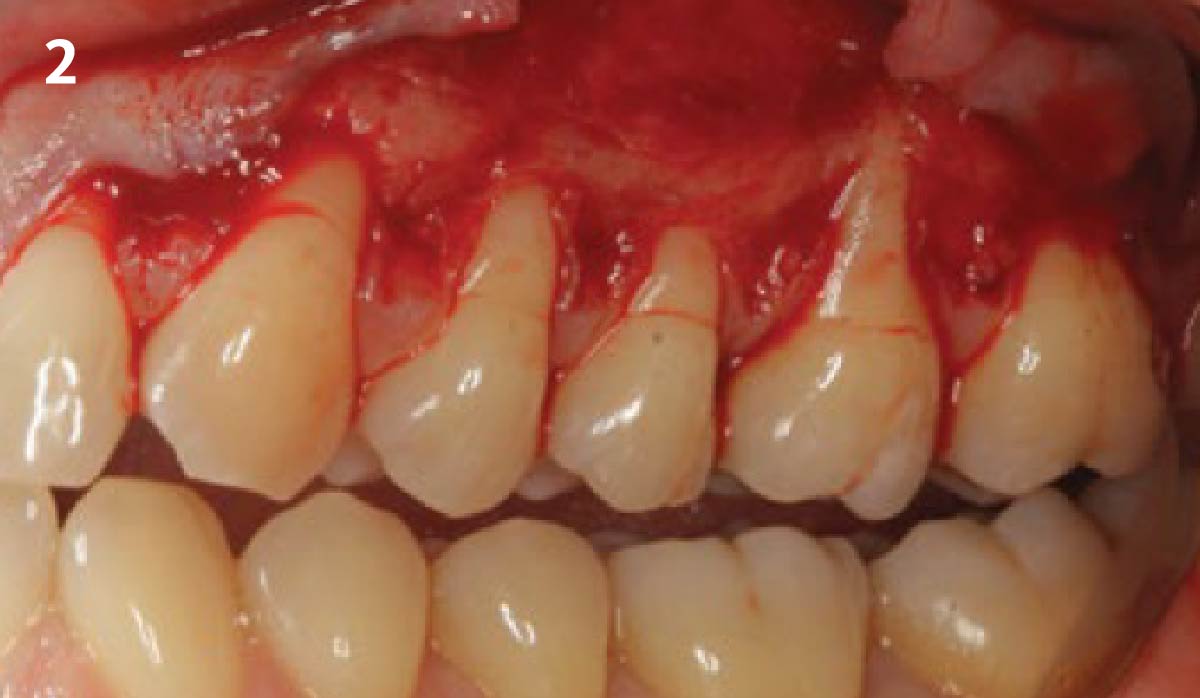
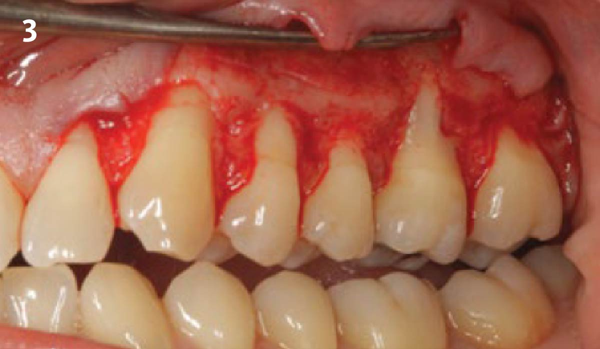
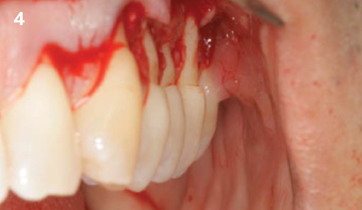
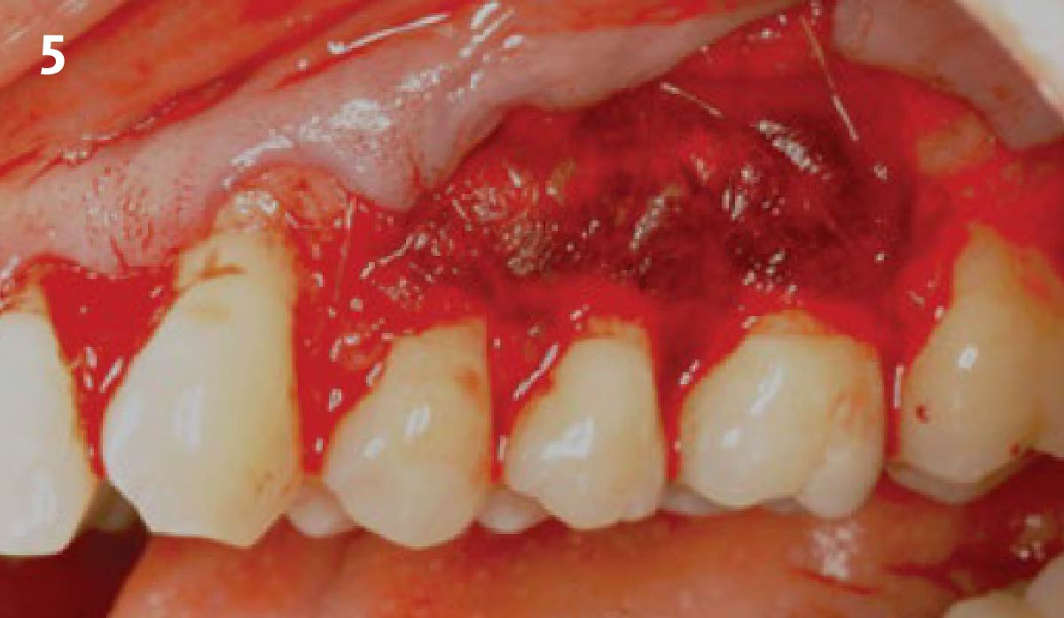
Pre-operative view of recession defects that ranges from 2-5 mm. There is minimal keratinized tissue on #14. Pocket depths are within limits with no loss of interproximal tissue. A 15c blade is used to make sulcular incisions with scooping incisions at the level of the CEJ. A combination flap consisting of full-thickness coronally followed by a partial-thickness dissection apically is reflected. The papillary tissue is de-epithelialized with a football diamond bur on a rotary hand piece. This exposes a vascular bed for the graft and intended coronal positioning of the flap. Side-view of the recession defects. It is clearly visible how deep the recession defects are. Geistlich Mucograft® is trimmed and positioned to extend beyond the root surfaces. A combination of simple interrupted sutures at its coronal edge and mattress sutures extending over the entire graft are used to adapt the graft to the recipient site. The flap is then advanced and coronally positioned with horizontal mattress sutures to release tension and simple interrupted sutures to approximate the flap edges to the de-epithelialized papillas. Follow-up after 1 week: note that the flap margins appear stable. Erythema and edema evident with maturation of the tissue beginning. Follow-up after 3 months: maturation of the tissue evident with complete root coverage. An increase in the zone of keratinized tissue by #14 is also visible. Follow-up after 1 year: stability of the graft is evident, complete coverage and a healthy and maintainable gingival situation have been achieved.
Bassam Kinaia, DDS, MS, DICOI
Sterling Heights, MI
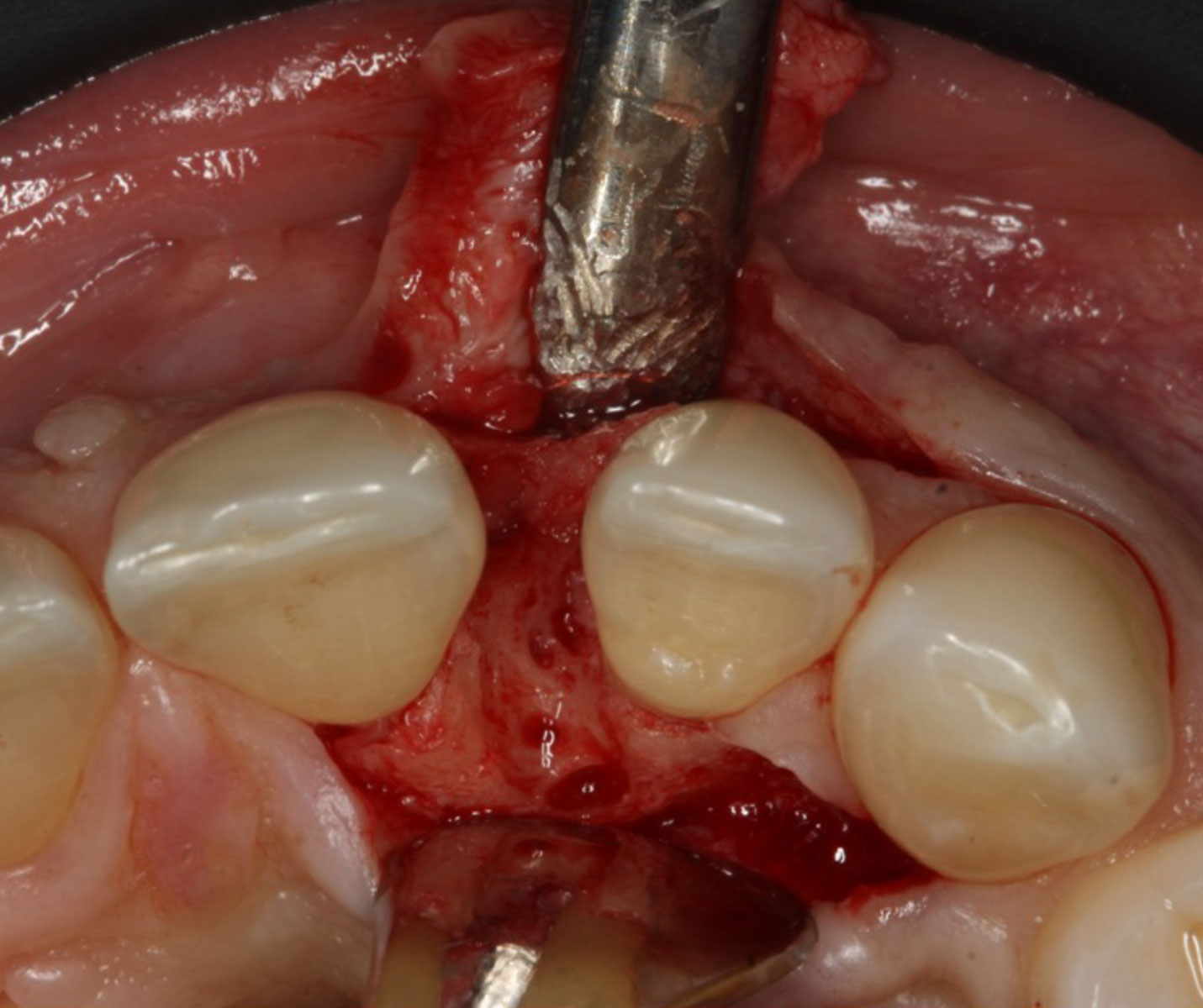
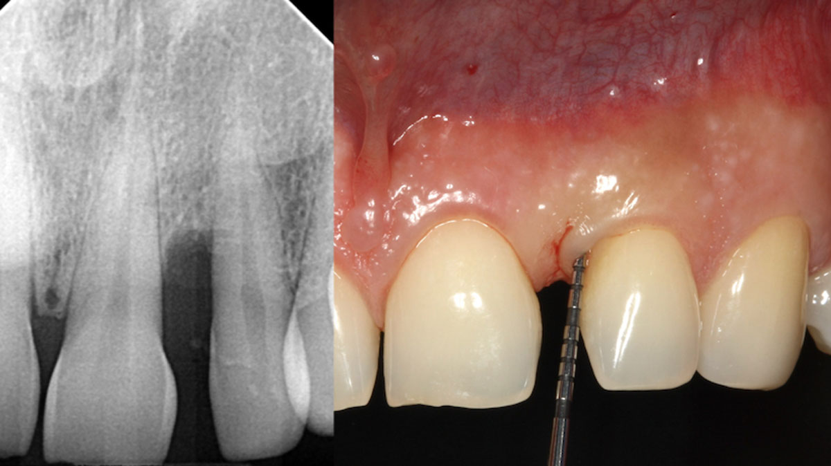
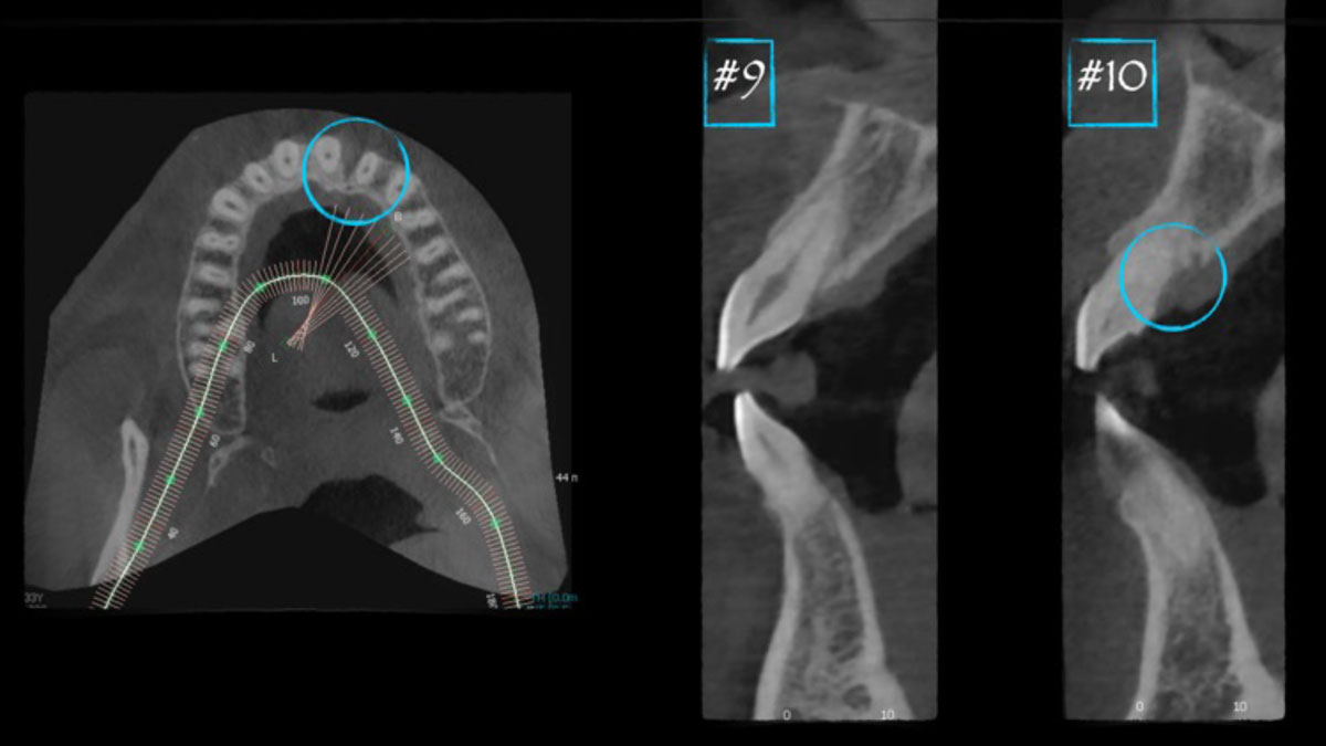
Guided Tissue Regeneration in the Esthetic Zone of a 34-Year-Old Male
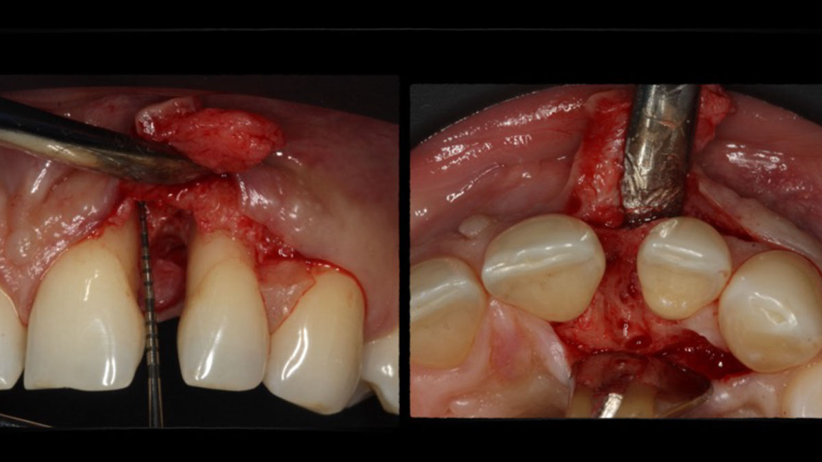
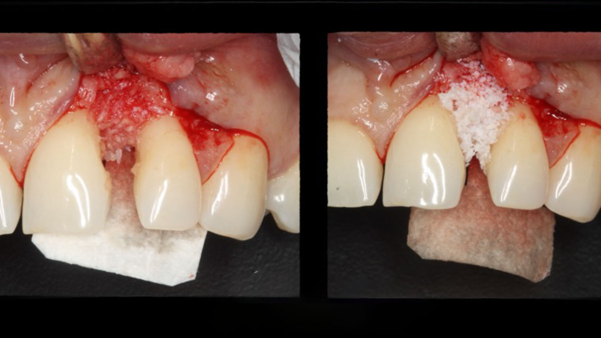
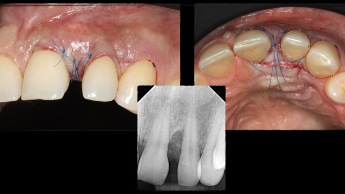
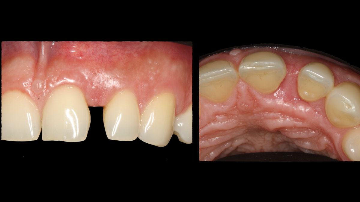
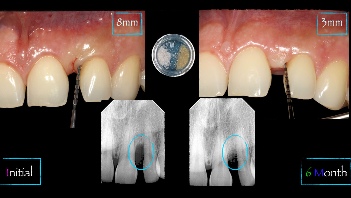
Initial clinical and radiographic presentation showing vertical bone loss #9-10 areaInitial CBCT presentation showing bone loss between #9-10 wrapping palatallyOcclusal clinical views showing sulcular incision with paracrestal incision #9-10Facial and occlusal clinical views showing full thickness flap reflection with wide 1-2 bony wall defect measuring ~7mm vertical bone lossFacial clinical views showing full thickness flap reflection and GTR procedure using Geistlich vallomix™ bone graft (allograft as an internal first layer and xenograft as an outside second layer) and collagen membraneFacial and occlusal clinical views showing primary closure using 6-0 prolene sutures with immediate post-surgical bone additionFacial and occlusal clinical views showing healing at 10 days with tissue granulating inFacial and occlusal clinical views showing healing at 6 weeksPre-operative vs post-surgical clinical and radiographic views showing adequate bone fill and reduction in probing depths