BIOBRIEF

Lateral and Vertical Bone Regeneration with Simultaneous Soft Tissue Augmentation
Arnd Lohmann, MSc

THE SITUATION

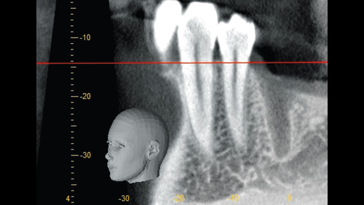
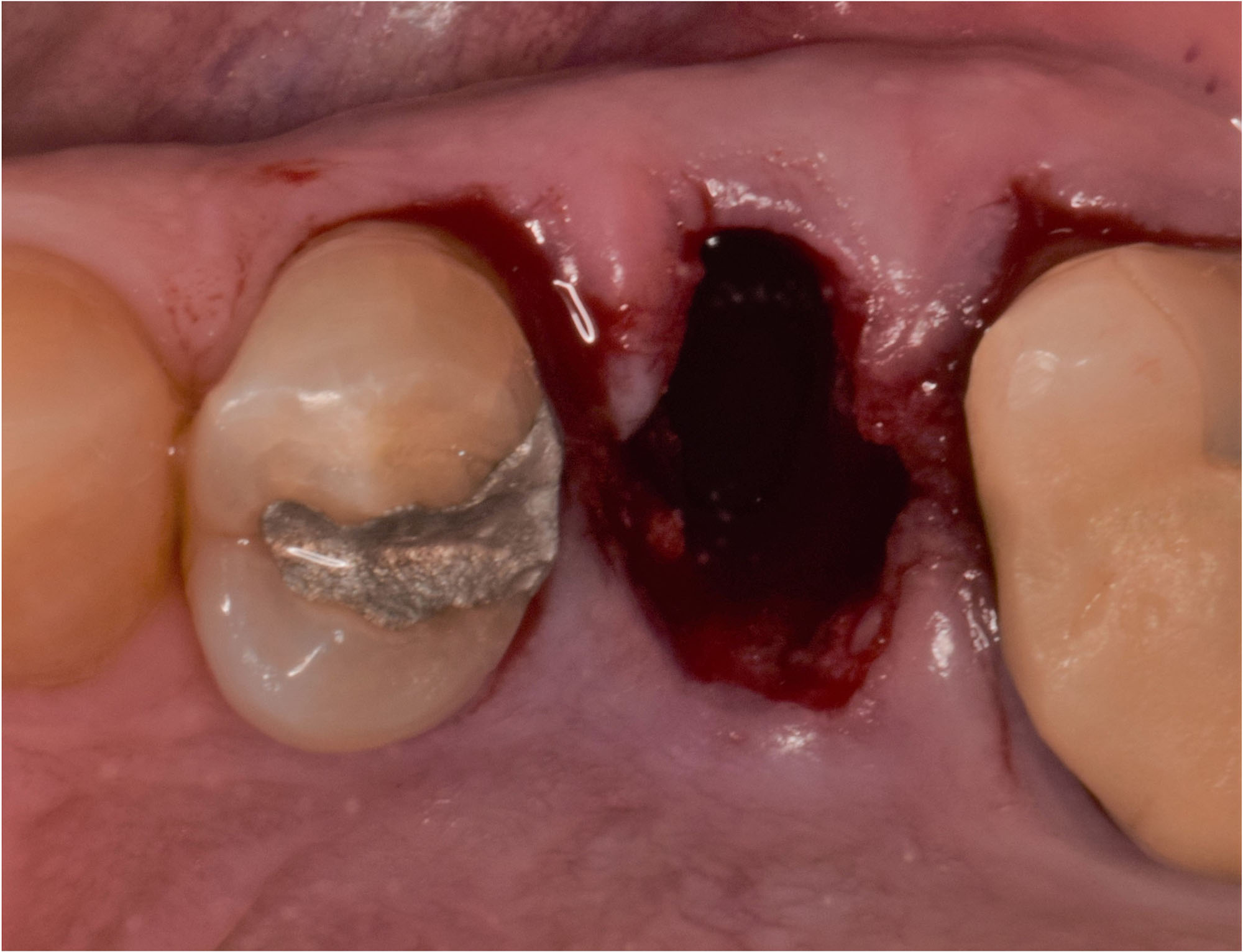
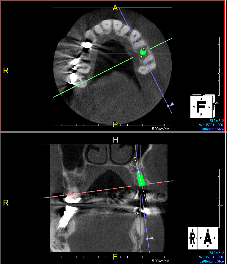
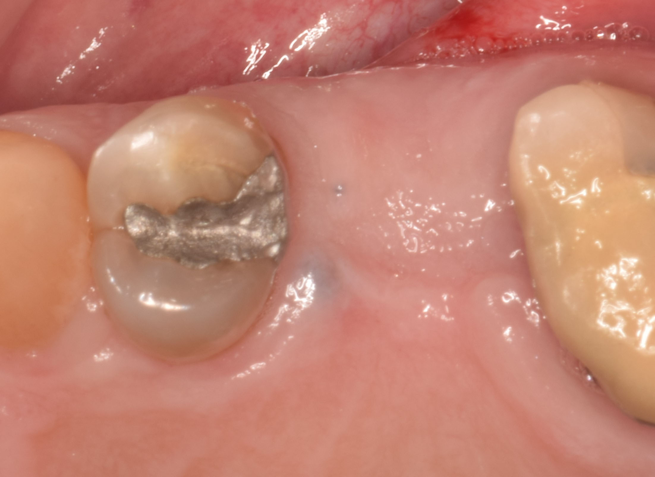
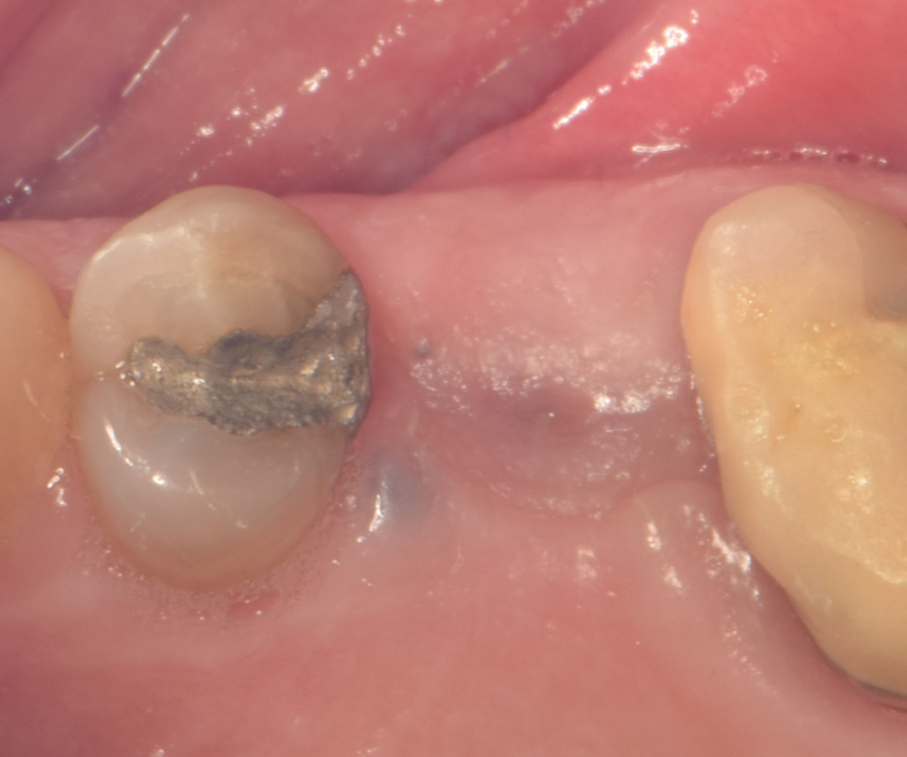
After extraction of the periodontally damaged tooth #20 the preoperative Cone-Beam Computed Tomography (CBCT) imaging shows reduced vertical bone volume in the area of tooth #s 18 – 20. A lateral and vertical bone  regeneration was necessary.

The goal of treatment was a late implant placement after bone regeneration and creation of stable periimplant soft tissue for long-term implant preservation.

THE RISK PROFILE

Low RiskMedium RiskHigh Risk
Patient’s healthIntact immune systemLight smokerImpaired immune system 
Patient’s esthetic requirementsLowMediumHigh
Gingival biotypeThick – “low scalloped”Medium – “medium scalloped”Thin – “high scalloped”
Infection at implant sightNoneChronicAcute
Bone height at adjacent tooth≤ 5 mm from contact point5.5 – 6.5 mm from contact point≥ 7 mm from contact point
Width of tooth gap1 tooth (≥ 7 mm)1 tooth (≤ 7 mm)2 teeth or more
Soft-tissue anatomyIntactCompromised
Bone anatomy of the alveolar ridgeNo defectHorizontal defectVertical defect

Additional Risk Factors: Roots were divergent, and intra-radicular bone (septal bone) was excellent, with more than 5 mm of remaining apical bone to achieve optimal primary stability.

THE APPROACH

A customized bone regeneration procedure utilizing Yxoss CBR®. Followed by coverage of the graft with Geistlich Bio-Gide® for the purpose of Guided Bone Regeneration (GBR). Soft tissue thickening using Geistlich Fibro-Gide®. Delayed implantation into the augmented tissue. A vestibuloplasty with Geistlich Mucograft® for the regeneration of keratinized mucosa.

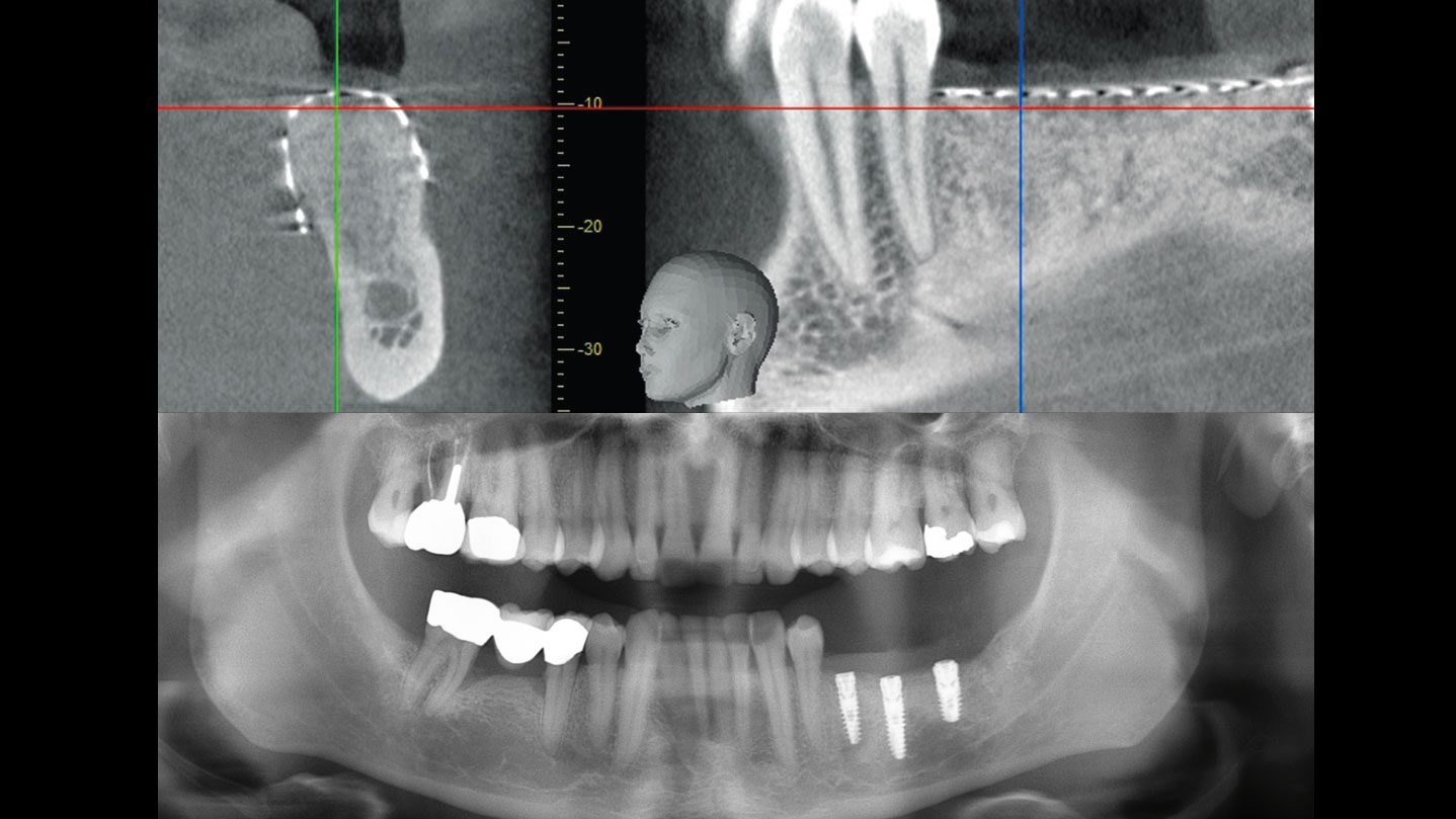
Baseline situation and planning: vertical bone deficit of 5.29mm in section 18-20.
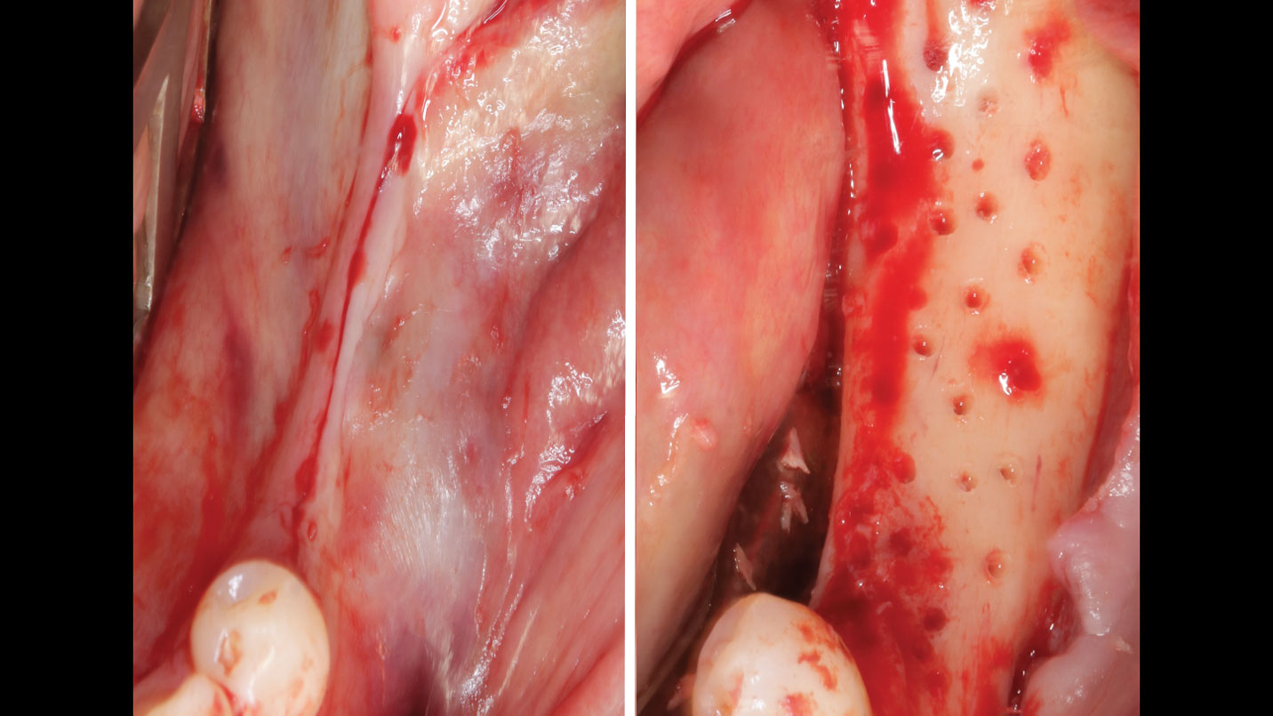
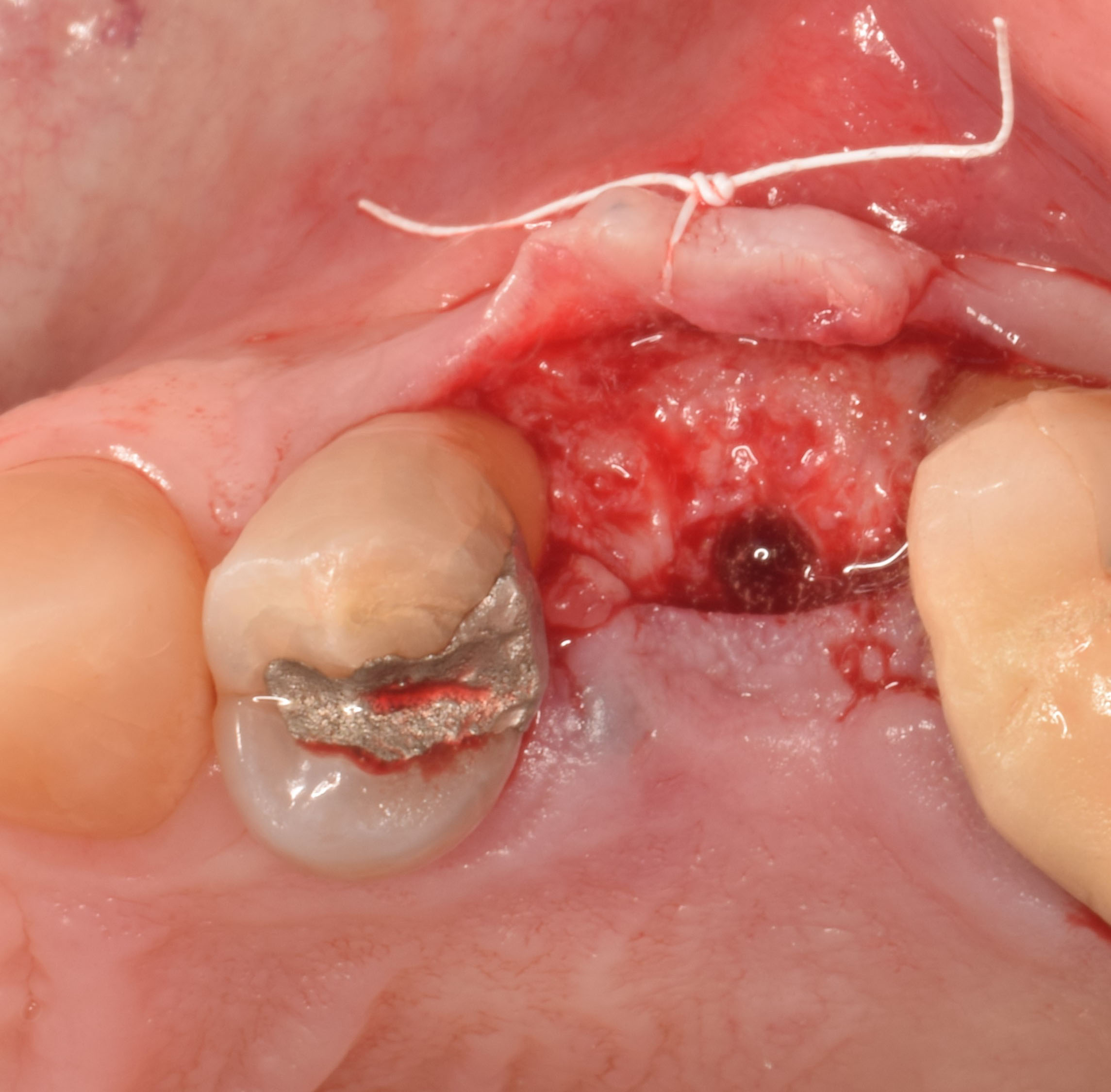
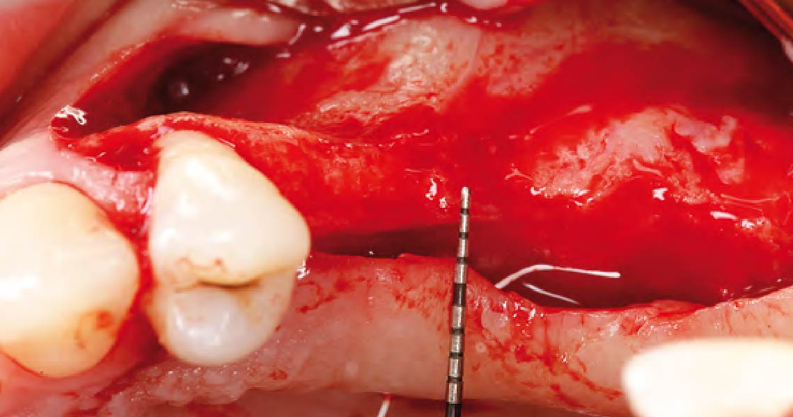
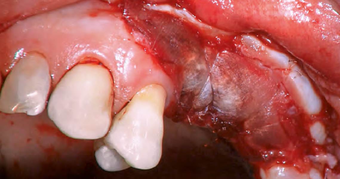
Central incision in the keratinized mucosa and perforated cortical bone
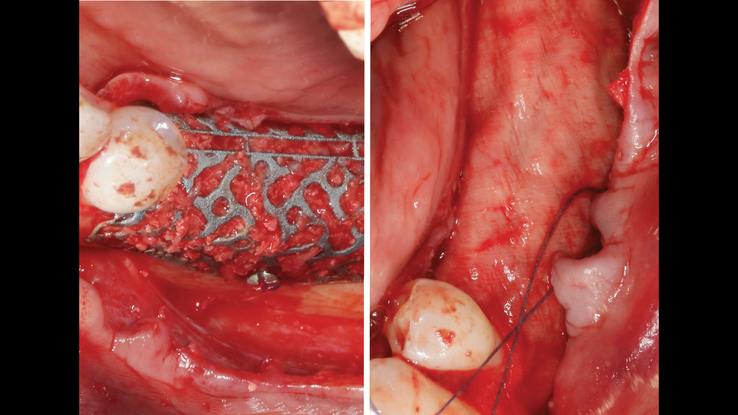
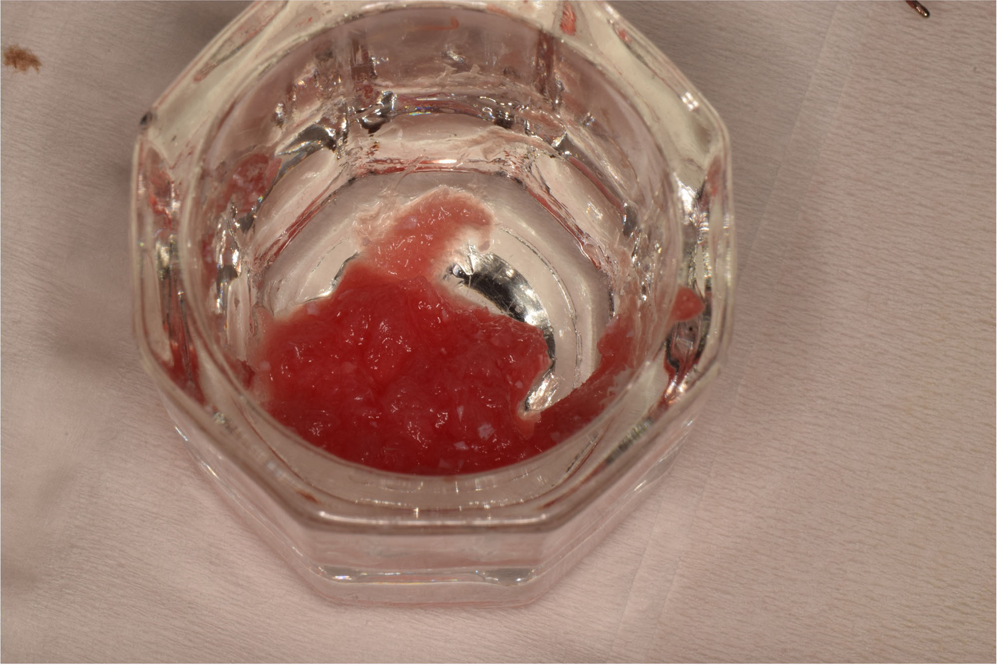
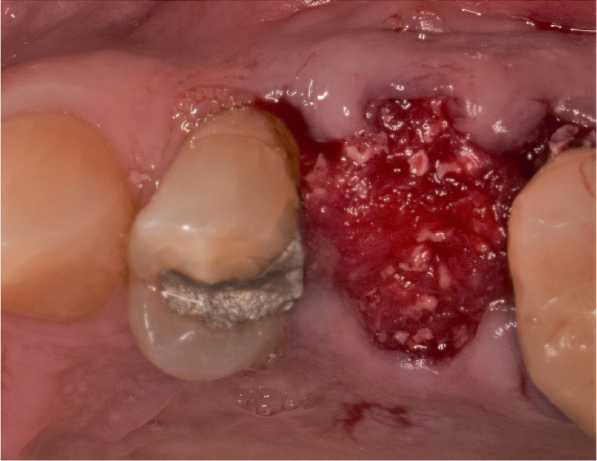
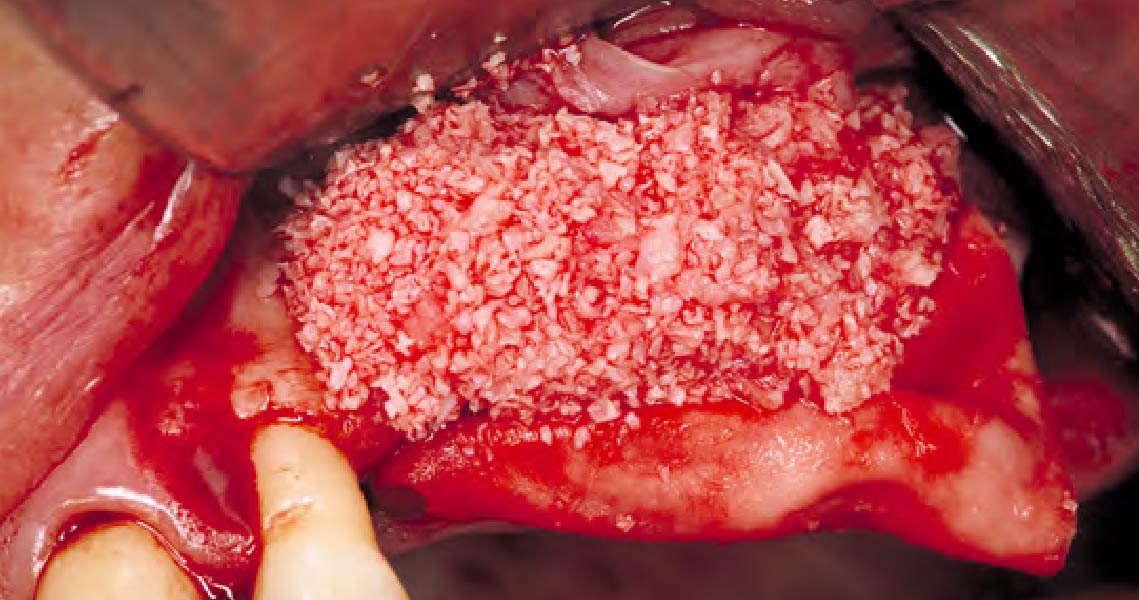
Yxoss CBR® filled with a 50:50 mix of autologous bone and Geistlich Bio-Oss®. Coverage of the titanium scaffold with Geistlich Bio-Gide®
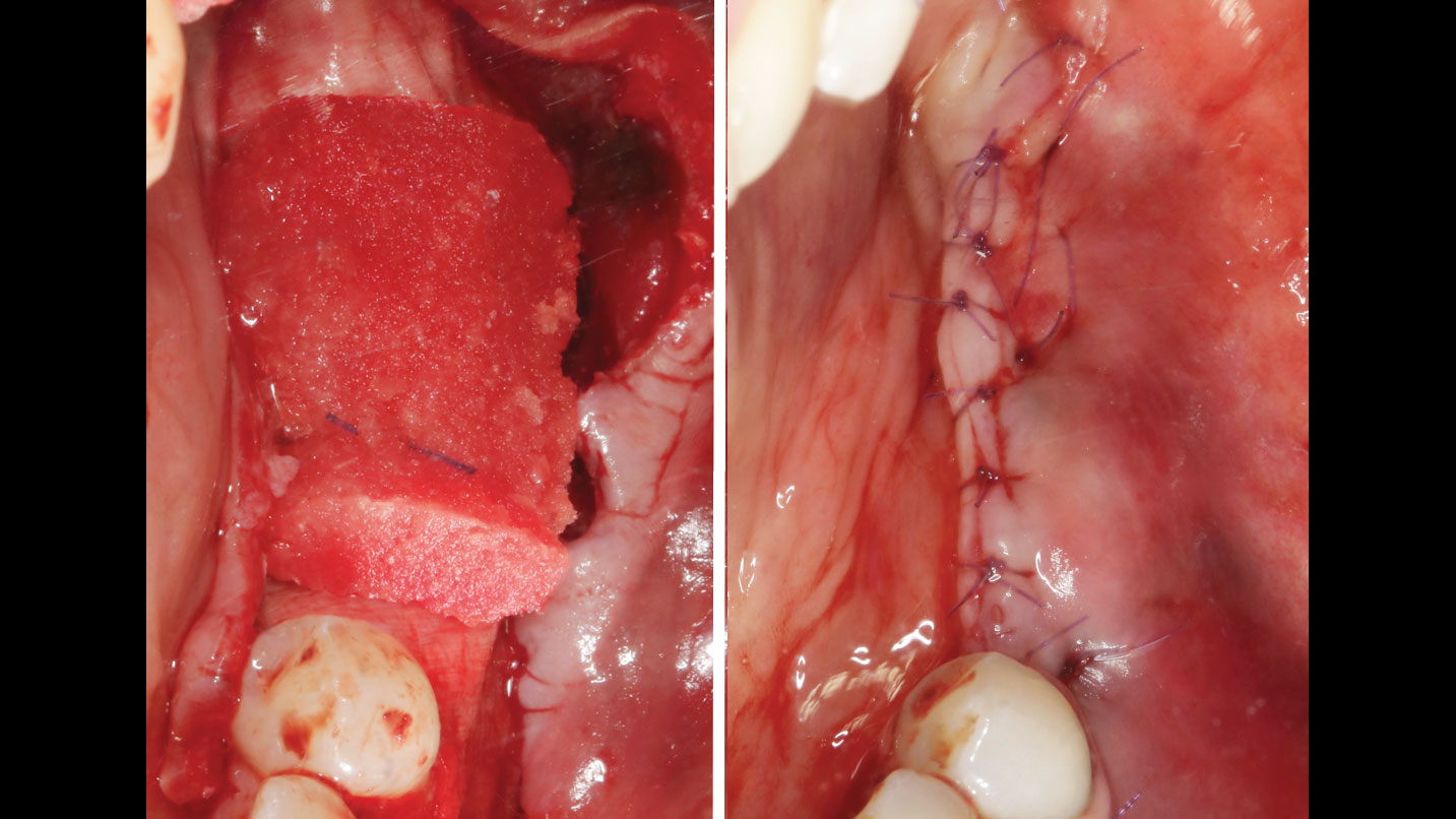
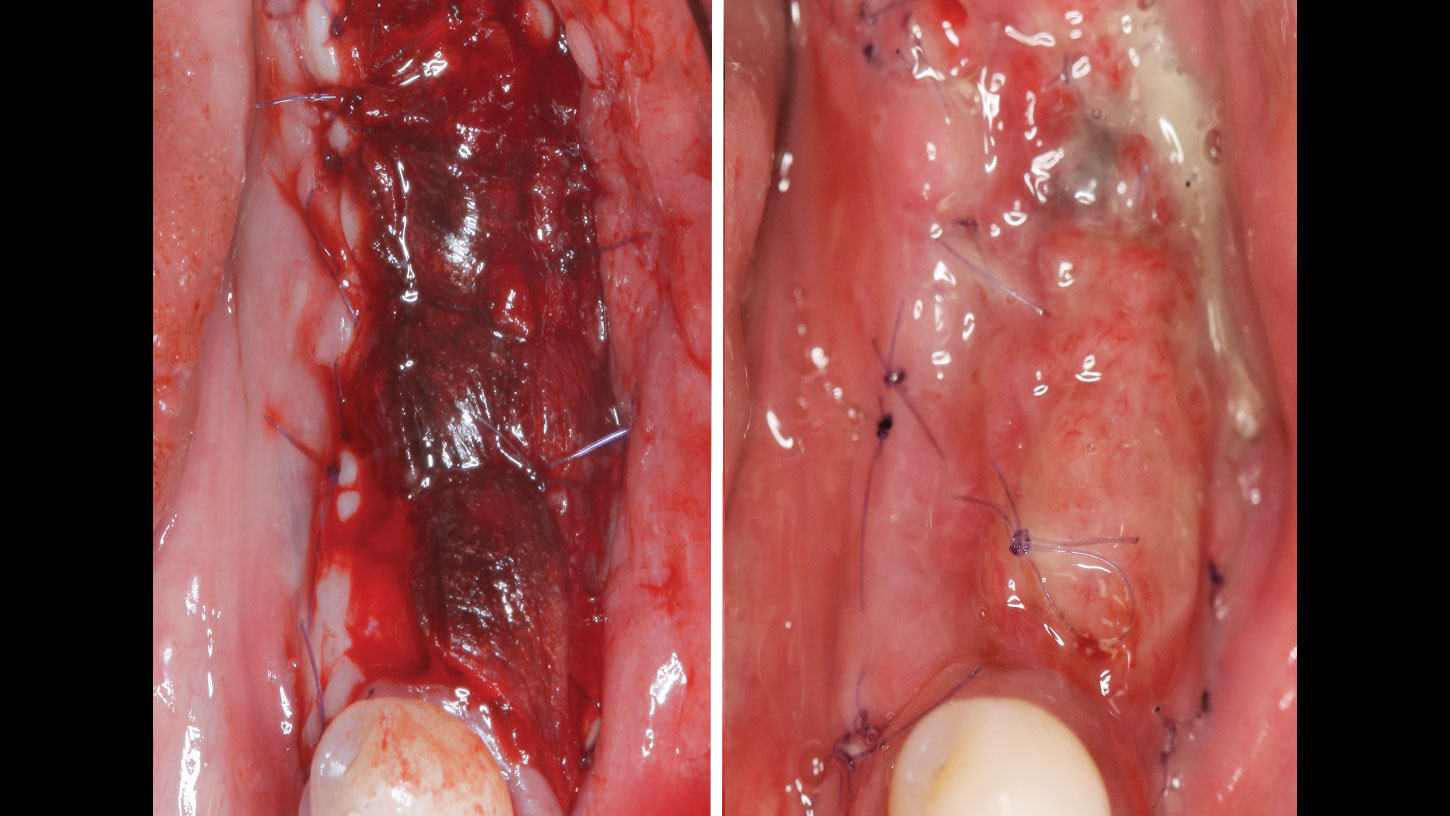
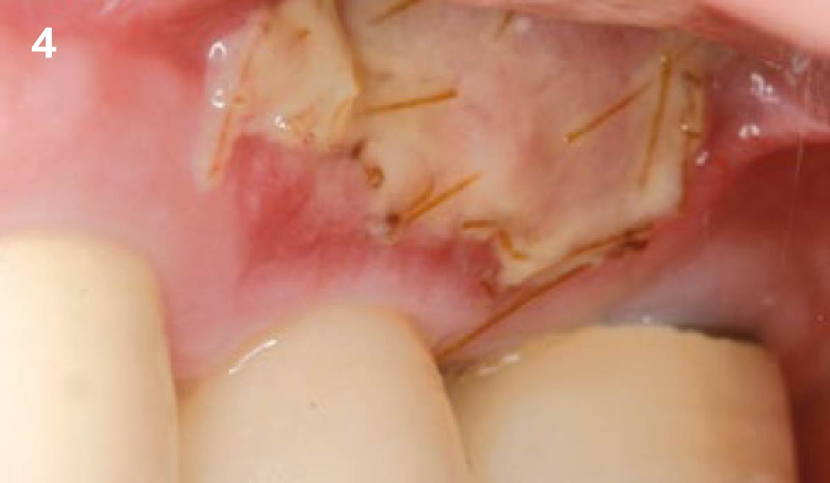
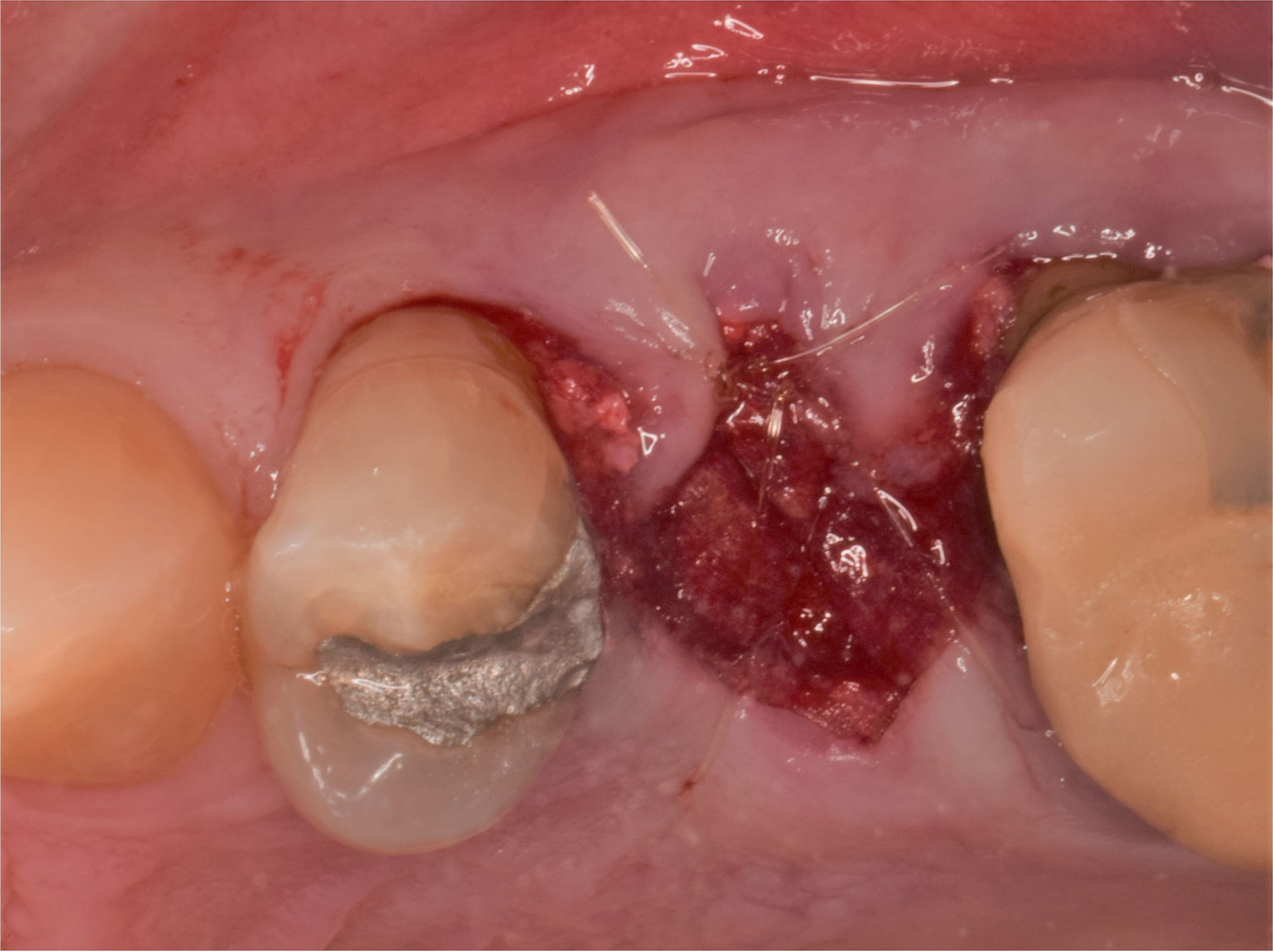
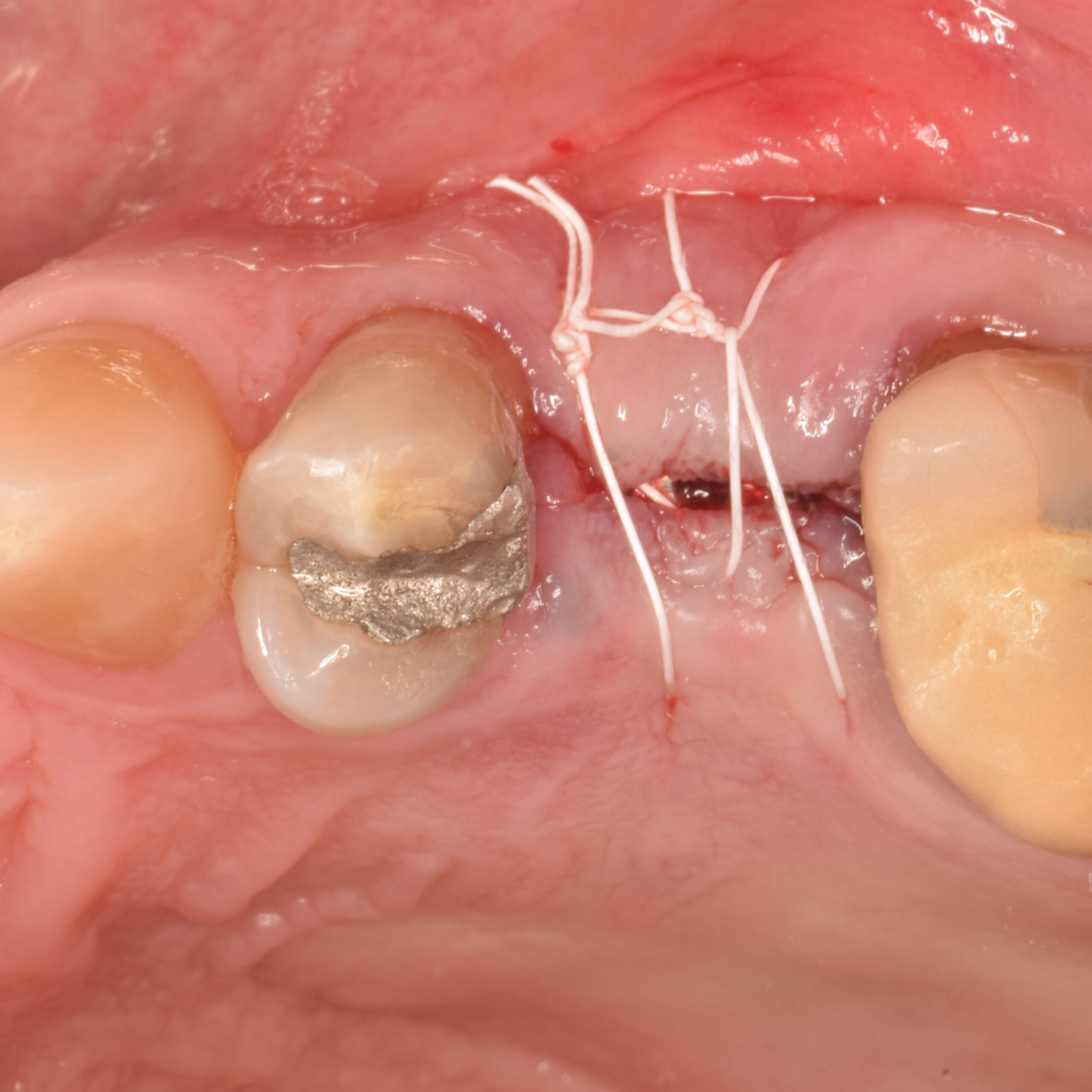
Geistlich Fibro-Gide® is tailored to the defect and a tension-free suture is done.
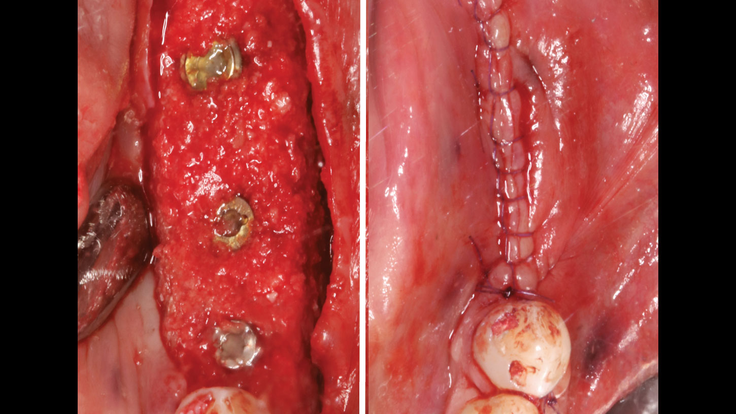
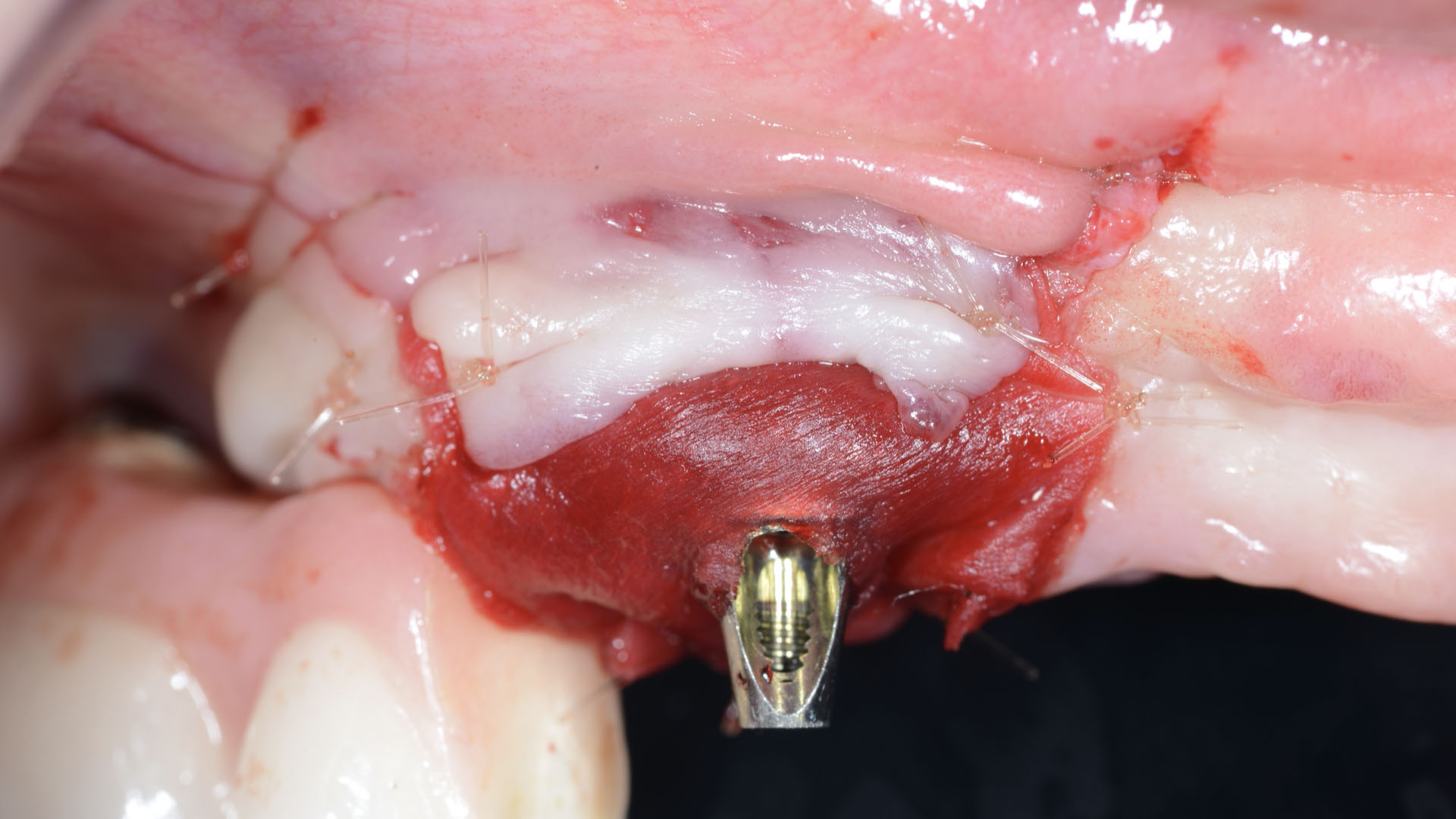
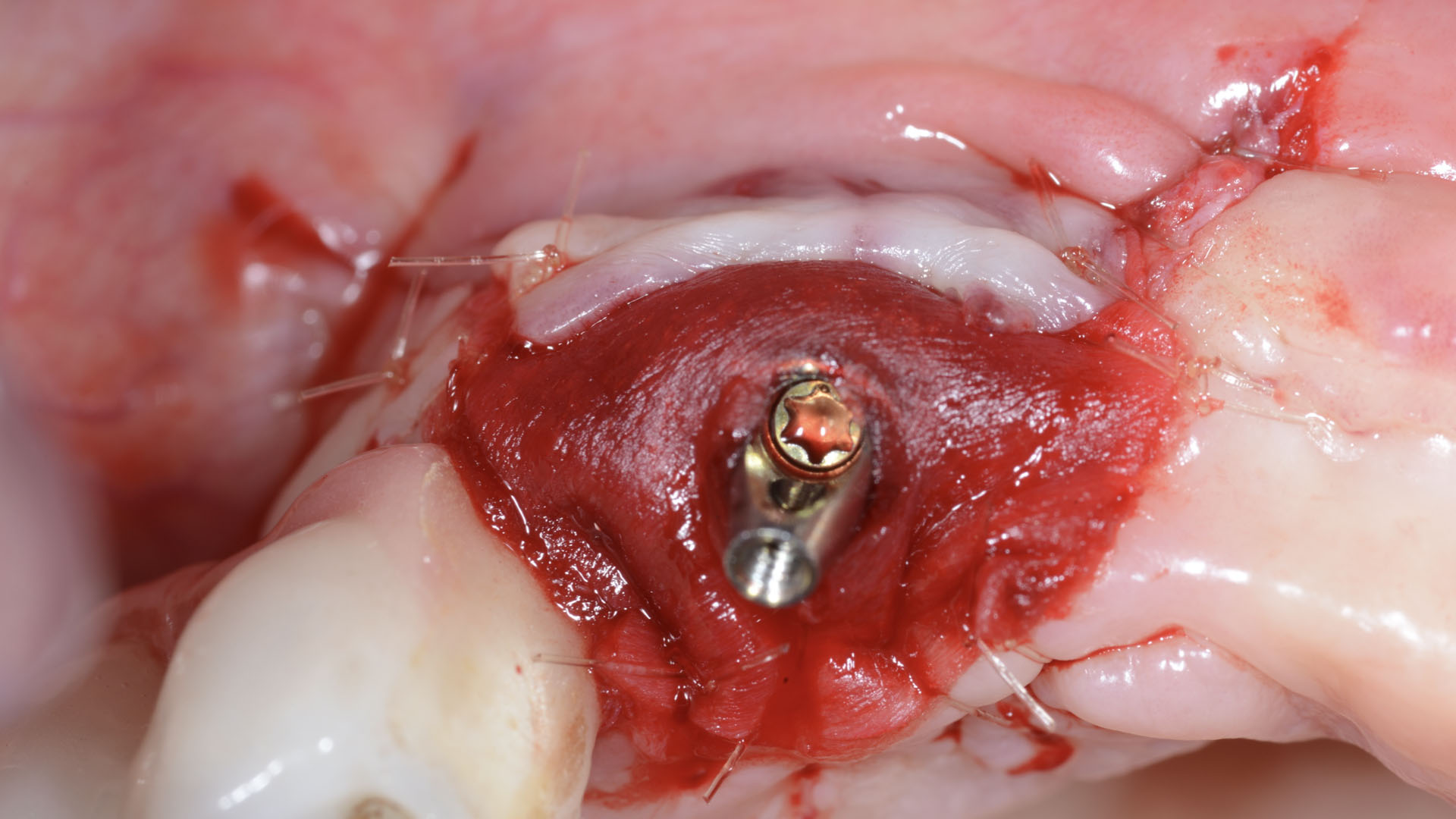
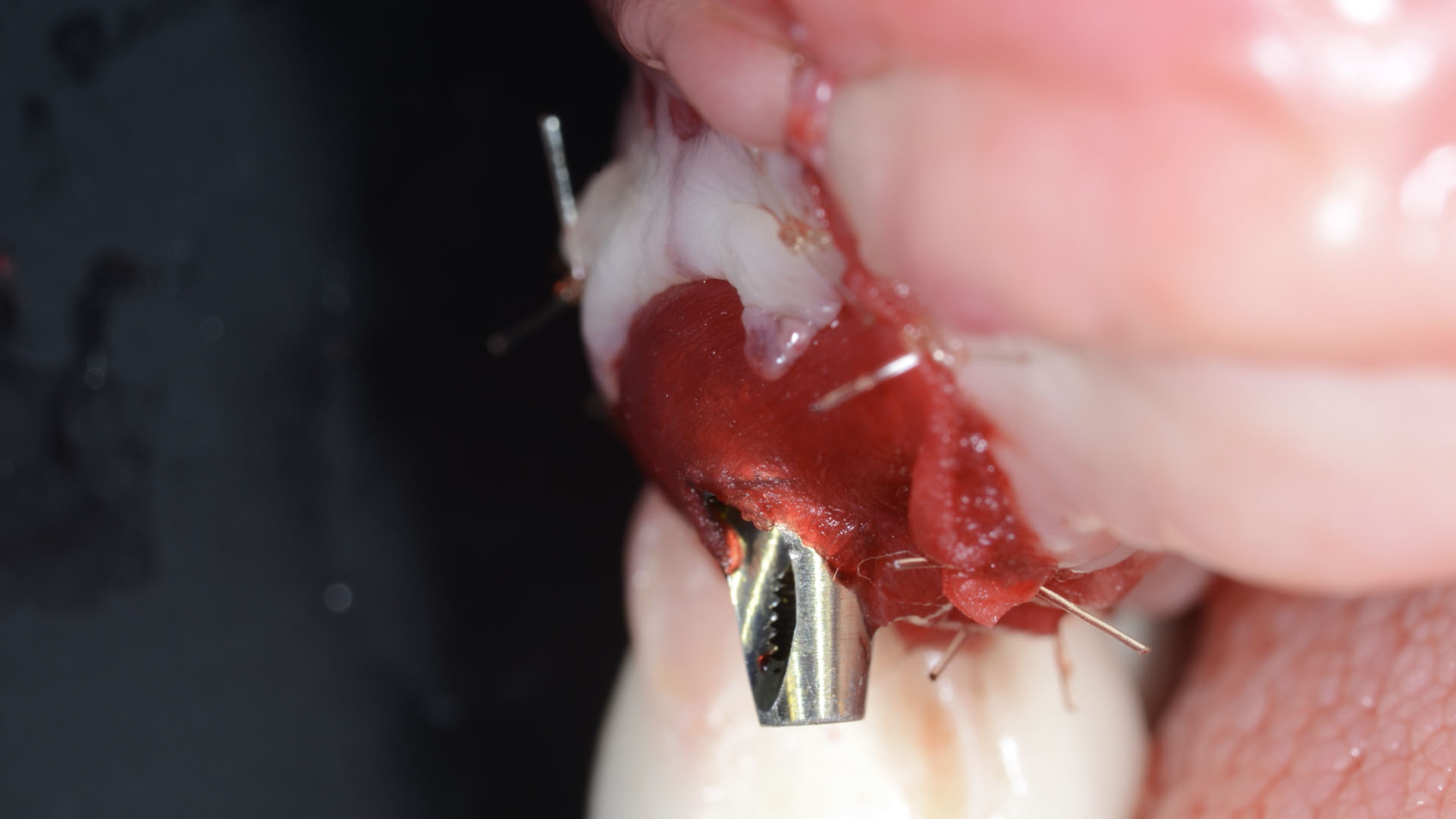
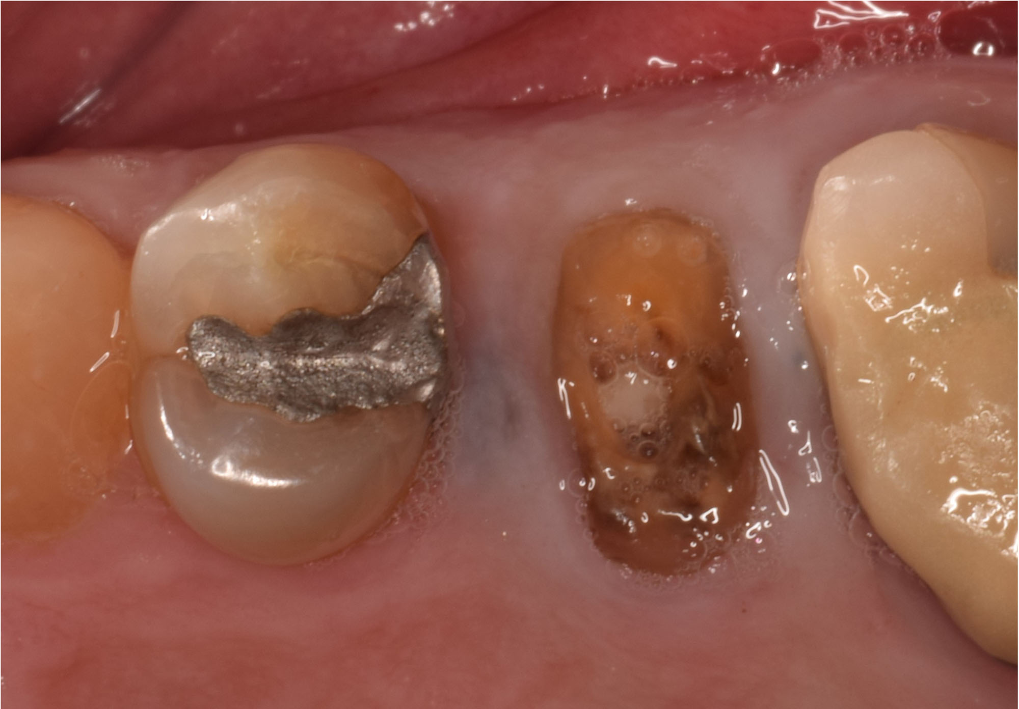
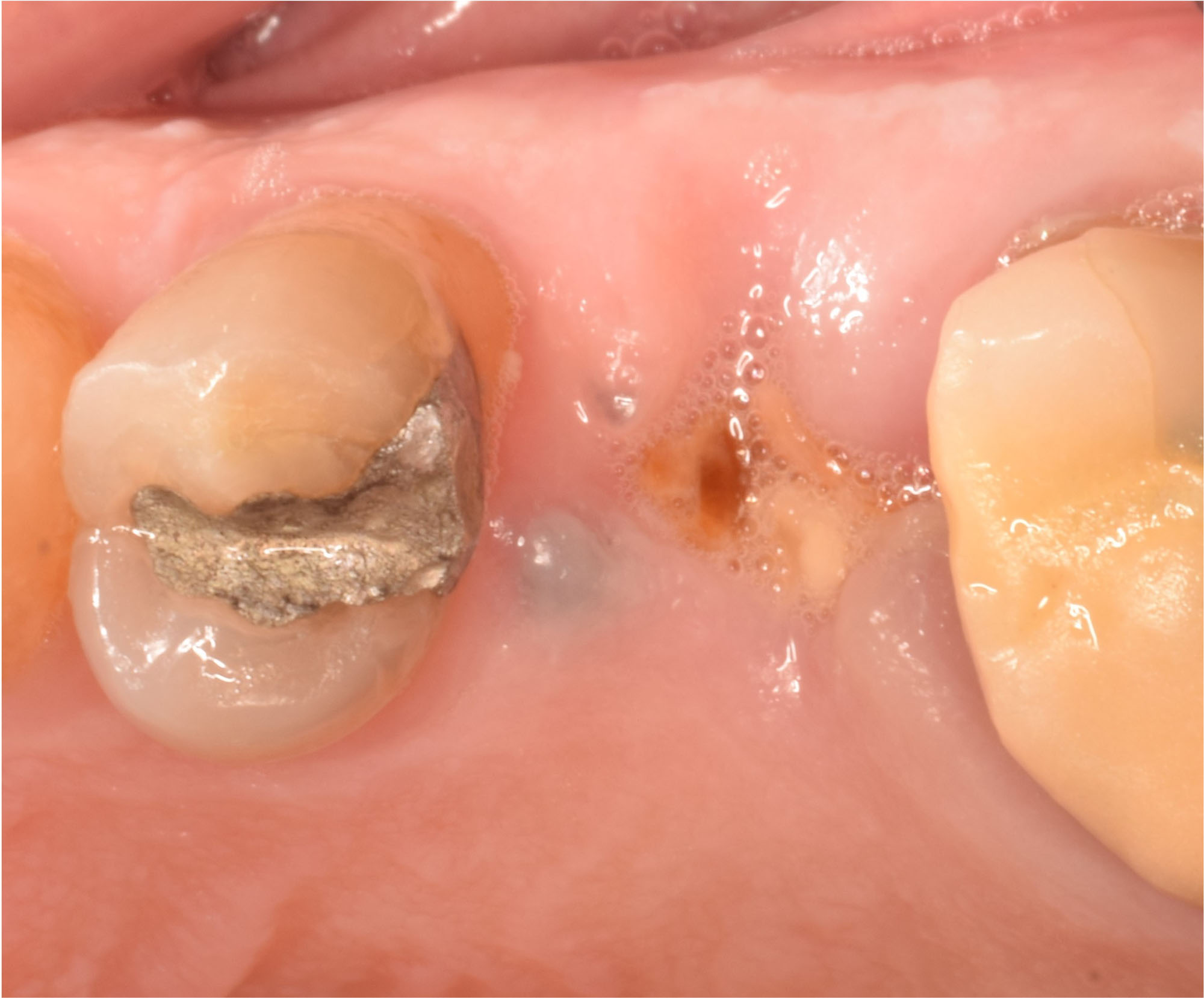
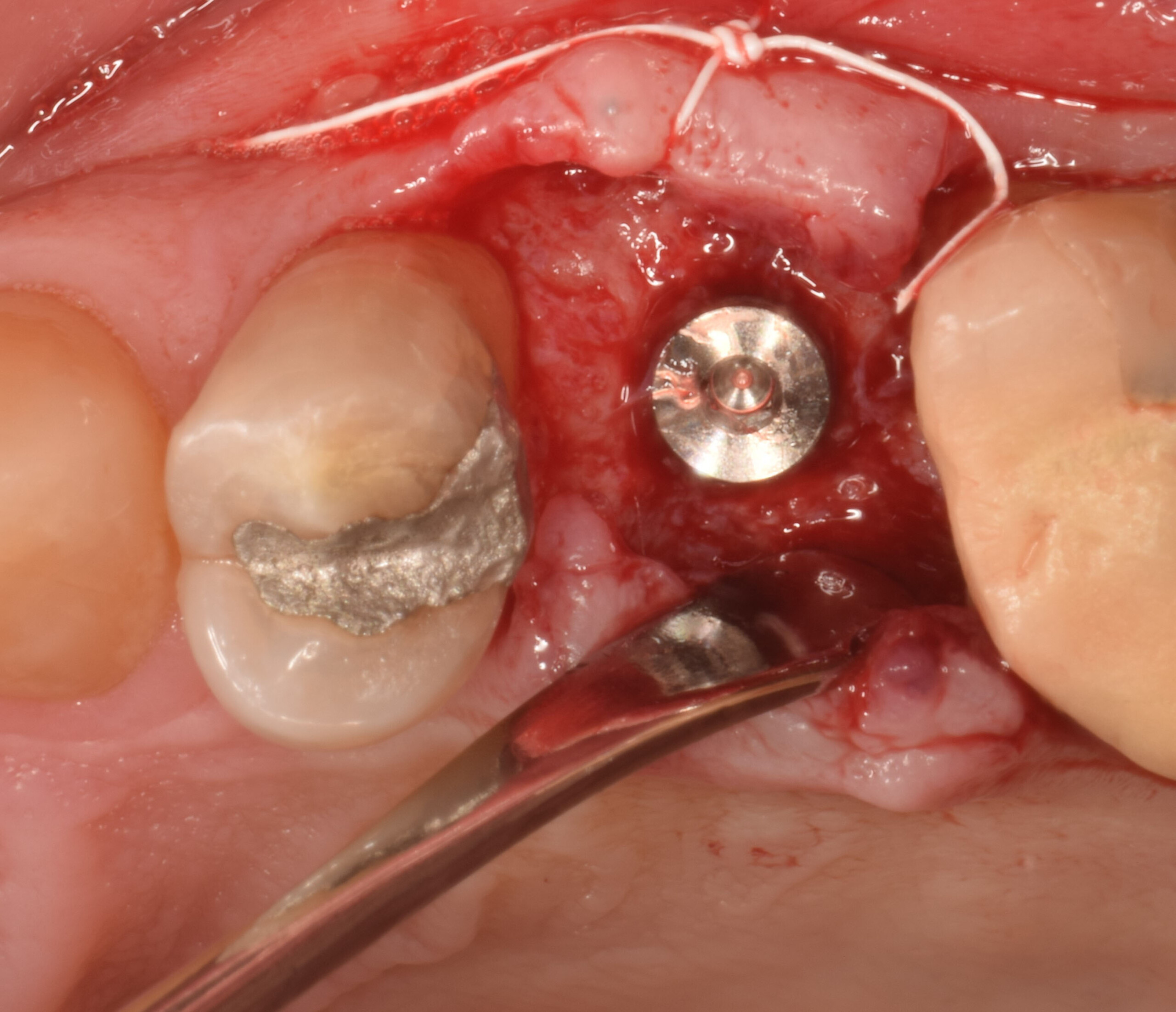
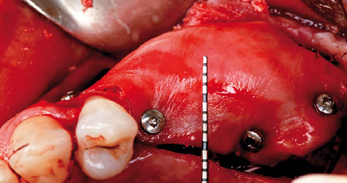
4 months after augmentation shows the bone situation after removal of the titanium scaffold and insertion of implants. Autologous bone chips are applied on top of the implants. Occlusal view of the mucosa level after implantation and wound closure.
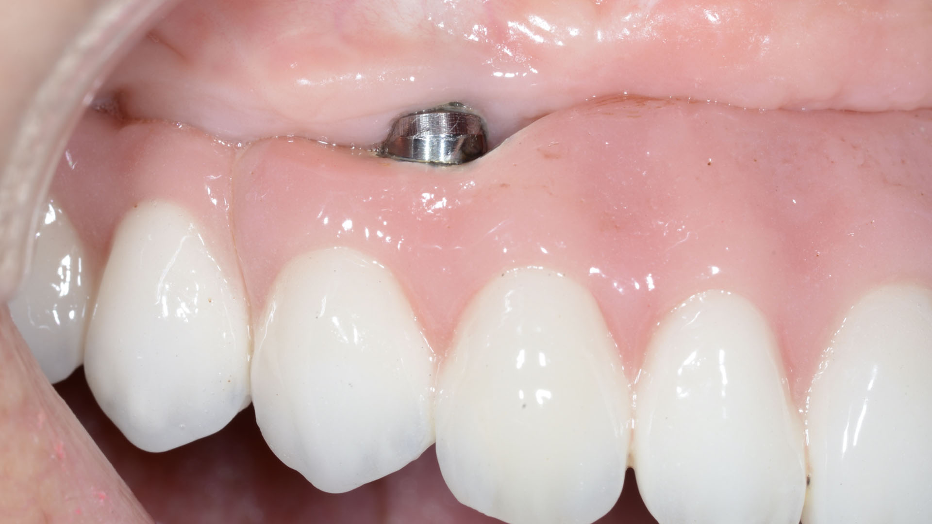
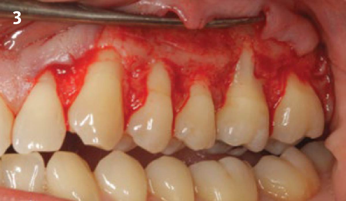
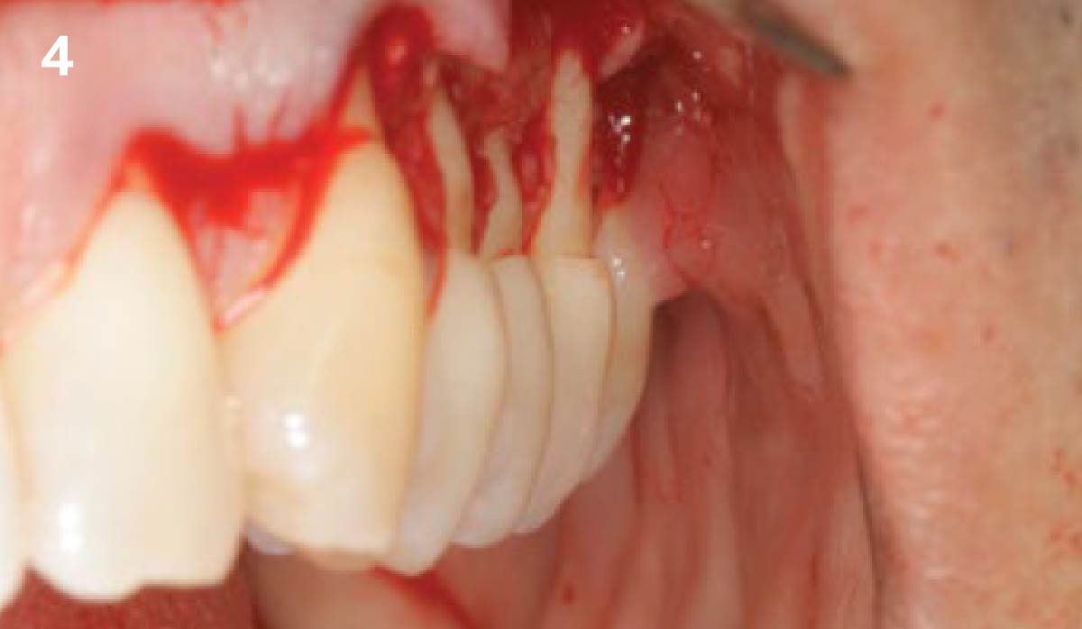
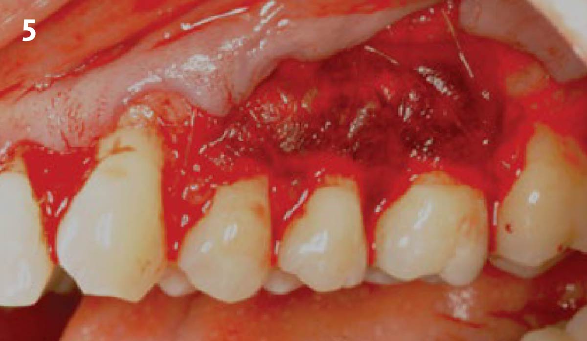
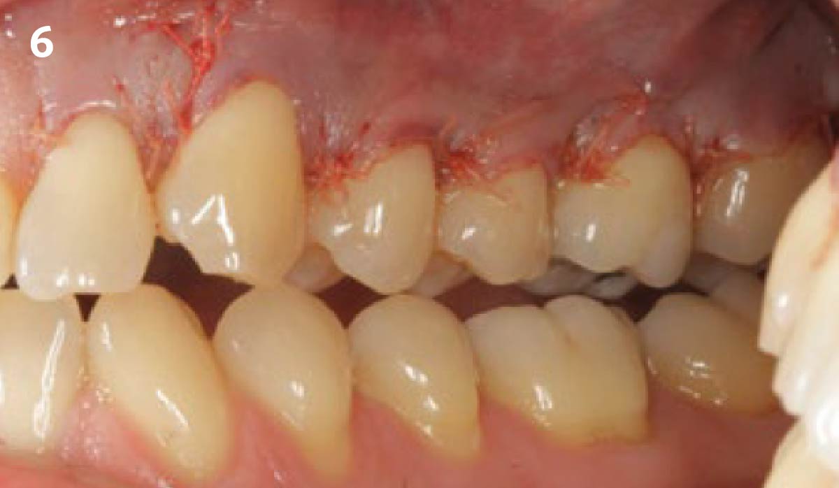
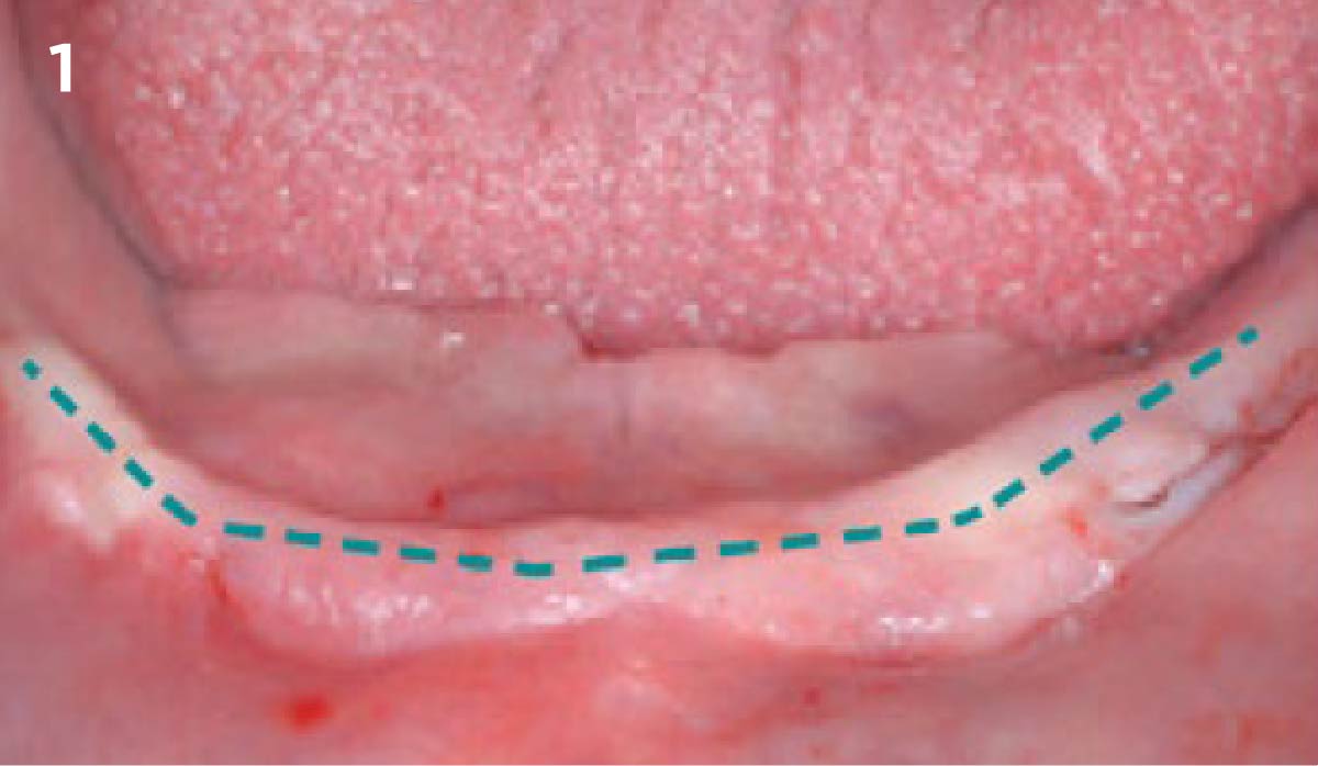
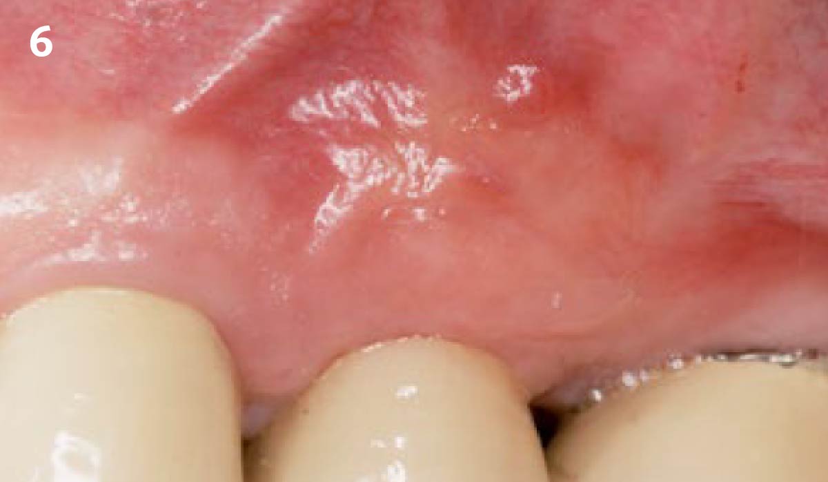
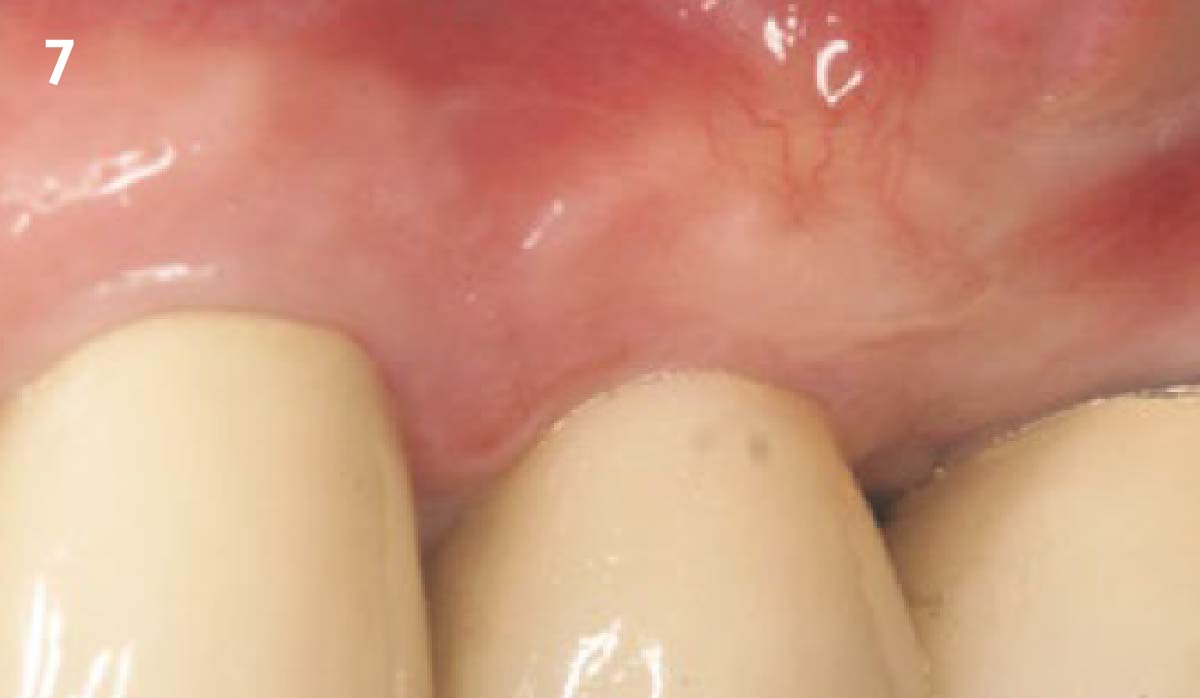
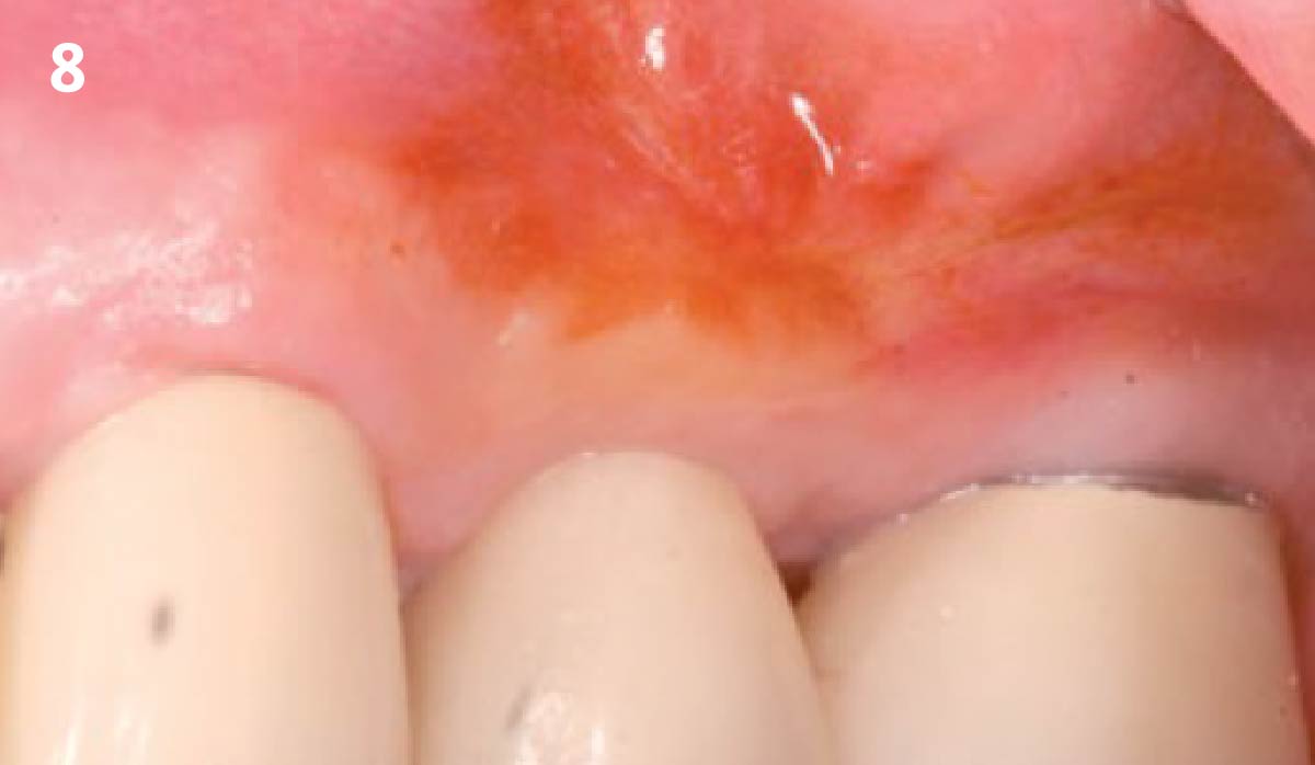
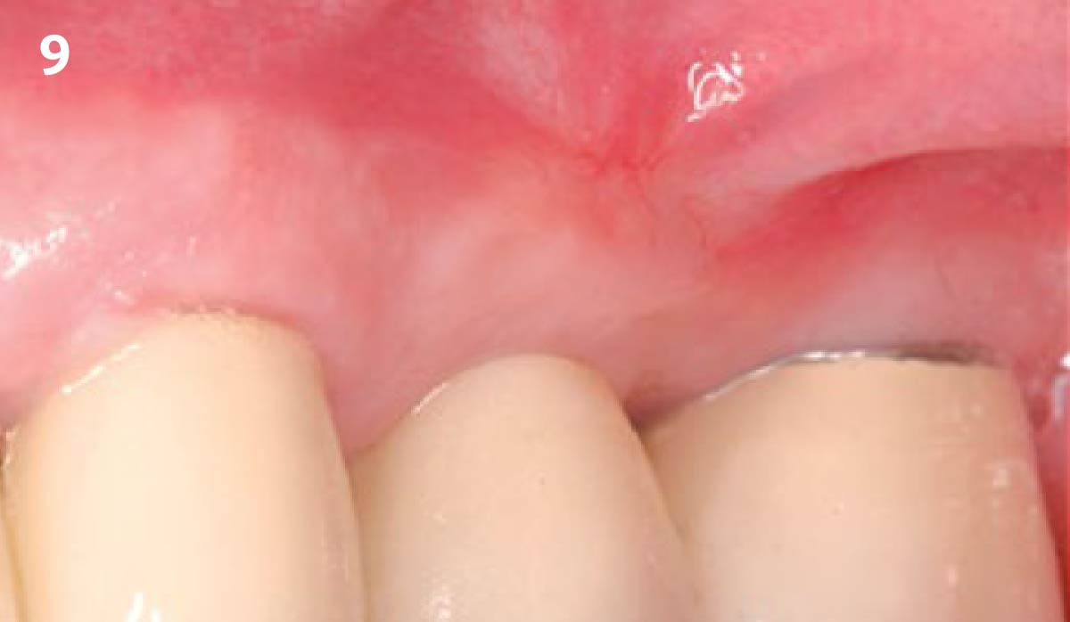
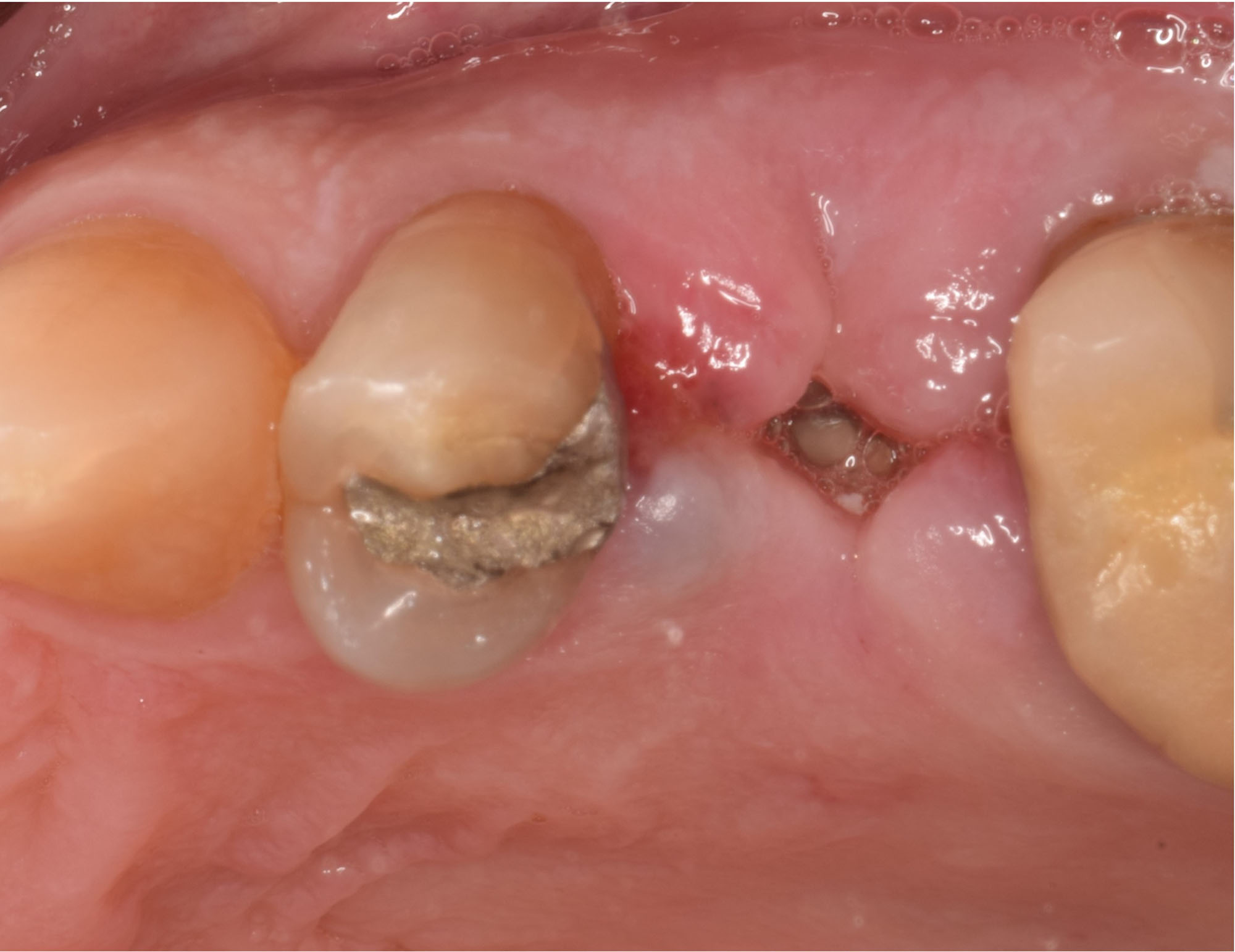
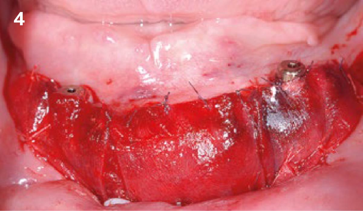
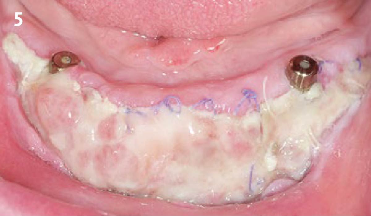
6 weeks after implantation and 5 1/2 months after augmentation. The mucosa is prepared apically; the preparation reaches the periosteum apically. The recipient site is fully surrounded by keratinized mucosa. Geistlich Mucograft® is sutured into the area.
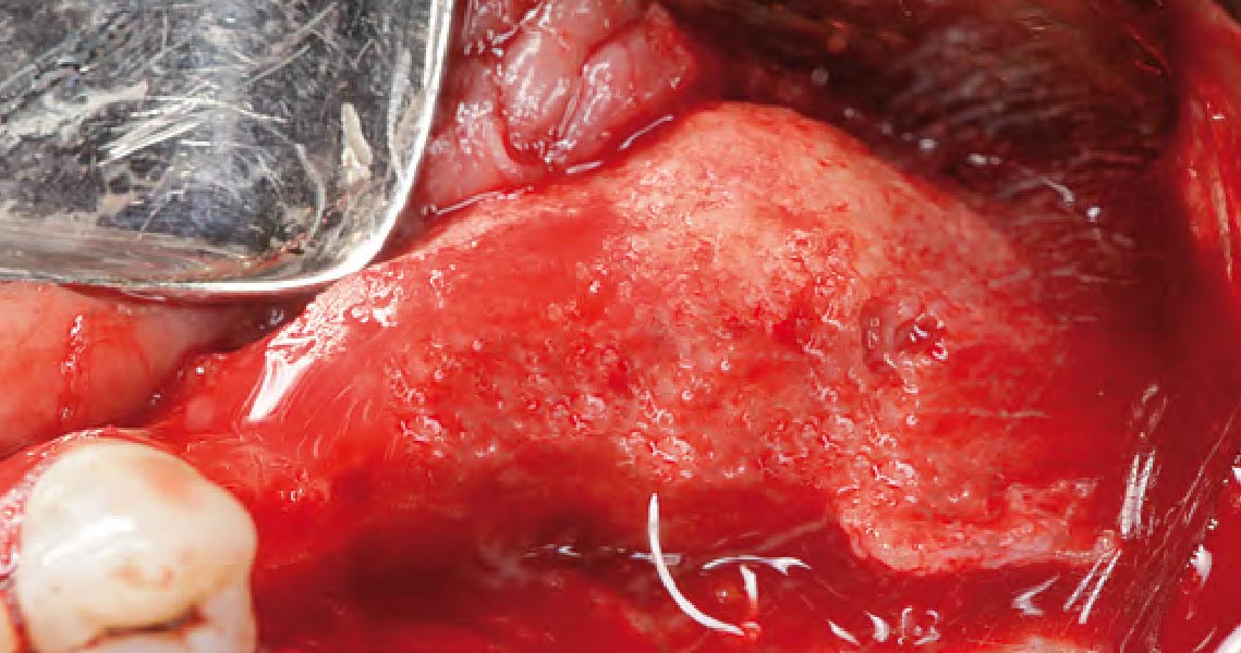
6 months after regeneration shows about 5mm of vertical bone regeneration. Panoram ic X-Ray after implant placement shows clearly recognizable mucosa shadows.
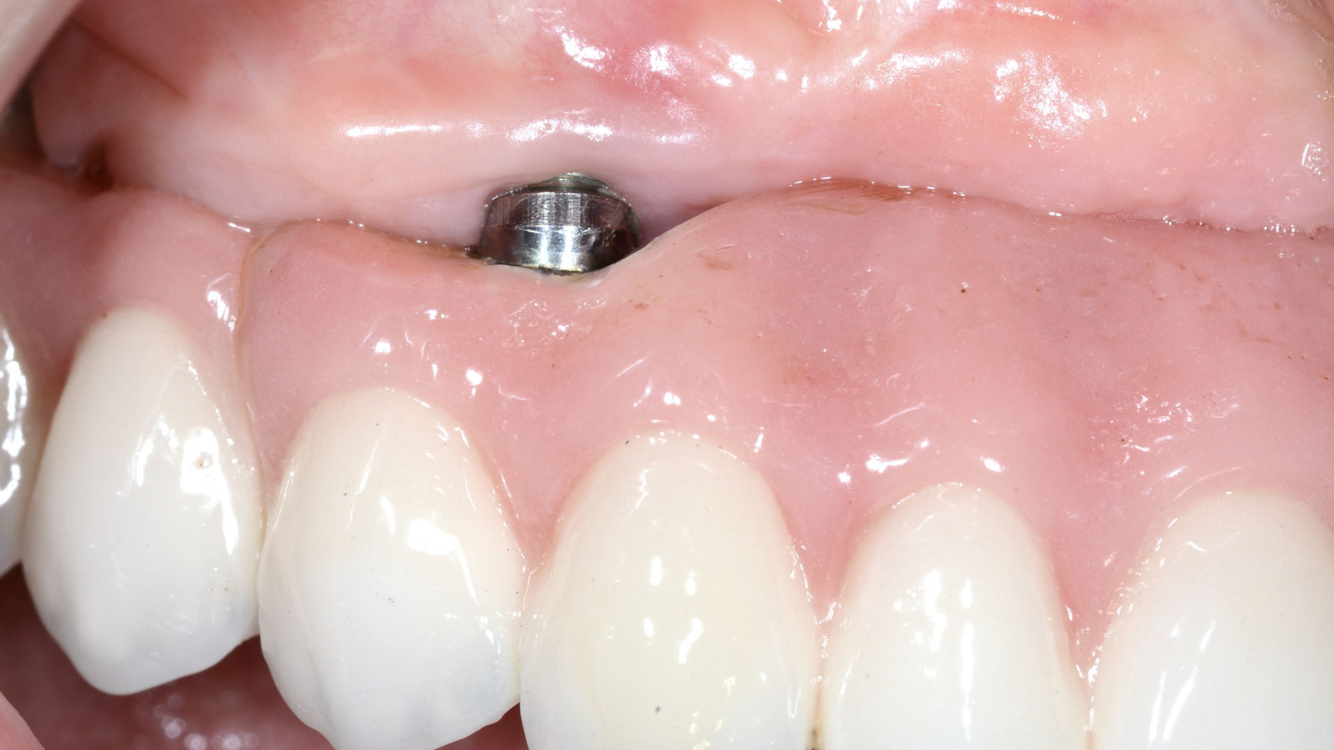
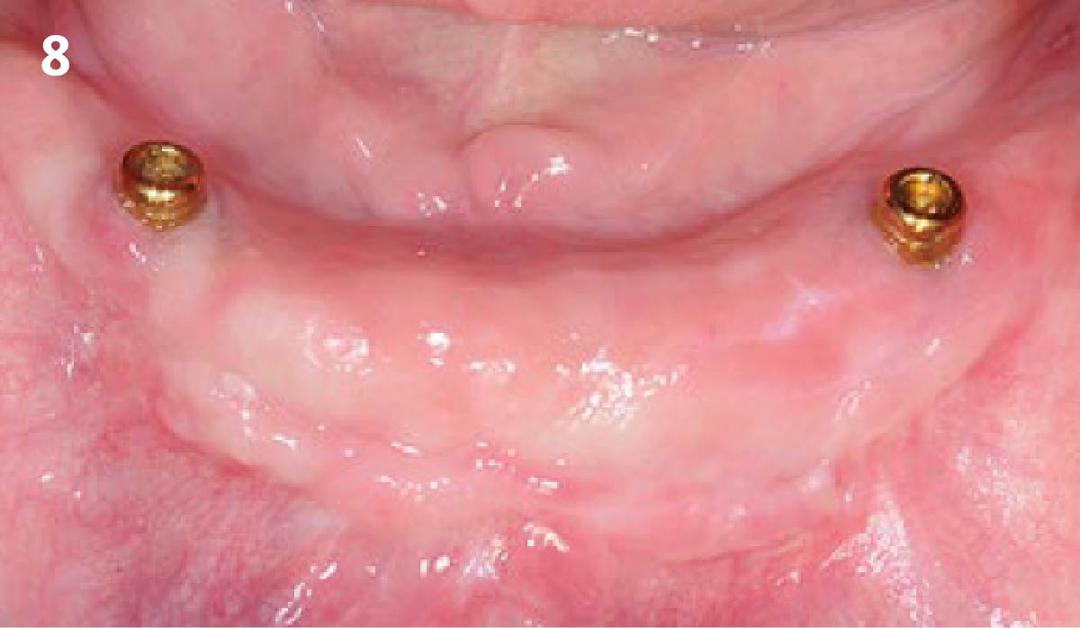
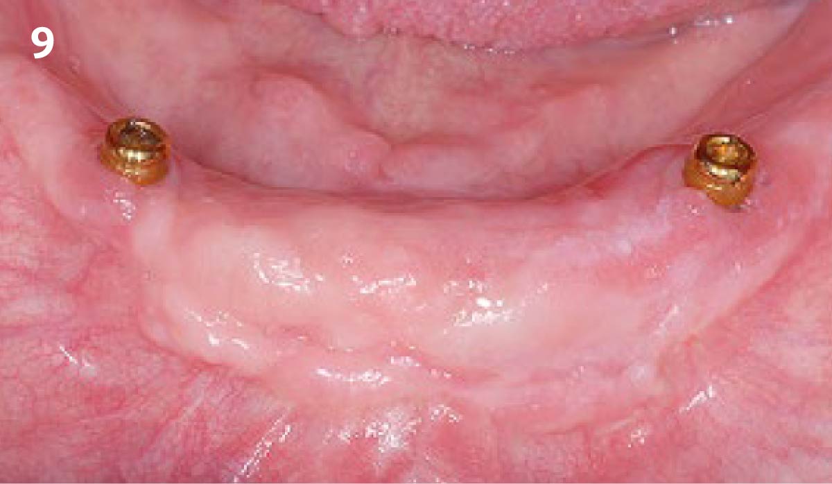
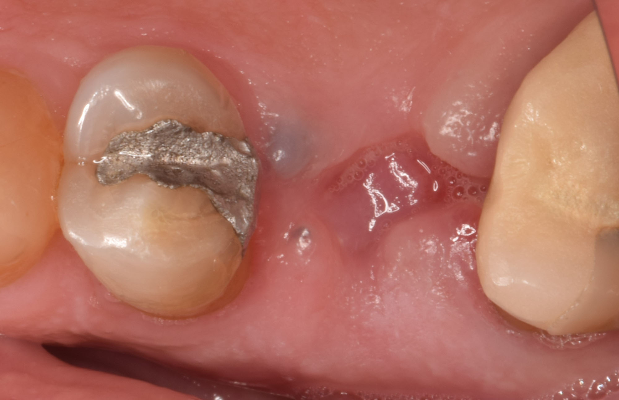
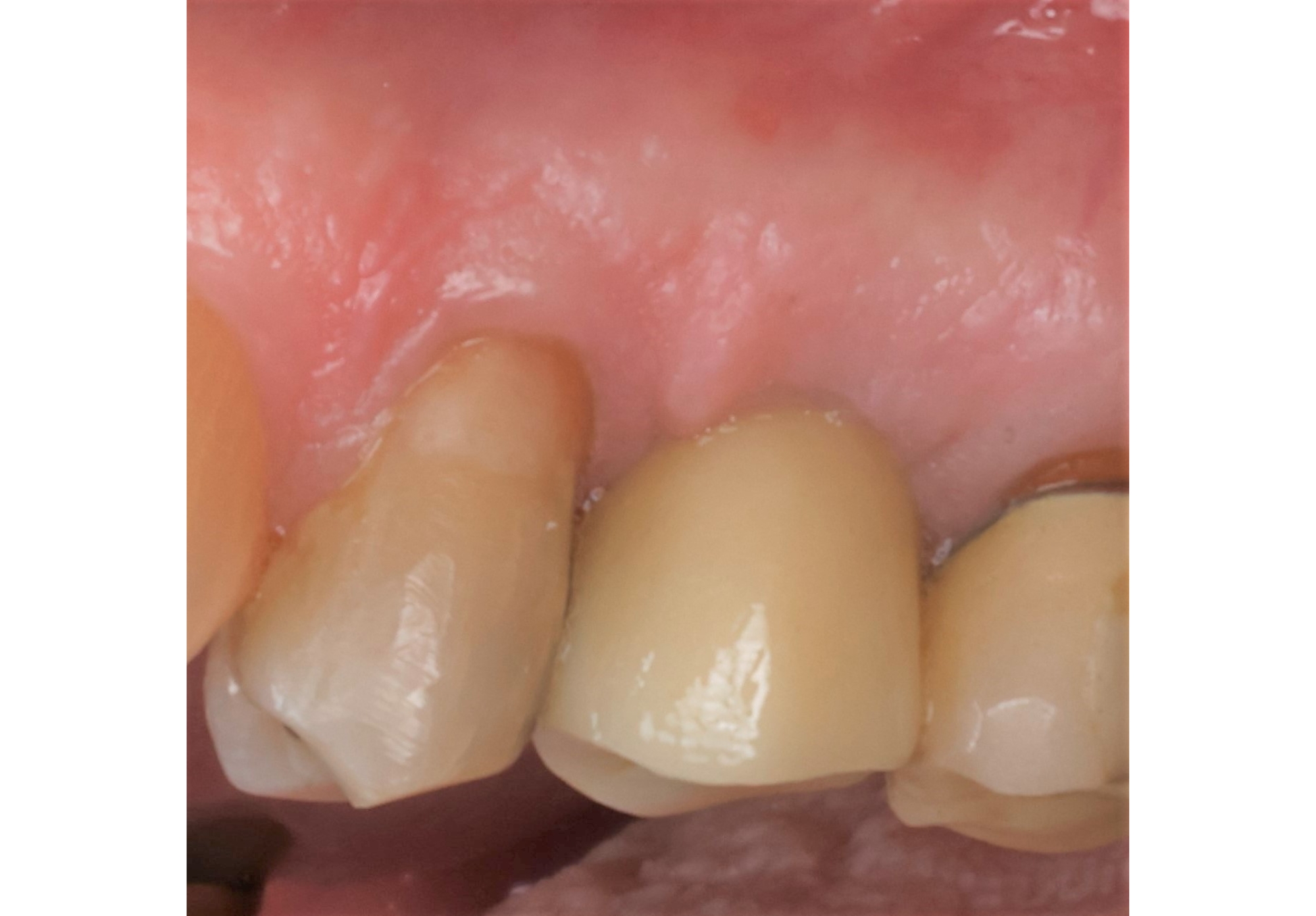
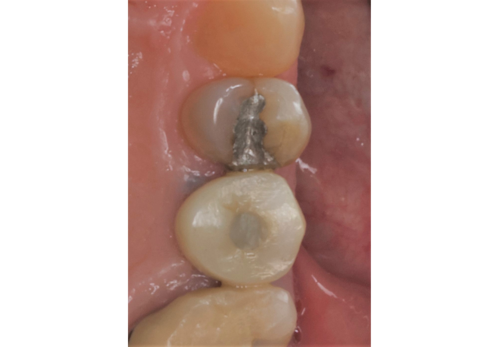
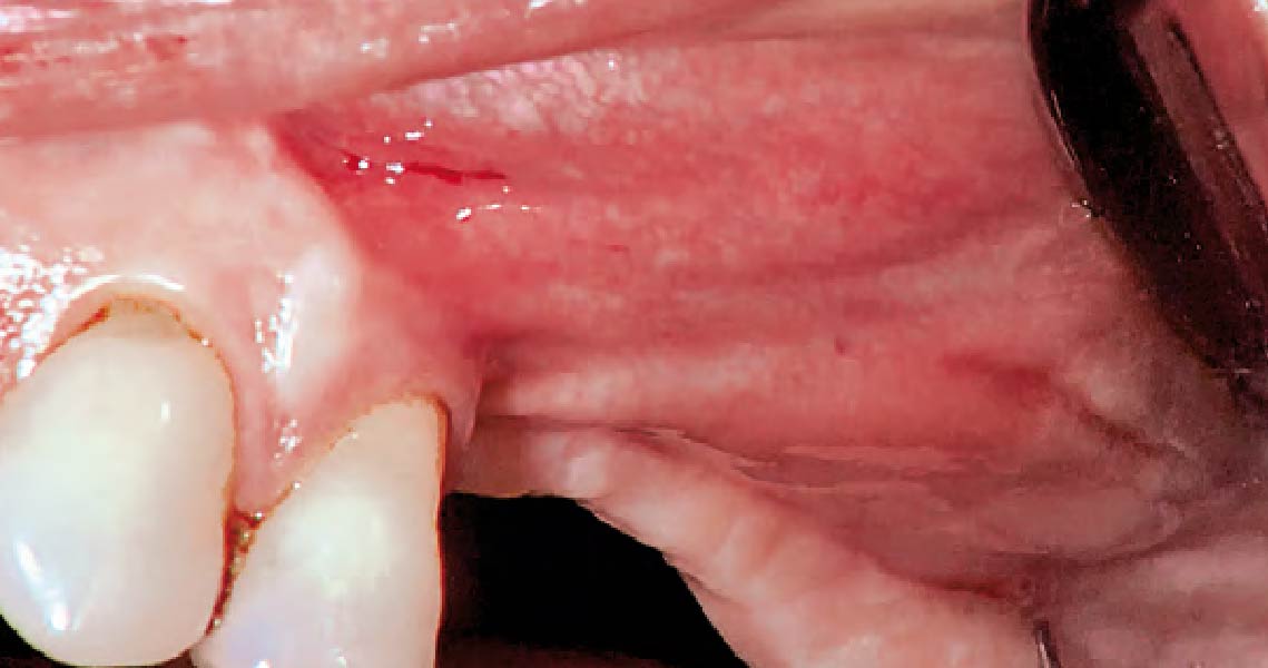
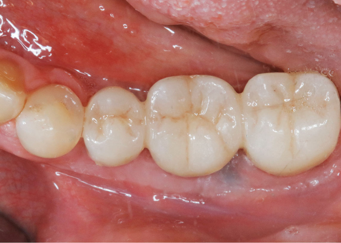
One year after augmentation shows the dental prosthesis inserted occlusally.

“Using the Geistlich Fibro-Gide® matrix enabled concurrent augmentation of hard
and soft tissues without any postoperative complications. At the same time, the soft
tissue thickening facilitated floor of the mouth surgery and vestibuloplasty.”

— Arnd Lohmann, MSc

THE OUTCOME

Treatment resulted in approximately 5 mm of vertical bone regeneration. The potential occurrence of a dehiscence associated with a wound opening and exposure of Yxoss CBR® was able to be prevented with Geistlich Fibro-Gide®.

On one hand, the quality of the peri-implant soft tissue was improved by the
soft tissue thickening with Geistlich Fibro‑Gide® and, on the other, by increasing the width of keratinized mucosa with Geistlich Mucograft®. The treatment method chosen resulted in a reduced invasiveness and morbidity by avoiding a donor site for sourcing a transplant.
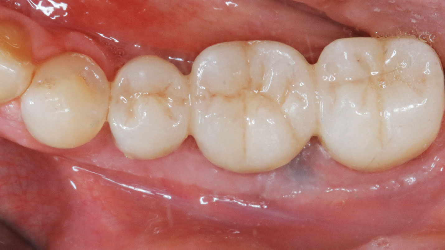
One year after augmentation shows the dental prosthesis inserted occlusally.

Implant therapy should restore the natural anatomical structures as closely as possible in order to avoid subsequent peri-implant problems.”

Arnd Lohmann, MSc

Arnd Lohmann, MSc

Dr. Arnd Lohmann is a recognized specialist in implantology and periodontology. He earned his dental license in Hamburg in 2002, completed his doctorate in 2003, and has been a partner at a private practice in Bremen since then.

With a Master of Science in Implantology (2007), he specializes in dental implantology and bone augmentation. He is an active speaker at national and international congresses, leads the Bremen study group of the German Society of Oral Implantology (DGOI), and is a member of DGOI, DGZI, and DGI. His practice is equipped with state-of-the-art technology, ensuring high-quality patient care.

BIOBRIEF

Clinical Efficacy of Geistlich Mucograft® in Regeneration of Oral Mucosa Combined with the Surgical Treatment of Peri-implantitis in Implants with Lack of Keratinized Tissue

Dr. Alberto Ortiz-Vigón
Dr. Erik Regidor Correa

THE SITUATION

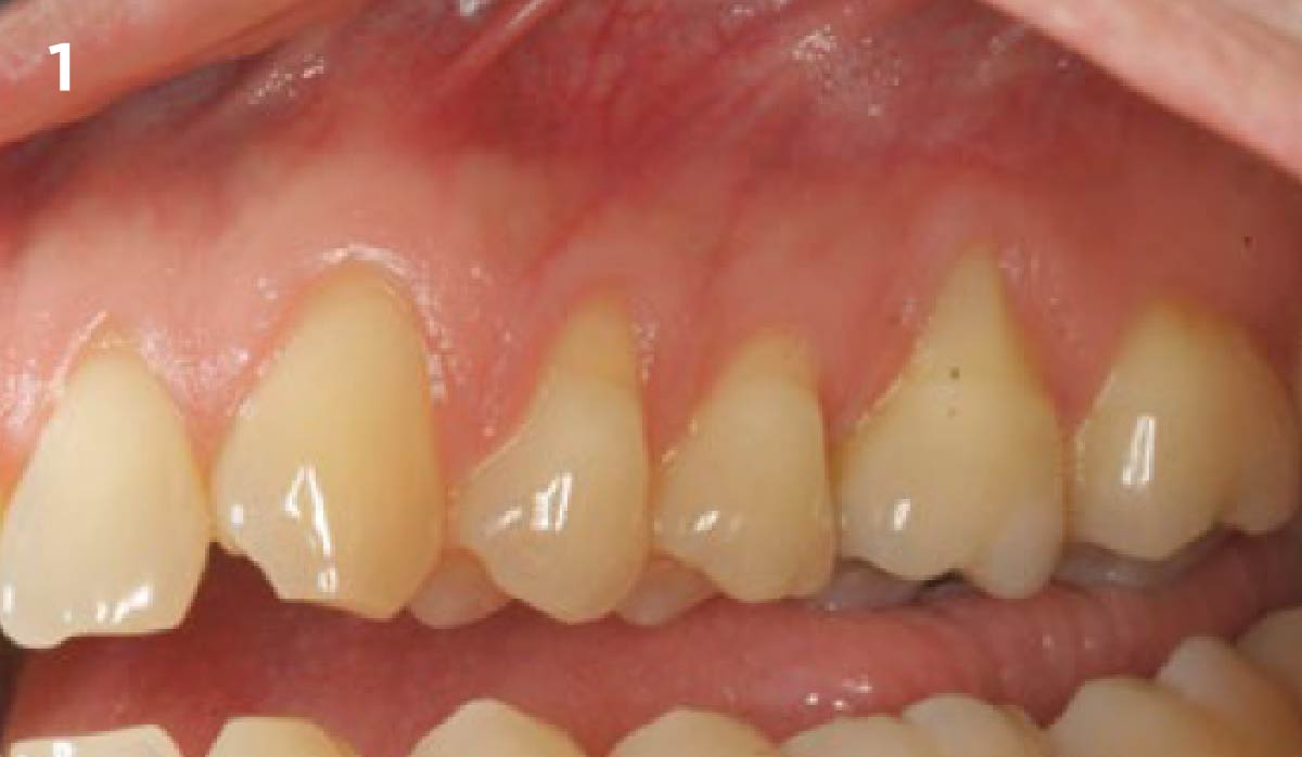
Adult patient, non-smoker and without relevant systemic history, attends to clinic referring peri-implant tissue inflammation, bleeding and brushing discomfort around her implant in the upper jaw. Clinically peri-implant pocket depth > 5 mm, bleeding and suppuration on probing were observed. Furthermore, the implant presented < 2 mm of keratinized mucosa and radiographic horizontal bone loss.

THE RISK PROFILE

Low RiskMedium RiskHigh Risk
Patient’s healthIntact immune system 
Non-smoker
Light smokerImpaired immune system 
Heavy smoker
Patient’s esthetic requirementsLowMediumHigh
Height of smile lineLowMediumHigh
Gingival biotypeThick – “low scalloped”Medium – “medium scalloped”Thin – “high scalloped”
Shape of dental crownsRectangularTriangular
Infection at implant sightNoneChronicAcute
Bone height at adjacent tooth site≤ 5 mm from contact point5.5 – 6.5 mm from contact point≥ 7 mm from contact point
Restorative status of adjacent toothIntactRestored
Width of tooth gap1 tooth (≥ 7 mm)1 tooth (≤ 7 mm)2 teeth or more
Soft-tissue anatomyIntactCompromised
Bone anatomy of the alveolar ridgeNo defectHorizontal defectVertical defect
watch video download pdf

THE APPROACH

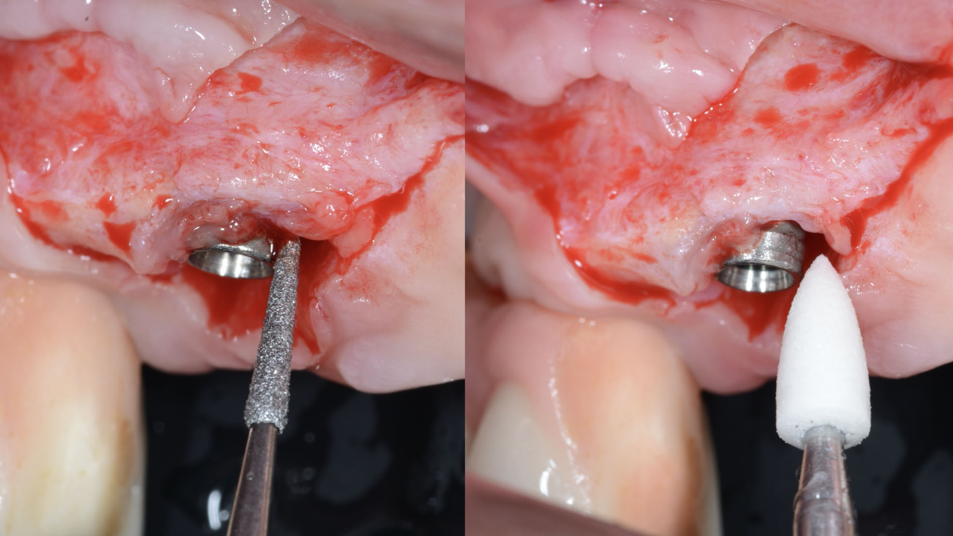
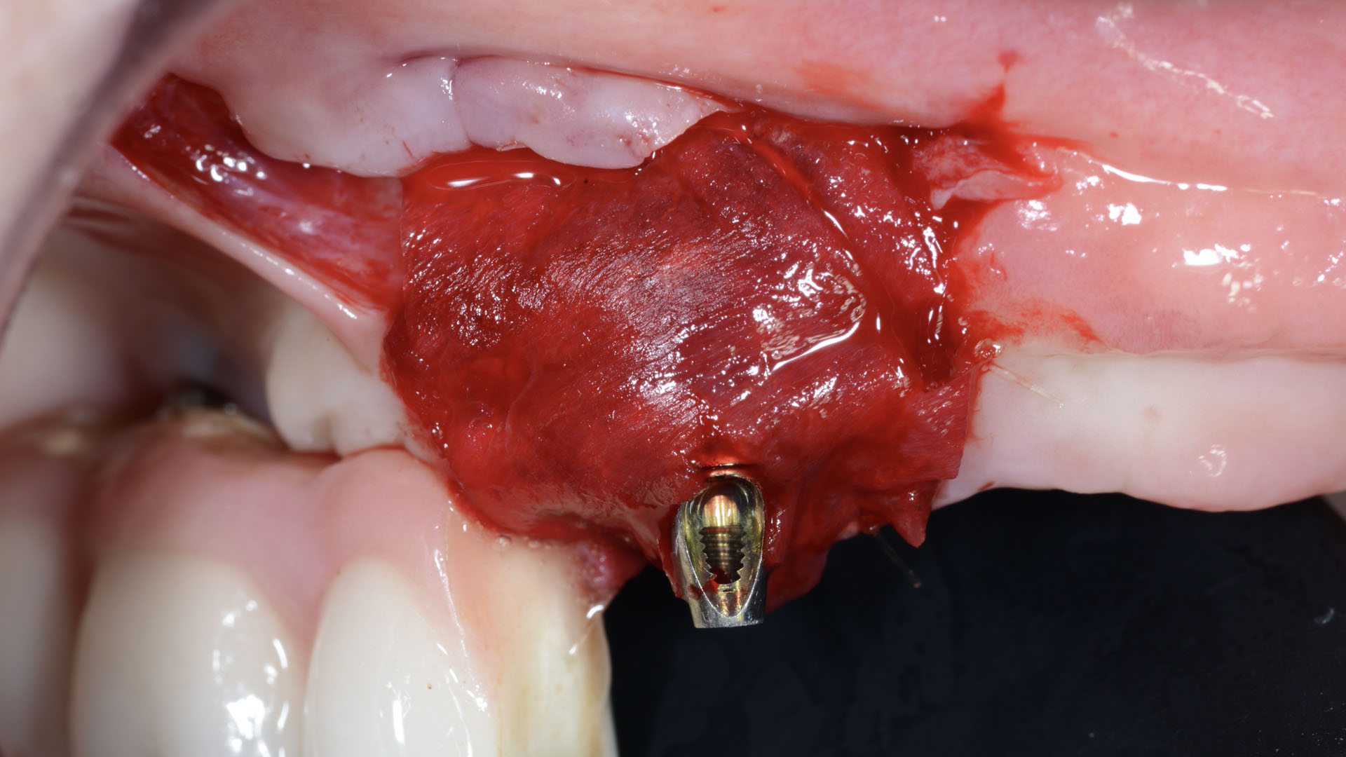
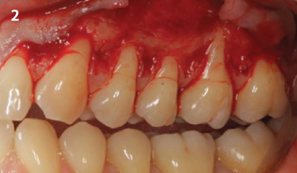
Intrasulcular incision was made and a mucosal partial thickness flap was raised. The recipient site was prepared by sharp disection in order to create a periosteal bed free of any muscle attachment. Peri-implant granulation tissue was removed and implantoplasty was performed. Finally, Geistlich Mucograft® was used to support the gain of keratinized tissue. Thus, the collagen matrix was sutured with the resulting flap apically at the base of the newly created vestibulum.
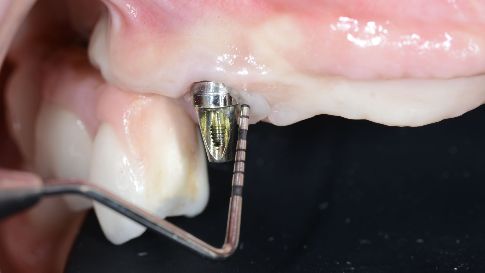
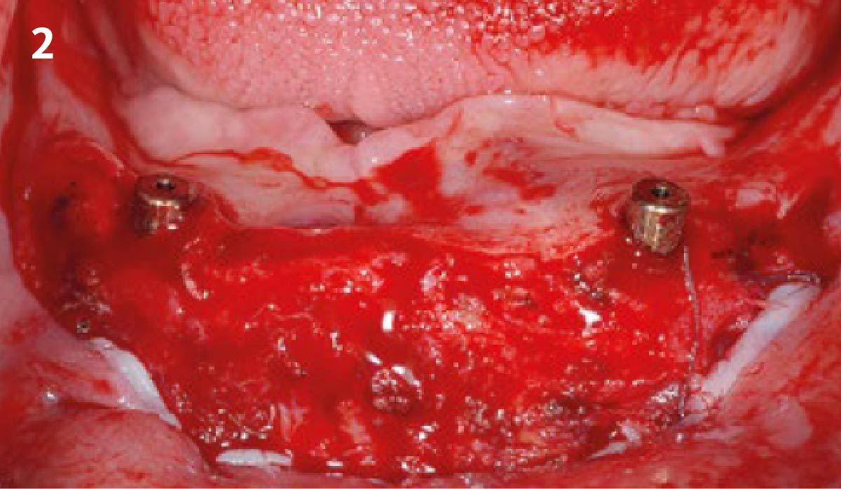
Pathological peri-implant pocket depth combined with bleeding on probing.
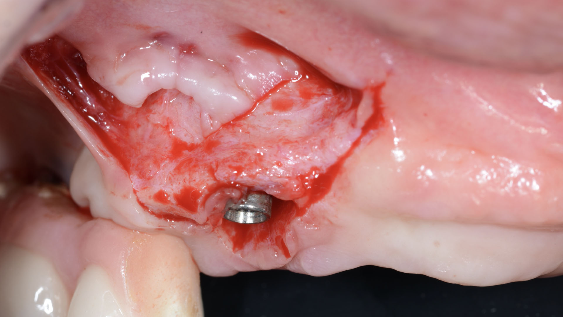
Partial thickness flap in order to create a periosteal bed free of any muscle attachment and apically positioned.
Implantoplasty of the exposed rough implant surface using burs and silicon carbide polishers.
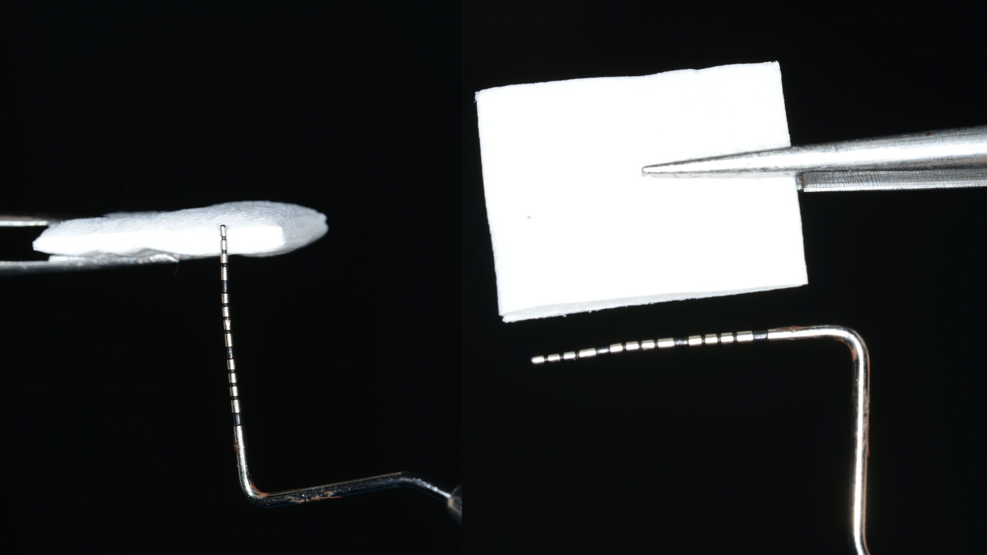
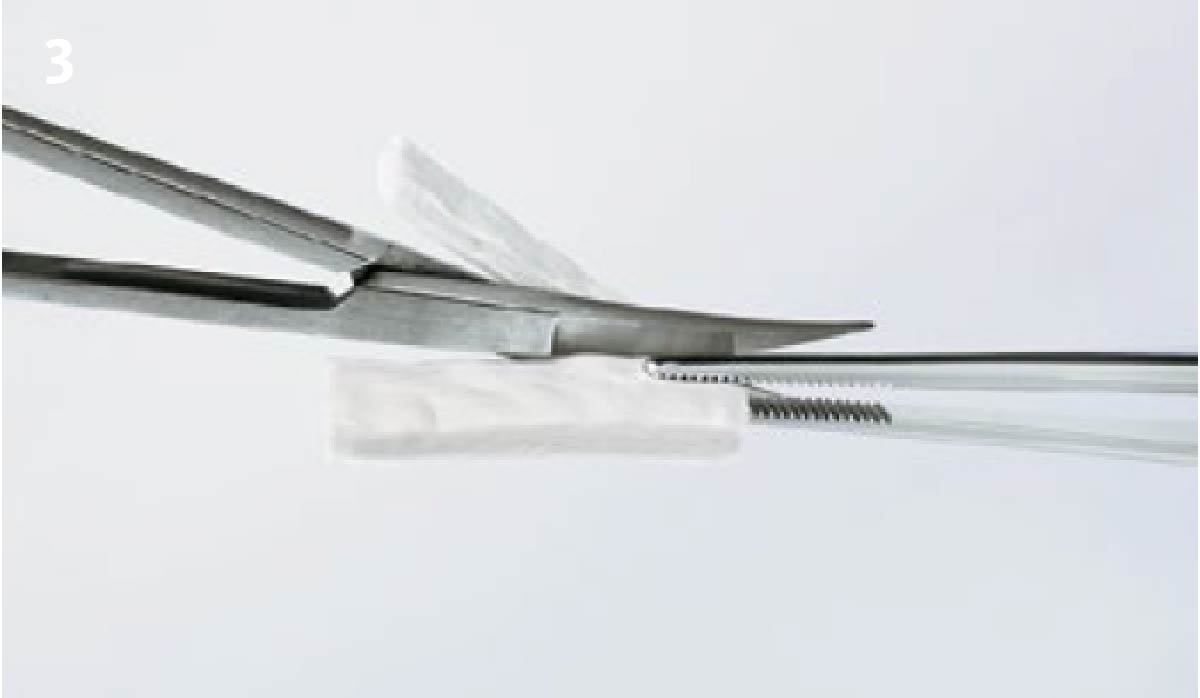
Xenogeneic collagen matrix structure (Geistlich Mucograft®).
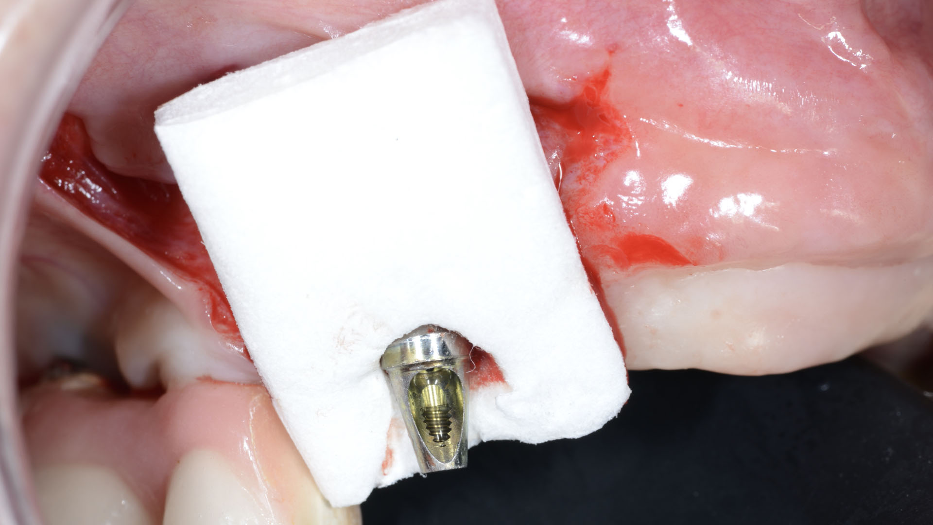
Future position of the xenogeneic collagen matrix facilitated by prosthodontic abutment.
Suture of xenogeneic collagen matrix around the abutment and over the recipient bed.
Buccal view of xenogeneic collagen matrix and apically positioned flap.
Occlusal view of xenogeneic collagen matrix and apically positioned flap.
Lateral view of xenogeneic collagen matrix and apically positioned flap.
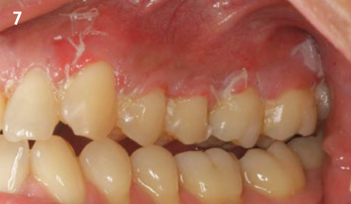
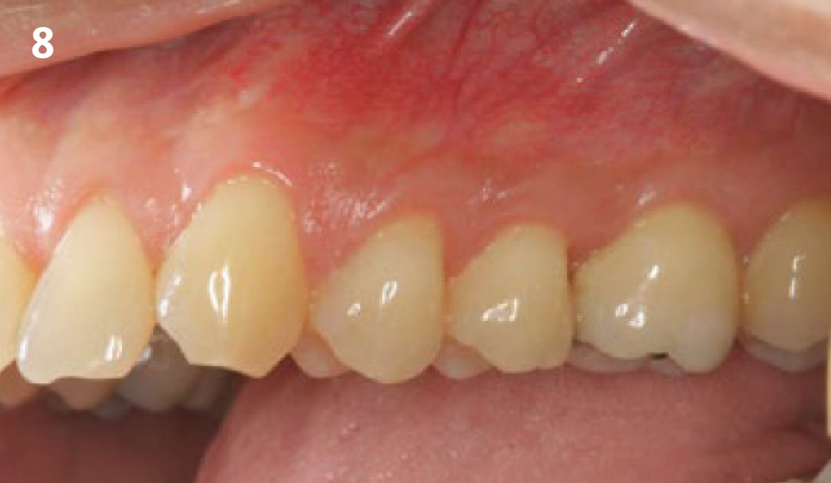
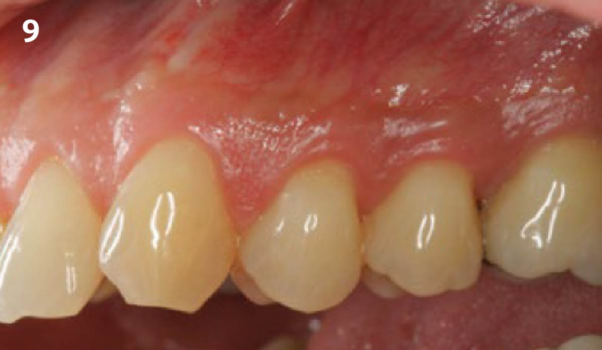
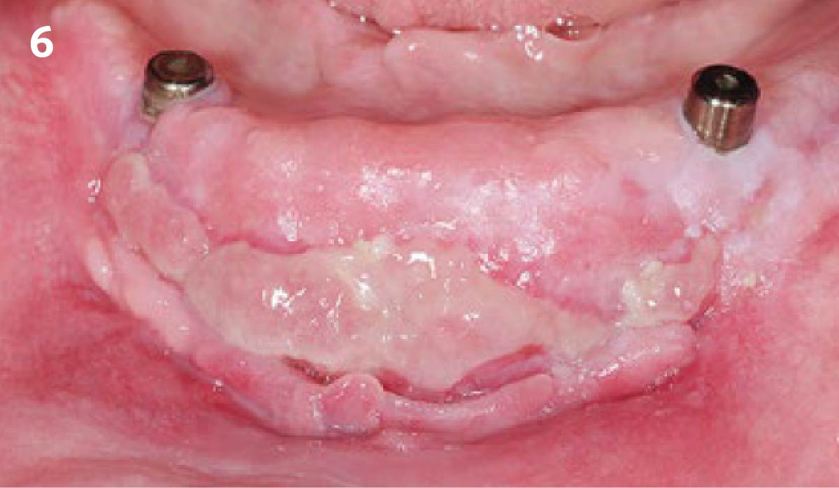
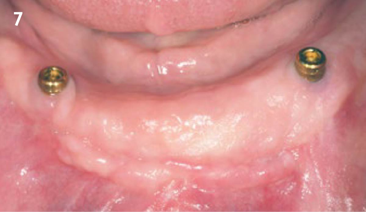
Peri-implant tissue health and maintenance of keratinized tissue after one year of surgical treatment.
Periimplant tissue health and maintenance of keratinized tissue after 2 years

Absence of > 2 mm of keratinized mucosa was associated with peri-implant soft-tissue inflammation, bleeding and discomfort on brushing.

THE OUTCOME

After two years follow-up, the successful outcome can be observed in terms of clinical peri-implant parameters, gain of keratinized mucosa without significant graft shrinkage and stability of vertical position of the mucosal margin.

The use of Geistlich Mucograft® xenogeneic collagen matrix for regeneration of oral mucosa, combined with the surgical respective approach to peri-implantitis provides an improvement in clinical parameters and increase of the peri-implant keratinized mucosa minimizing the risk of recession in the esthetic area.”

Dr. Erik Regidor Correa & Dr. Alberto Ortiz-Vigón

The use of soft-tissue substitutes may play an important role in patient perception and satisfaction without jeopardizing the final clinical outcome.”

Dr. Erik Regidor Correa & Dr. Alberto Ortiz-Vigón

Dr. Alberto Ortiz-Vigón

  • DDS from the University of the Basque Country
  • MSc and PhD in bone regeneration from the University Complutense of Madrid (UCM)
  • Master in Periodontology and Implant dentistry from the EFP
  • Research fellowship at the University of Gothenburg
  • MBA from the Deusto Business School
  • Assistant professor and clinical researcher at UCM and ThinkingPerio Research
  • PerioCentrum Clinic in Bilbao
  • Co-founder of ARC Healthtech Innovation Holding
  • Socially engaged & NGO co-founder of Smile is a Foundation

Dr. Erik Regidor Correa

  • DDS from the University of the Basque Country
  • MSc from the U. of the Basque Country
  • Master in Periodontology and Implant Dentistry U. of the Basque Country
  • PhD student in the U. of the Basque Country
  • Assistant professor and clinical researcher ThinkingPerio Research

CLINICAL CASE

CLINICAL CASE

CLINICAL CASE

CLINICAL CASE

CLINICAL CASE

CONCLUSIONS:

  • Geistlich Mucograft® with a keratinized tissue strip was utilized to increase vestibular depth and gain additional keratinized tissue.
  • Augmentation of severely atrophied alveolar ridge provided sufficient bone for implant placement 8 months following augmentation.