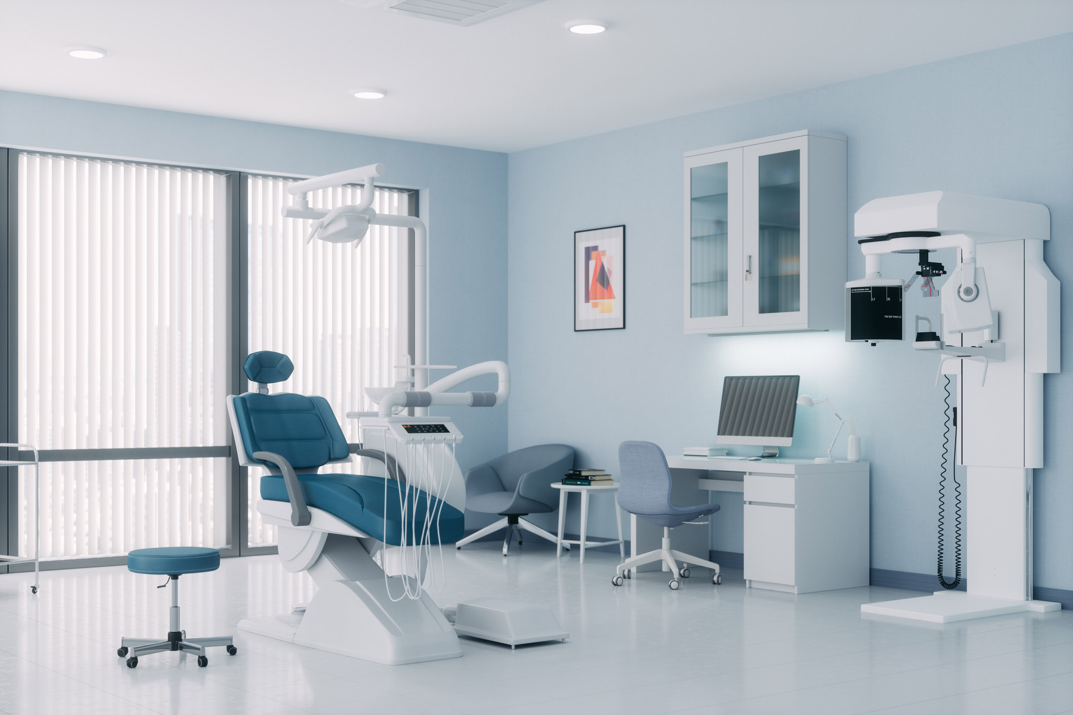
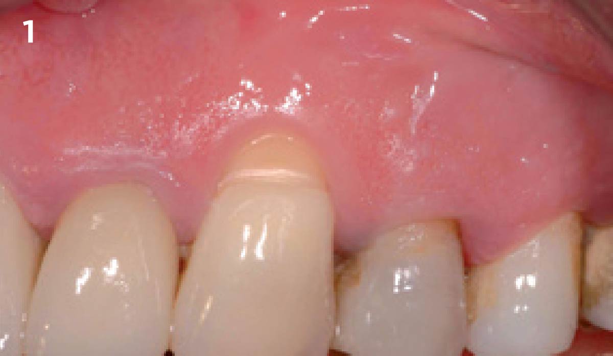
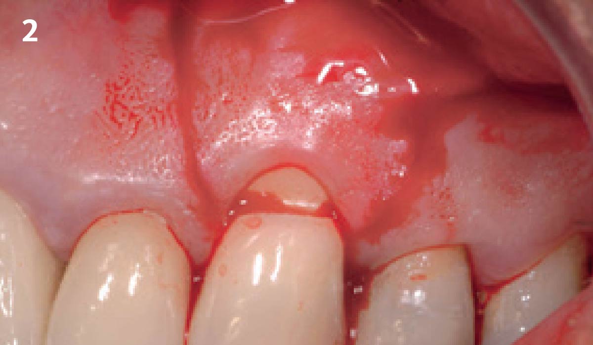
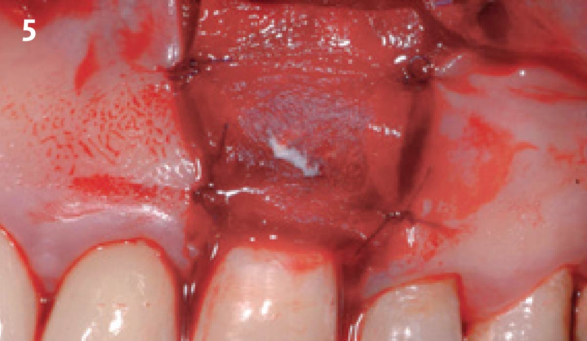
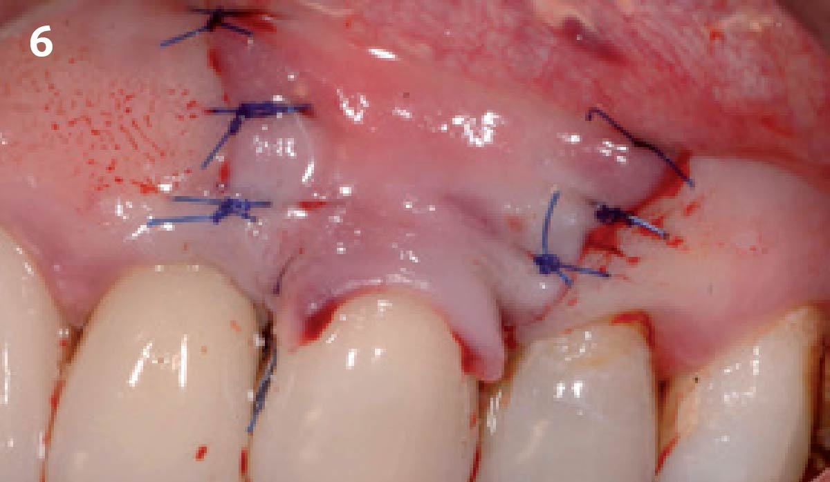
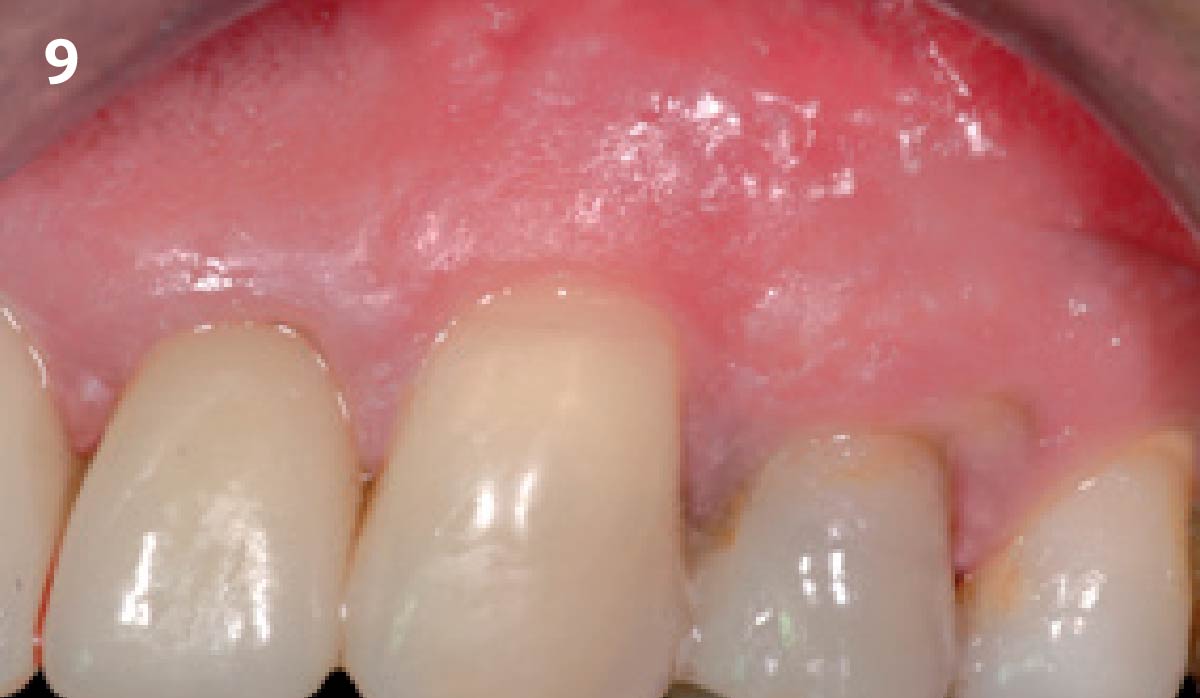
CLINICAL CASE 1 – Before preparation of the flap the exposed root portion is cleaned with a scraper and is wiped with EDTA (or similar). 2 – After measuring the dimension of the recession defect using a periodontal probe, the incisions for raising the flap are cut. 3 – A split-full-split thickness flap is elevated and coronally mobilized. 4 – The area of the papillae is de-epithelialized to allow anchorage of the flap coronal to the cemento-enamel junction. 5 – Geistlich Mucograft®is applied dry to the defect and is fixed with 4 single sutures. 6 – The coronally advanced flap is sutured over Geistlich Mucograft®. 7 – Nice, uneventful healing 15 days post-operatively at suture removal. 8 – Soft tissue situation immediately after suture removal. 9 – Complete root coverage 7 months after surgery. Note the excellent color match.