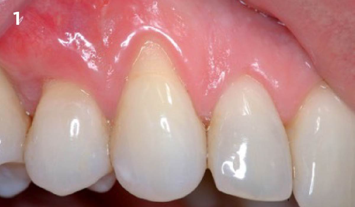
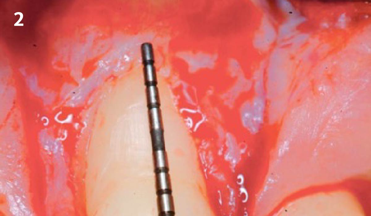
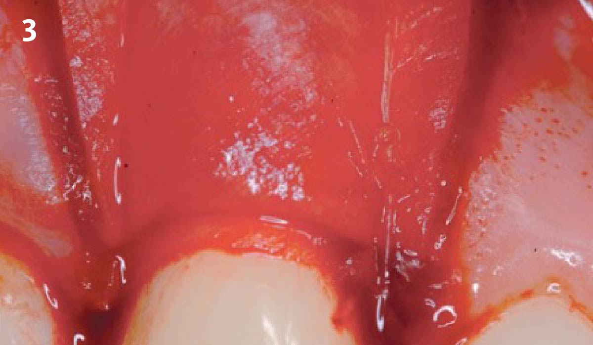
CLINICAL CASE
New Product! Buy 5 Geistlich Bio-Gide® Forte, Get 1 Free! Use code FORTE. Check it out!
Mix & Match! Buy 5 Products, Get 1 Free! Use code B5G1. Get Started!

NEW EBOOK: How to Master Bone Regeneration with Digital Innovation. Download Today!
GEM 21S®, the first recombinant growth factor product for use in oral regenerative surgery. NEW EBOOK!


