Buy products 5, get 1 free. Use code b5g1. Check it out!
Clinical Resources
Use the search and filter form below to find informative clinical content by format, therapeutic areas, or product.

FEATURED RESOURCE
BIOBRIEF
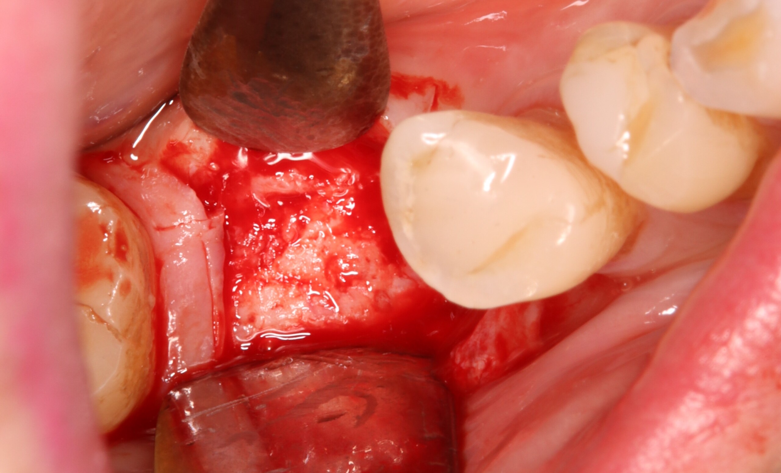
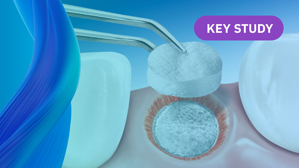
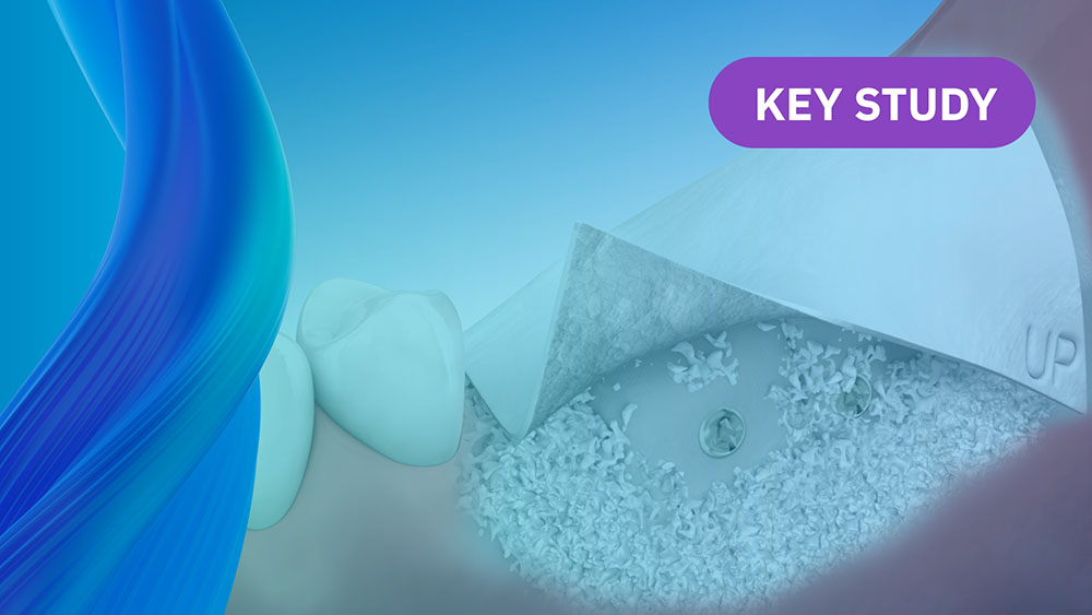
Alveolar Ridge Preservation with vallos® Mineralized Cortico-Cancellous Allograft
Hanae Saito, DDS, MS, CCRC
, Andrew Tong, DDS
On-Demand Webinars
Learn on your own Time and Earn CE
Geistlich is committed to educating our clinician partners in regenerative dentistry through in-person and virtual sessions. We look forward to growing with you on this journey.
Randomized controlled clinical study comparing a volume-stable collagen matrix to autogenous connective tissue grafts for soft tissue augmentation at implant sites: linear volumetric soft tissue changes up to 3 months
Randomized controlled clinical study evaluating effectiveness and safety of a volume-stable collagen matrix compared to autogenous connective tissue grafts for soft tissue augmentation at implant sites
Randomized, controlled clinical trial to evaluate a xenogeneic collagen matrix as an alternative to free gingival grafting for oral soft tissue augmentation: 6 to 8 year follow-up
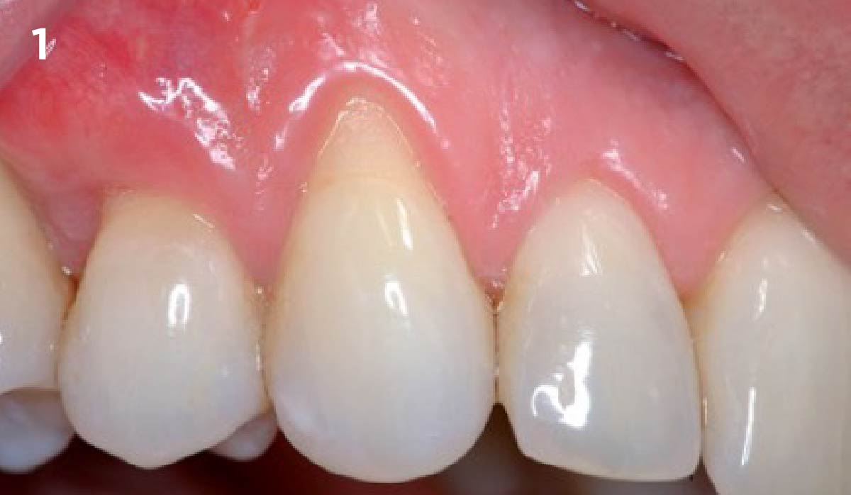
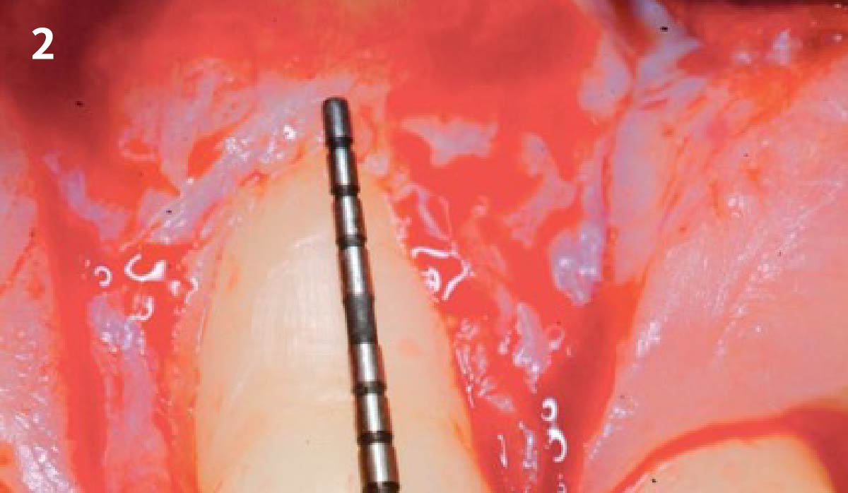
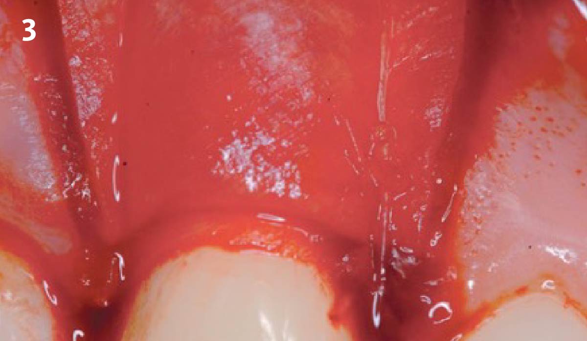
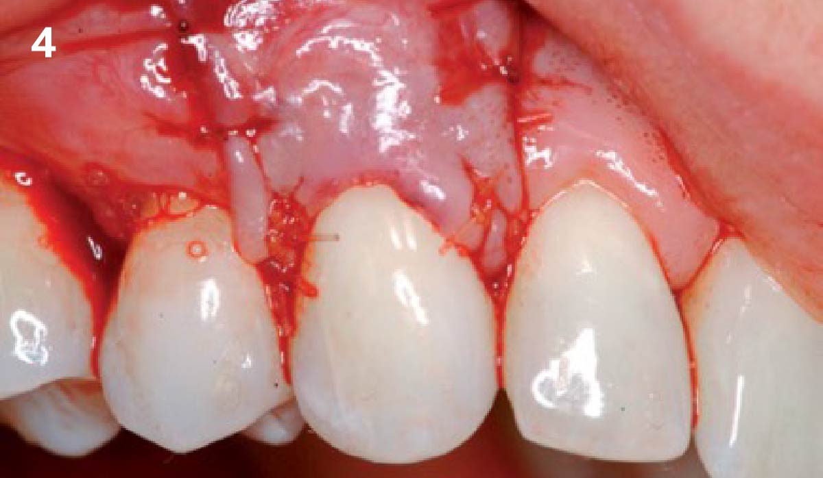
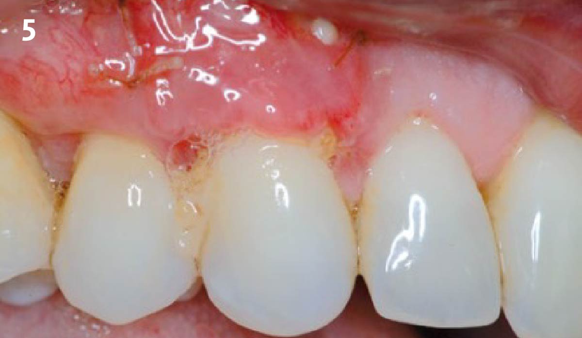
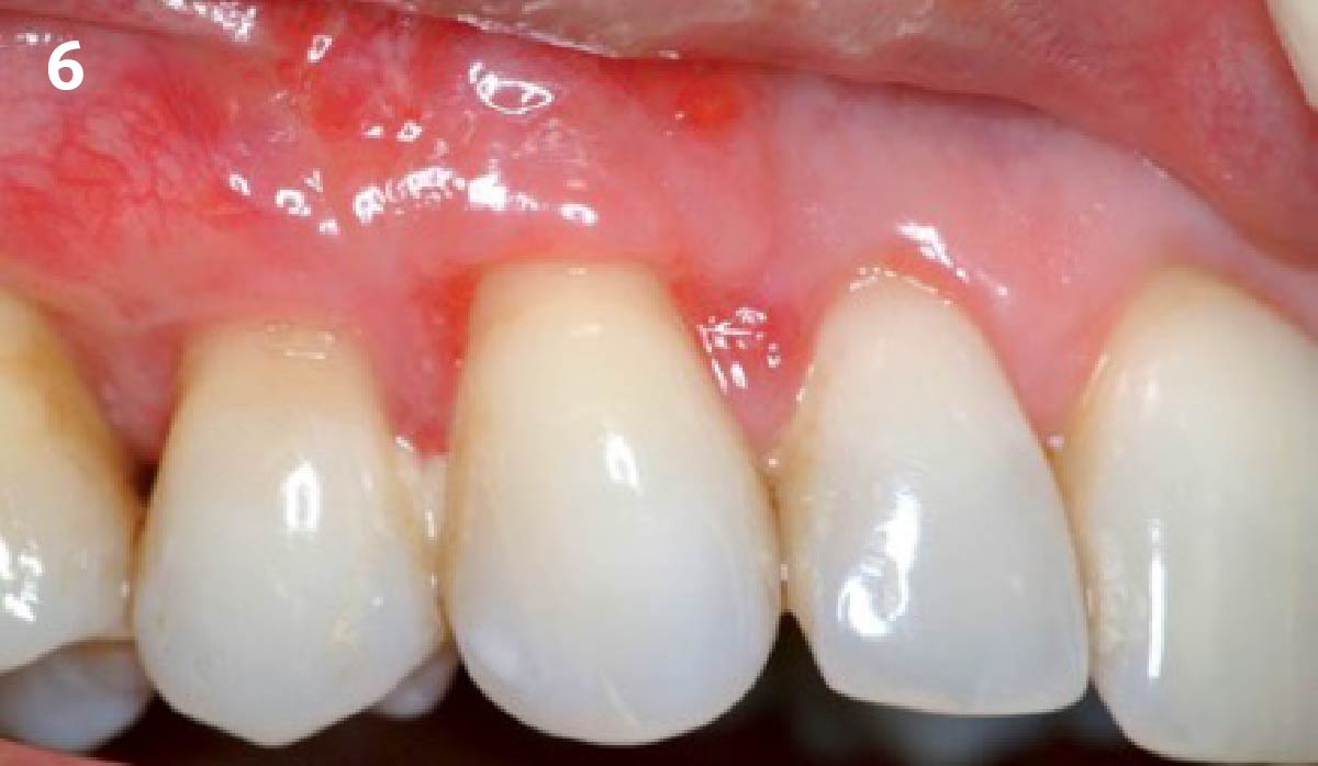
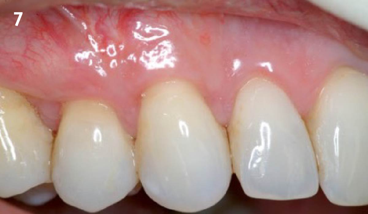
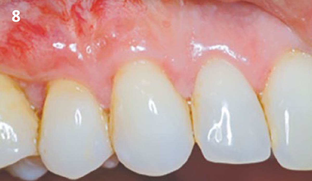
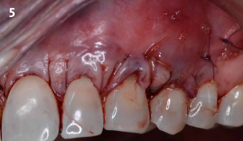
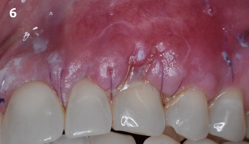
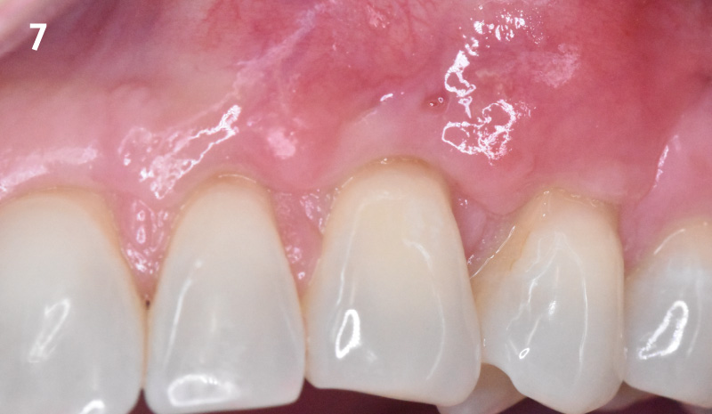
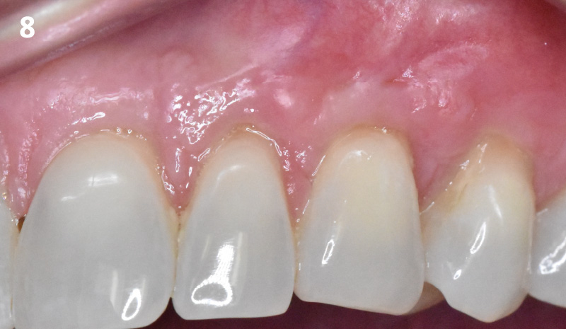
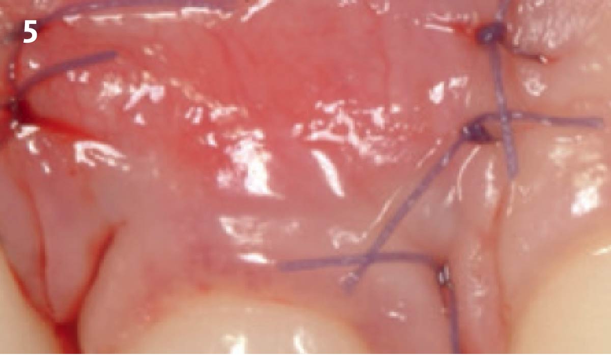
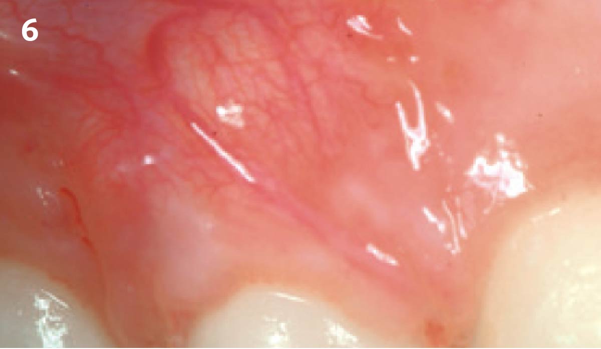
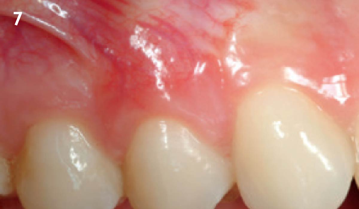
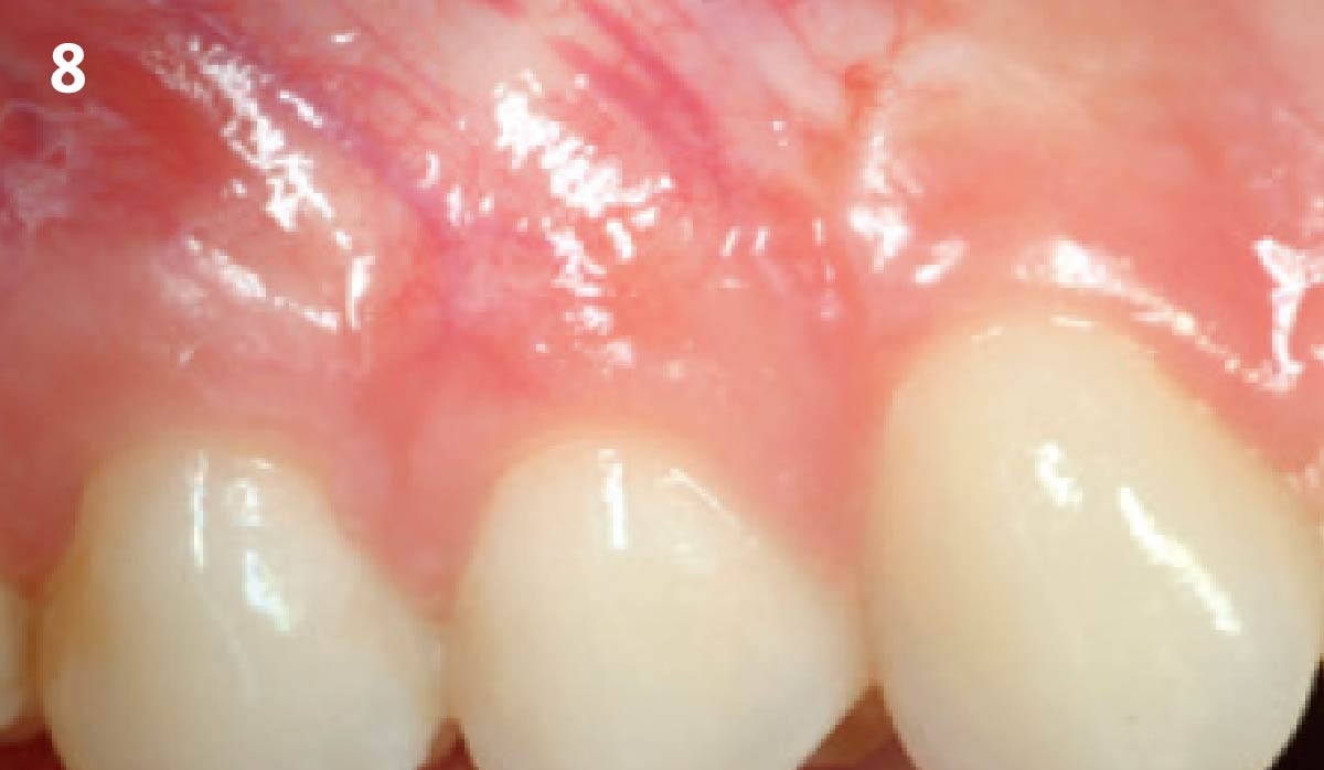
1 - Pre-operative image showing the recession defect on tooth #6. 2 - After elevation of a partial thickness flap, the interdental papillae are de-epithelialized. 3 - Geistlich Mucograft® is placed over the defect and sutured to the papillae. 4 - The flap is coronally advanced and sutured completely covering the matrix. 5 - Follow-up after 1 week: healing of the surgical site. 6 - Follow-up situation after 4 weeks. 7 - Follow-up after 3 months. 8 - Follow-up after 6 months: note the natural appearance of the soft tissue achieved with Geistlich Mucograft®. 9 - Follow-up after 1 year.
Regenerative materials in site preparation for implant placement: A clinically validated histological perspective
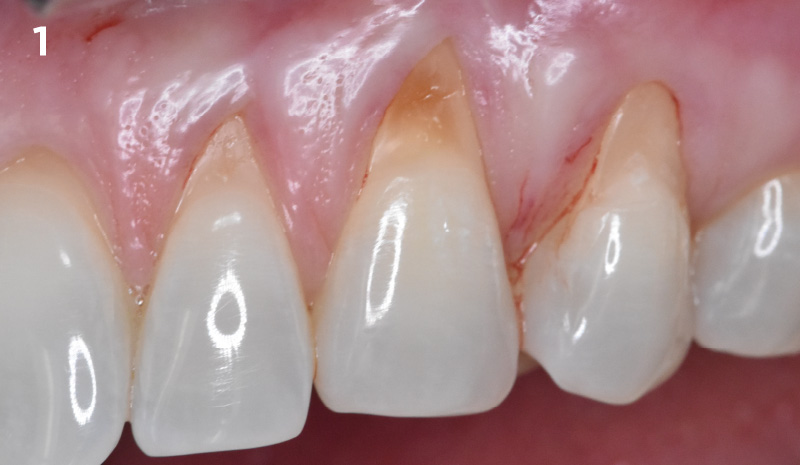
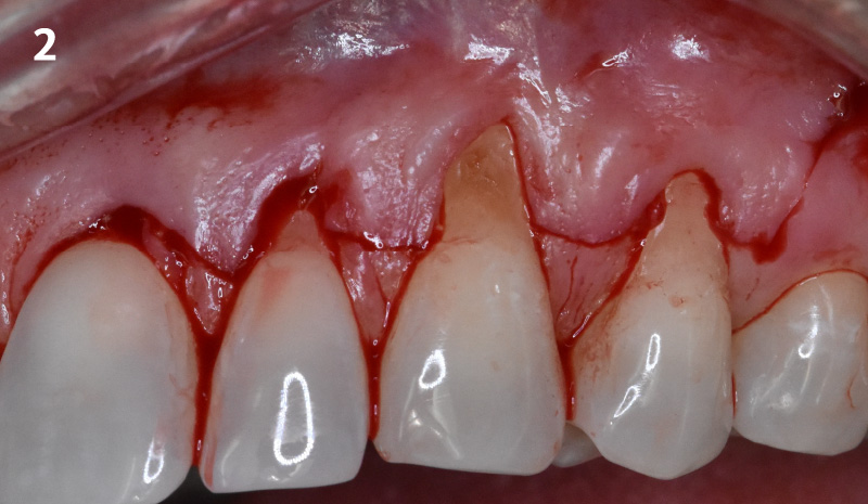
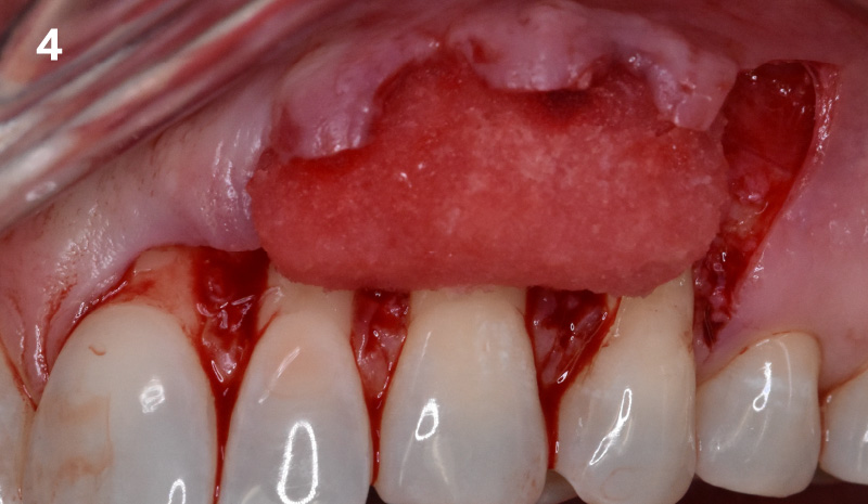
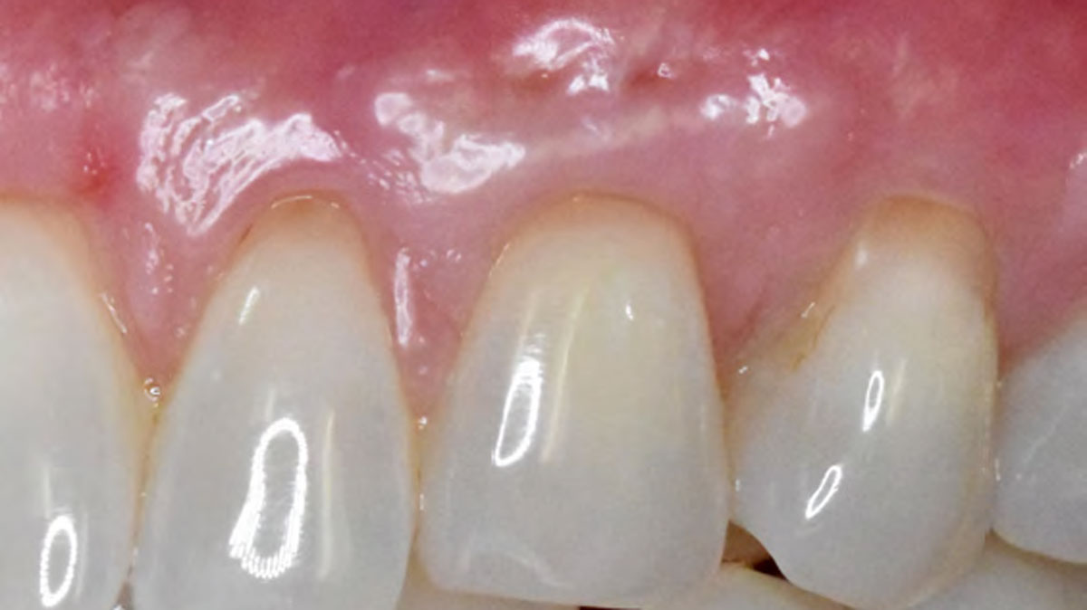
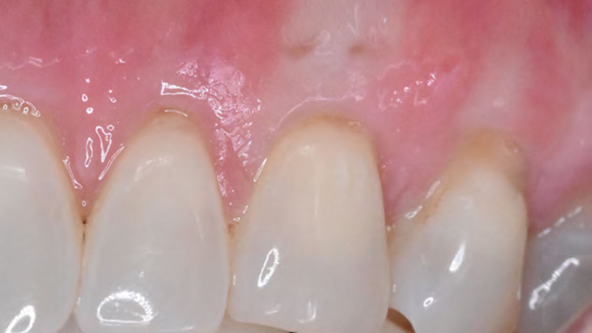
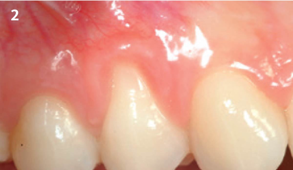
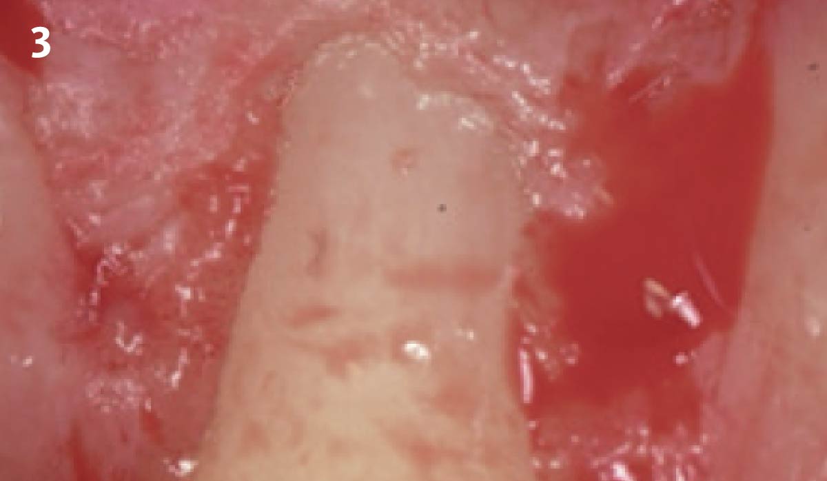
Pre-operative condition: Note that the gingival recession of 3-4 mm is evident as is the cervical root abrasions. The interdental papillae completely fills the embrasure space. Incision design showing the sulcular incisions with horizontal incisions across the interdental regions ending with a remote oblique vertical releasing incision distal to the first bicuspid tooth.The interdental papillae were de-epithelialized and Geistlich Fibro-Gide®was placed over the exposed roots extending onto the bone. Geistlich Fibro-Gide® was not secured with sutures. Internal periosteal releasing incision was made to allow tension-free coronal advancement of the buccal flap to completely cover Geistlich Fibro-Gide®.The flap was secured with 5-0 Monocryl® sutures. 1-week post-operative visit: the healing looks good and sutures are intact. There was a small soft-tissue dehiscence at the buccal margin of the canine tooth. Healing progressed well at 2 months post-operatively and the dehiscence defect seen at 1 week appears to be healing. Soft-tissue thickness is also evident at this stage. At 6 months, 100% root coverage has been achieved. Note the increase in keratinized gingiva at the canine tooth where there was previously delayed healing. The patient is happy with the esthetic and functional outcome. At 1 year, we can see root coverage
has been sustained. Complete root coverage is not seen for the upper left bicuspid, not surprising given the tooth had an older restoration which was removed prior to grafting and the CEJ on the proximal surface is visible. Partial coverage was achieved however and is much more pleasing to the patient.At 1.5 years, the tissue looks stable, healthy and esthetic. The patient is very happy with results thus far both from esthetic and functional standpoints. She is still free of sensitivity.
Vinay Bhide, DDS, MSc, FRCD(C)
Aurora, Ontario
Root Coverage for Multiple Adjacent Teeth in the Maxilla with Geistlich Fibro-Gide®
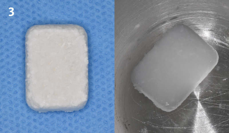
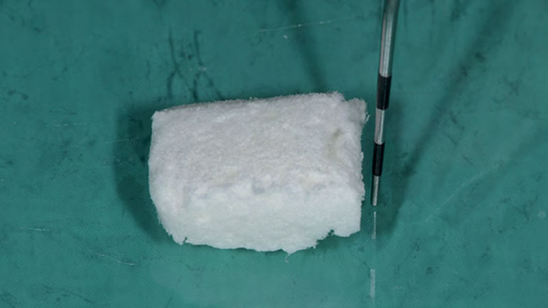
Pre-operative condition: Note that the gingival recession of 3-4 mm is evident as is the cervical root abrasions. The interdental papillae completely fills the embrasure space. Incision design showing the sulcular incisions with horizontal incisions across the interdental regions ending with a remote oblique vertical releasing incision distal to the first bicuspid tooth.Geistlich Fibro-Gide® is trimmed in a dry state to 10 x 15 x 6mm. The corners were trimmed for better adaptation and the matrix was then hydrated in sterile saline solution.The interdental papillae were de-epithelialized and Geistlich Fibro-Gide®was placed over the exposed roots extending onto the bone. Geistlich Fibro-Gide® was not secured with sutures. Internal periosteal releasing incision was made to allow tension-free coronal advancement of the buccal flap to completely cover Geistlich Fibro-Gide®.The flap was secured with 5-0 Monocryl® sutures. 1-week post-operative visit: the healing looks good and sutures are intact. There was a small soft-tissue dehiscence at the buccal margin of the canine tooth. Healing progressed well at 2 months post-operatively and the dehiscence defect seen at 1 week appears to be healing. Soft-tissue thickness is also evident at this stage. At 6 months, 100% root coverage has been achieved. Note the increase in keratinized gingiva at the canine tooth where there was previously delayed healing. The patient is happy with the esthetic and functional outcome.
Pier Gallo, DDS, PhD
Bogotá, Colombia
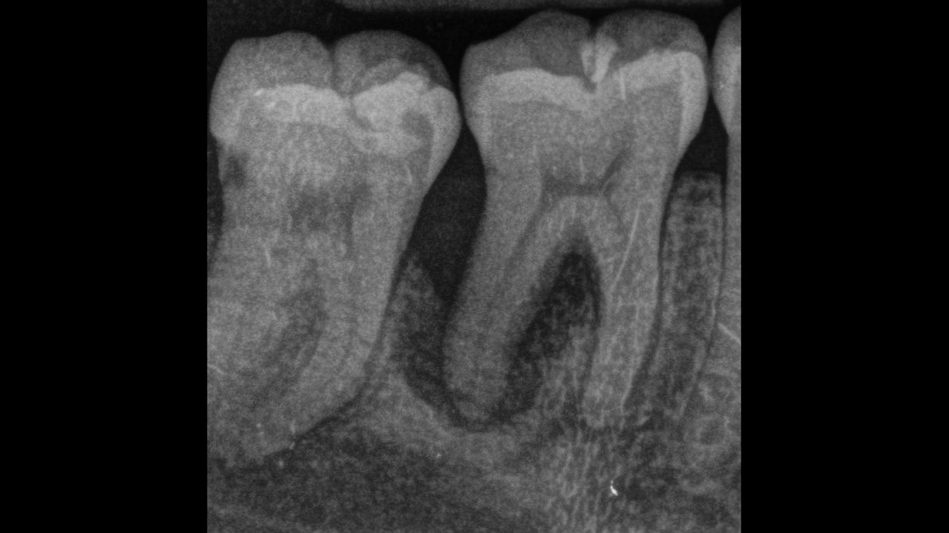
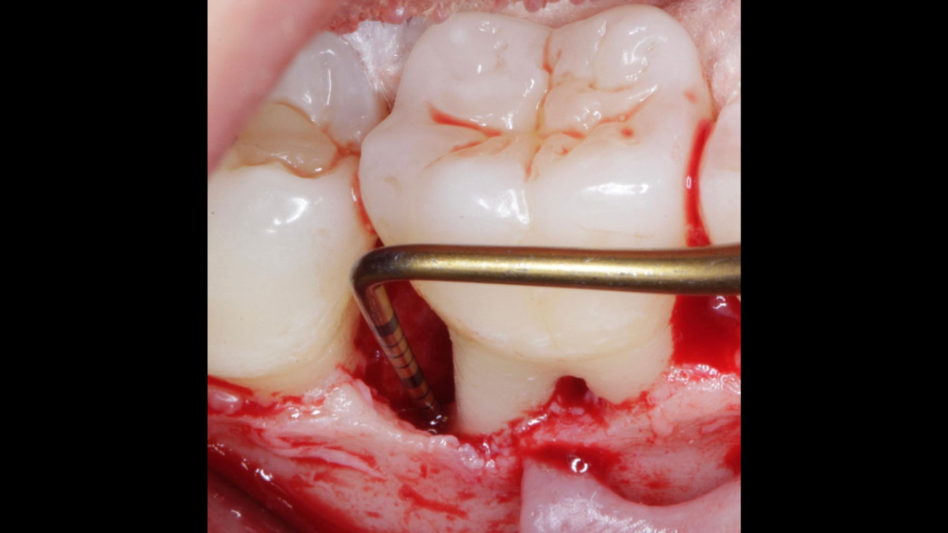
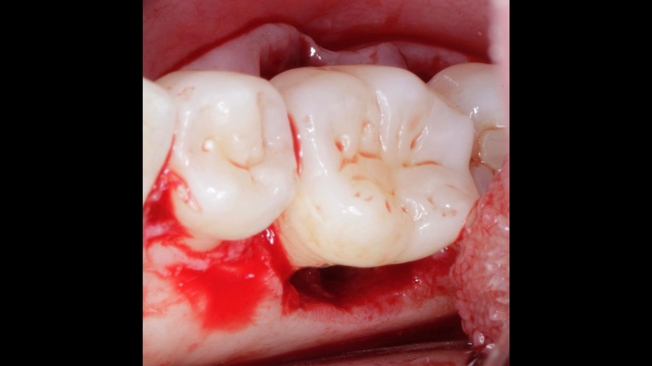
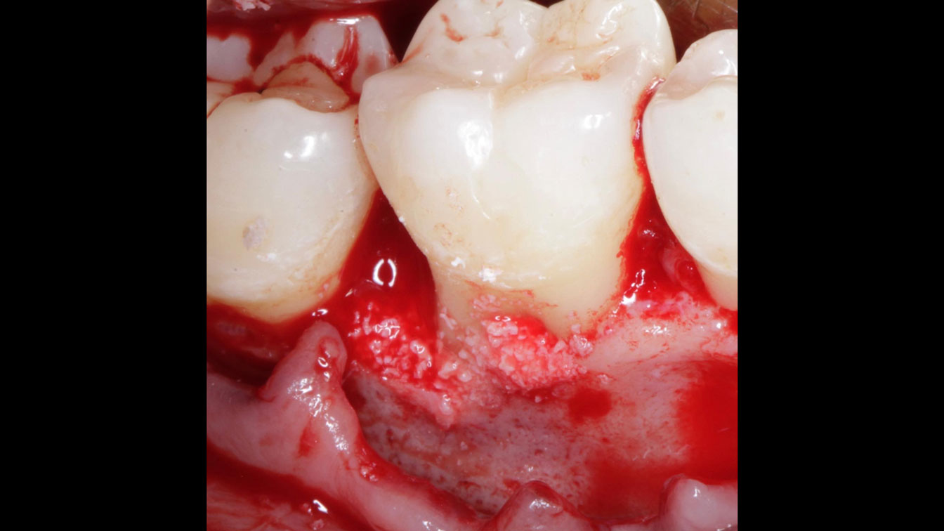
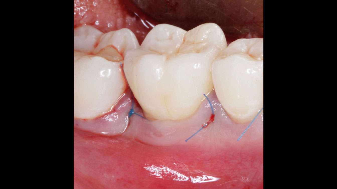
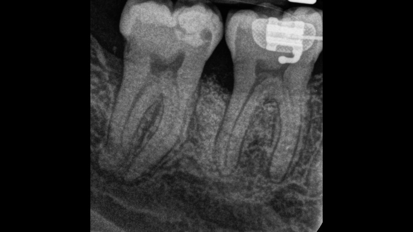
Saving a Hopeless Molar with Grade II Furcation Involvement with rhPDGF-BB
Baseline RadiographFlap reflection showing the defect tooth #30
Defect dimensions from the lingual aspect
Defect filled and graft is extended beyond margins, 1-2mm on the buccal aspect
Suturing with 5/0 polypropylene sutures. Primary closure is attained.
Radiograph 1-year post-op showing almost complete bone fill
Prof. Dr. Giovanni Zucchelli
Bologna, Italy
Single recession coverage with coronally advanced flap in thin biotype
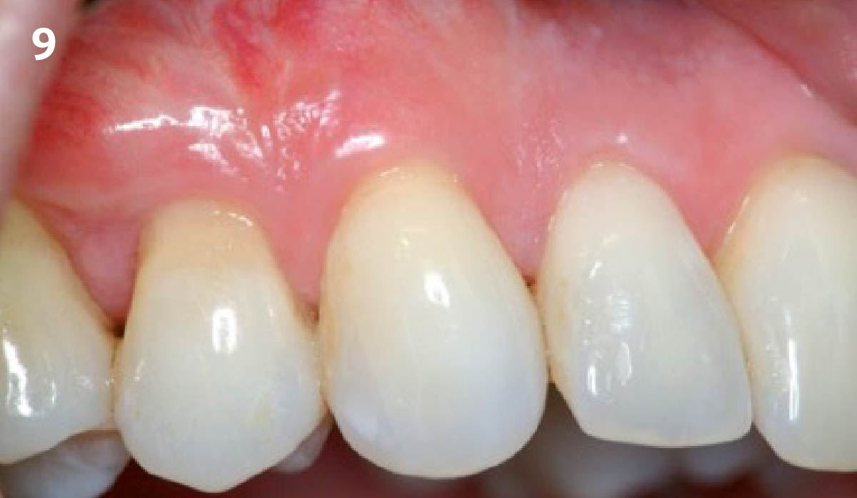
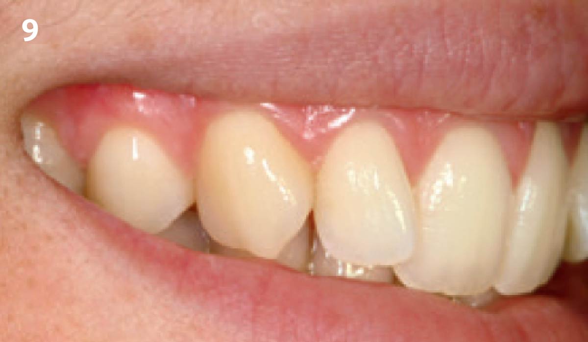
1 - Pre-operative lateral smile showing the recession defect of tooth 5. 2 - Pre-operative image of recession defect (tooth 5). 3 - After elevation of split-full-split flap the interdental papillae are de-epithelialized. 4 - Geistlich Mucograft® is placed over the root and sutured to the papillae5 - The flap is mobilized, coronally advanced and sutured completely covering the Geistlich Mucograft®. 6 - Healing of the surgical site 2 weeks after surgery. 7 - Surgical site 6 months after surgery. 8 - Outcome 1 year after treatment. 9 - Lateral smile 1 year after surgery showing the optimal esthetic outcome.
Dr. Tomas Linkevičius
Soft Tissue Management to Prevent Tissue Loss / Prevent Complications
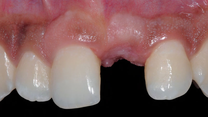
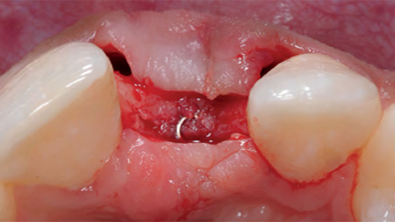
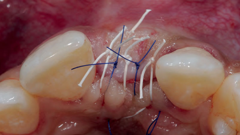
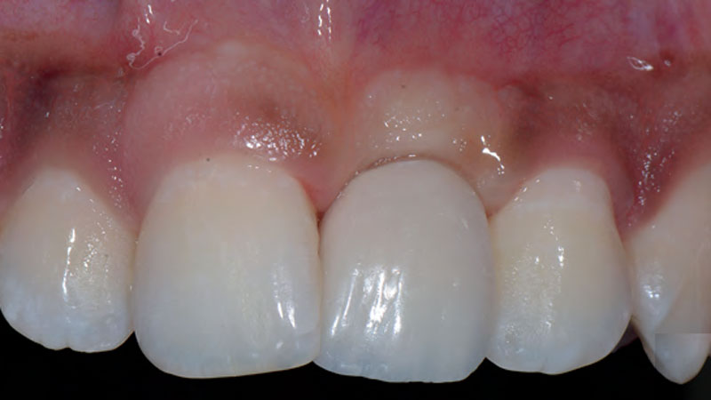
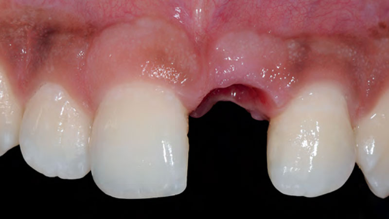
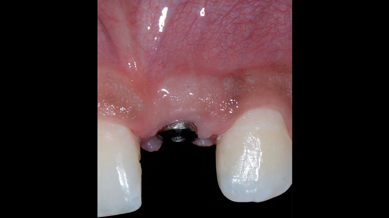
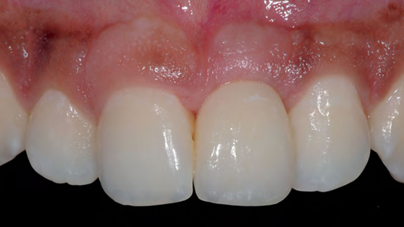
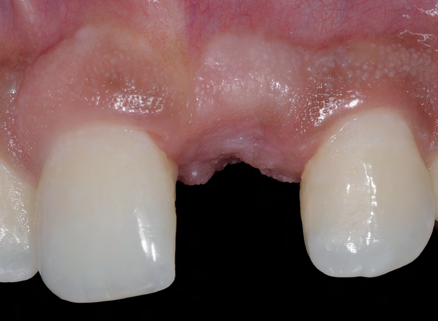
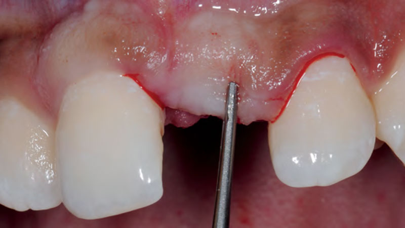
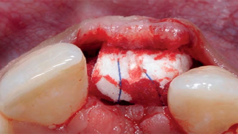
A young male patient was referred to the clinic with a missing central incisor, #9 following trauma.Preparation of a split-thickness flap (buccal pouch).Due to releasing incisions within the periosteum, the tissues can be advanced more coronally.The dimension and shape of Geistlich
Fibro-Gide® with a maximal thickness
(5mm) at the transition between the
buccal and occlusal aspect.Geistlich Fibro-Gide® inserted and immobilized with a horizontal cross-suture to the palatal flap.Primary wound closure.A provisional reconstruction is inserted; blanching of the tissues can be observed.Final emergence profile established
with a provisional reconstruction.8 weeks healing: Abutment connection is performedThe clinical situation at 1-year follow-up.
Stability of Root Coverage Outcomes After Soft-Tissue Augmentation With a Collagen Matrix With or Without rhPDGF-BB: A 3-Year Triple-Blinded, Randomised, Placebo-Controlled Trial